STED microscopy
STED microscopy
Main page
1806687

STED microscopy

logo
Community Hub0 subscribers
Read side by side
from Wikipedia
Stimulated emission depletion (STED) microscopy provides significant resolution improvements over those possible with Confocal microscopy.

Stimulated emission depletion (STED) microscopy is one of the techniques that make up super-resolution microscopy. It creates super-resolution images by the selective deactivation of fluorophores, minimizing the area of illumination at the focal point, and thus enhancing the achievable resolution for a given system.[1] It was developed by Stefan W. Hell and Jan Wichmann in 1994,[2] and was first experimentally demonstrated by Hell and Thomas Klar in 1999.[3] Hell was awarded the Nobel Prize in Chemistry in 2014 for its development. In 1986, V.A. Okhonin[4] (Institute of Biophysics, USSR Academy of Sciences, Siberian Branch, Krasnoyarsk) had patented the STED idea.[5] This patent was unknown to Hell and Wichmann in 1994.

STED microscopy is one of several types of super resolution microscopy techniques that have recently been developed to bypass the diffraction limit of light microscopy to increase resolution. STED is a deterministic functional technique that exploits the non-linear response of fluorophores commonly used to label biological samples in order to achieve an improvement in resolution, that is to say STED allows for images to be taken at resolutions below the diffraction limit. This differs from the stochastic functional techniques such as photoactivated localization microscopy (PALM) and stochastic optical reconstruction microscopy (STORM) as these methods use mathematical models to reconstruct a sub diffraction limit from many sets of diffraction limited images.

Background

[edit]
Ernst Abbe's formula for the diffraction limit, set in stone at a monument in Jena.
Jablonski diagram showing the redshift of the stimulated photon. This redshift allows the stimulated photon to be ignored.
Diagram of the design of a STED device. The double laser design allows for excitation and stimulated emission to be used together for STED.

In traditional microscopy, the resolution that can be obtained is limited by the diffraction of light. Ernst Abbe developed an equation to describe this limit. The equation is:
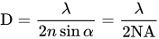
where D is the diffraction limit, λ is the wavelength of the light, and NA is the numerical aperture, or the refractive index of the medium multiplied by the sine of the angle of incidence. n describes the refractive index of the specimen, α measures the solid half‐angle from which light is gathered by an objective, λ is the wavelength of light used to excite the specimen, and NA is the numerical aperture. To obtain high resolution (i.e. small D values), short wavelengths and high NA values (NA = n sinα) are optimal.[6] This diffraction limit is the standard by which all super resolution methods are measured. Because STED selectively deactivates the fluorescence, it can achieve resolution better than traditional confocal microscopy. Normal fluorescence occurs by exciting an electron from the ground state into an excited electronic state of a different fundamental energy level (S0 goes to S1) which, after relaxing back to the vibrational ground state (of S1), emits a photon by dropping from S1 to a vibrational energy level on S0. STED interrupts this process before the photon is released. The excited electron is forced to relax into a higher vibration state than the fluorescence transition would enter, causing the photon to be released to be red-shifted as shown in the image to the right.[7] Because the electron is going to a higher vibrational state, the energy difference of the two states is lower than the normal fluorescence difference. This lowering of energy raises the wavelength, and causes the photon to be shifted farther into the red end of the spectrum. This shift differentiates the two types of photons, and allows the stimulated photon to be ignored.

To force this alternative emission to occur, an incident photon must strike the fluorophore. This need to be struck by an incident photon has two implications for STED. First, the number of incident photons directly impacts the efficiency of this emission, and, secondly, with sufficiently large numbers of photons fluorescence can be completely suppressed.[8] To achieve the large number of incident photons needed to suppress fluorescence, the laser used to generate the photons must be of a high intensity. Unfortunately, this high intensity laser can lead to the issue of photobleaching the fluorophore. Photobleaching is the name for the destruction of fluorophores by high intensity light.

Process

[edit]
Comparison of confocal microscopy and STED microscopy. This shows the improved resolution of STED microscopy over traditional techniques.
Excitation spot (2D, left), doughnut-shape de-excitation spot (center) and remaining area allowing fluorescence (right).

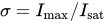
STED functions by depleting fluorescence in specific regions of the sample while leaving a center focal spot active to emit fluorescence. This focal area can be engineered by altering the properties of the pupil plane of the objective lens.[9][10][11] The most common early example of these diffractive optical elements, or DOEs, is a torus shape used in two-dimensional lateral confinement shown below. The red zone is depleted, while the green spot is left active. This DOE is generated by a circular polarization of the depletion laser, combined with an optical vortex. The lateral resolution of this DOE is typically between 30 and 80 nm. However, values down to 2.4 nm have been reported.[12] Using different DOEs, axial resolution on the order of 100 nm has been demonstrated.[13] A modified Abbe's equation describes this sub diffraction resolution as:
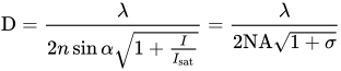
Where is the refractive index of the medium, is the intracavity intensity and is the saturation intensity. Where is the saturation factor expressing the ratio of the applied (maximum) STED intensity to the saturation intensity, .[6][14]

To optimize the effectiveness of STED, the destructive interference in the center of the focal spot needs to be as close to perfect as possible. That imposes certain constraints on the optics that can be used.

Dyes

[edit]

Early on in the development of STED, the number of dyes that could be used in the process was very limited. Rhodamine B was named in the first theoretical description of STED.[2] As a result, the first dyes used were laser emitting in the red spectrum. To allow for STED analysis of biological systems, the dyes and laser sources must be tailored to the system. This desire for better analysis of these systems has led to living cell STED and multicolor STED, but it has also demanded more and more advanced dyes and excitation systems to accommodate the increased functionality.[7]

One such advancement was the development of immunolabeled cells. These cells are STED fluorescent dyes bound to antibodies through amide bonds. The first use of this technique coupled MR-121SE, a red dye, with a secondary anti-mouse antibody.[8] Since that first application, this technique has been applied to a much wider range of dyes including green emitting, Atto 532,[15][16][17] and yellow emitting, Atto 590,[18] as well as additional red emitting dyes. In addition, Atto 647N was first used with this method to produce two-color STED.[19]

Applications

[edit]

Over the last several years, STED has developed from a complex and highly specific technique to a general fluorescence method. As a result, a number of methods have been developed to expand the utility of STED and to allow more information to be provided.

Structural analysis

[edit]

From the beginning of the process, STED has allowed fluorescence microscopy to perform tasks that had been only possible using electron microscopy. As an example, STED was used for the elucidation of protein structure analysis at a sub-organelle level. The common proof of this level of study is the observation of cytoskeletal filaments. In addition, neurofilaments, actin, and tubulin are often used to compare the resolving power of STED and confocal microscopes.[20][21][22]

Using STED, a lateral resolution of 70 – 90 nm has been achieved while examining SNAP25, a human protein that regulates membrane fusion. This observation has shown that SNAP25 forms clusters independently of the SNARE motif's functionality, and binds to clustered syntaxin.[23][24] Studies of complex organelles, like mitochondria, also benefit from STED microscopy for structural analysis. Using custom-made STED microscopes with a lateral resolution of fewer than 50 nm, mitochondrial proteins Tom20, VDAC1, and COX2 were found to distribute as nanoscale clusters.[25][26] Another study used a homemade STED microscopy and DNA binding fluorescent dye, measured lengths of DNA fragments much more precisely than conventional measurement with confocal microscopy.[27]

Correlative methods

[edit]

Due to its function, STED microscopy can often be used with other high-resolution methods. The resolution of both electron and atomic force microscopy is even better than STED resolution, but by combining atomic force with STED, Shima et al. were able to visualize the actin cytoskeleton of human ovarian cancer cells while observing changes in cell stiffness.[28]

Multicolor

[edit]

Multicolor STED was developed in response to a growing problem in using STED to study the dependency between structure and function in proteins. To study this type of complex system, at least two separate fluorophores must be used. Using two fluorescent dyes and beam pairs, colocalized imaging of synaptic and mitochondrial protein clusters is possible with a resolution down to 5 nm [18]. Multicolor STED has also been used to show that different populations of synaptic vesicle proteins do not mix of escape synaptic boutons.[29][30] By using two color STED with multi-lifetime imaging, three channel STED is possible.

Live-cell

[edit]

Early on, STED was thought to be a useful technique for working with living cells.[13] Unfortunately, the only way for cells to be studied was to label the plasma membrane with organic dyes.[29] Combining STED with fluorescence correlation spectroscopy showed that cholesterol-mediated molecular complexes trap sphingolipids, but only transiently.[31] However, only fluorescent proteins provide the ability to visualize any organelle or protein in a living cell. This method was shown to work at 50 nm lateral resolution within Citrine-tubulin expressing mammalian cells.[32][33] In addition to detecting structures in mammalian cells, STED has allowed for the visualization of clustering YFP tagged PIN proteins in the plasma membrane of plant cells.[34]

Recently, multicolor live-cell STED was performed using a pulsed far-red laser and CLIPf-tag and SNAPf-tag expression.[35]

In the brain of intact animals

[edit]

Superficial layers of mouse cortex can be repetitively imaged through a cranial window.[36] This allows following the fate and shape of individual dendritic spines for many weeks.[37] With two-color STED, it is even possible to resolve the nanostructure of the postsynaptic density in life animals.[38]

STED at video rates and beyond

[edit]

Super-resolution requires small pixels, which means more spaces to acquire from in a given sample, which leads to a longer acquisition time. However, the focal spot size is dependent on the intensity of the laser being used for depletion. As a result, this spot size can be tuned, changing the size and imaging speed. A compromise can then be reached between these two factors for each specific imaging task. Rates of 80 frames per second have been recorded, with focal spots around 60 nm.[1][39] Up to 200 frames per second can be reached for small fields of view.[40]

Problems

[edit]

Photobleaching can occur either from excitation into an even higher excited state, or from excitation in the triplet state. To prevent the excitation of an excited electron into another, higher excited state, the energy of the photon needed to trigger the alternative emission should not overlap the energy of the excitation from one excited state to another.[41] This will ensure that each laser photon that contacts the fluorophores will cause stimulated emission, and not cause the electron to be excited to another, higher energy state. Triplet states are much longer lived than singlet states, and to prevent triplet states from exciting, the time between laser pulses needs to be long enough to allow the electron to relax through another quenching method, or a chemical compound should be added to quench the triplet state.[20][42][43]

See also

[edit]

References

[edit]
[edit]
Revisions and contributorsEdit on WikipediaRead on Wikipedia
from Grokipedia
Stimulated emission depletion (STED) microscopy is a super-resolution fluorescence microscopy technique that circumvents the Abbe diffraction limit of conventional light microscopy by using a shaped depletion laser beam to suppress fluorescence emission outside a central sub-diffraction-sized region within the excitation focus, enabling nanoscale resolution in biological imaging.[1] The term "STED scan" refers to the scanning process in STED microscopy. This super-resolution fluorescence microscopy technique is primarily used in biomedical research for the nanoscale visualization of cellular structures, proteins, and organelles, particularly in fields such as neuroscience and cell biology. STED microscopy is not a standard clinical radiology imaging modality (unlike computed tomography (CT) or magnetic resonance imaging (MRI) scans) but a specialized research tool in the medical and life sciences. Developed by Stefan W. Hell and colleagues, STED microscopy was first theoretically proposed in 1994 as a method to break the diffraction barrier through coordinated excitation and stimulated emission processes, marking a foundational advance in far-field optical nanoscopy.[1] The technique gained practical implementation in the early 2000s, with experimental demonstrations achieving resolutions below 70 nm in fixed cells, and Hell received the 2014 Nobel Prize in Chemistry for this innovation alongside super-resolution developments by others.[2] In STED, a diffraction-limited excitation beam illuminates the sample, while a concentric, doughnut-shaped STED beam—typically generated using a phase mask or spatial light modulator—overlaps it and drives stimulated emission in fluorophores at wavelengths longer than the excitation but shorter than spontaneous emission, effectively silencing them everywhere except at the intensity null in the center.[3] This nonlinear optical process confines the fluorescent spot size, with resolution improving as the square root of the STED beam intensity relative to a saturation threshold, theoretically allowing unlimited refinement limited only by photophysics and sample viability.[2] Early implementations relied on pulsed lasers for high peak powers, but continuous-wave (CW-STED) variants later reduced complexity and cost while maintaining resolutions of 50-100 nm using standard fluorophores like Alexa dyes or eGFP. STED microscopy has transformed biological research by enabling visualization of subcellular structures, such as synaptic vesicles, nuclear pores, and viral proteins, at 20-50 nm lateral resolution in live cells and tissues, often integrated with confocal scanning for optical sectioning.[3] Key advantages include compatibility with existing fluorescence labeling, multicolor imaging via sequential or simultaneous depletion, and adaptability to 3D and time-resolved studies, though challenges like photobleaching and high laser intensities necessitate optimized fluorophores and sample preparation. Recent advancements, including adaptive optics for aberration correction and hybrid STED with other super-resolution methods, have extended its utility to deeper tissue imaging and dynamic processes in neuroscience and cell biology.[3]

Introduction and History

Development and Key Milestones

The concept of STED microscopy was theoretically proposed in 1994 by Stefan W. Hell and Jan Wichmann, who described a method to break the diffraction barrier in far-field fluorescence microscopy by using stimulated emission to deplete fluorescence from the periphery of the excitation spot, enabling resolutions down to 20-30 nm in principle.[1] The first experimental demonstration of STED microscopy was achieved in 2000 by Thomas A. Klar and colleagues in Hell's group at the Max Planck Institute for Biophysical Chemistry, where they imaged fixed cells, including nuclear pore complexes, with resolutions improved to approximately 100-130 nm radially and 100 nm axially, a significant enhancement over the confocal limit of around 200-500 nm.[4] In recognition of his pioneering contributions to super-resolution techniques, including the development of STED microscopy, Stefan W. Hell shared the 2014 Nobel Prize in Chemistry with Eric Betzig and William E. Moerner for advancing fluorescence microscopy beyond the diffraction limit.[5] During the early 2000s, STED microscopy saw key implementations that expanded its utility, such as high-resolution imaging of synaptic proteins in fixed neurons at 67 nm resolution in 2006, enabling visualization of molecular clustering post-exocytosis,[6] and the introduction of two-color STED in 2007, which allowed simultaneous imaging of multiple fluorophores with sub-diffraction resolution.[7] Commercial availability of STED microscopy began in 2007 through Leica Microsystems, which released the TCS STED system under license from the Max Planck Society, making super-resolution imaging accessible for routine biomedical research with resolutions below 100 nm.[8] Post-2010 milestones included advancements in speed; video-rate STED nanoscopy was first demonstrated in 2008 for living neurons at 28 frames per second with ~62 nm resolution, facilitating the study of dynamic processes like synaptic vesicle movement without compromising spatial detail,[9] with comparative studies in 2010 confirming 65 nm resolution at similar video rates.[10] Following the 2014 Nobel Prize recognition, further developments included continuous-wave STED variants for reduced phototoxicity in live imaging (from 2011) and integration of adaptive optics for deeper tissue imaging (mid-2010s), enabling resolutions below 20 nm in specialized applications by 2020.[11]

Fundamental Concepts

Fluorescence microscopy relies on the use of fluorophores, which are fluorescent molecules or probes attached to or expressed within biological specimens, to generate contrast for imaging. These fluorophores absorb photons from an excitation light source at a specific wavelength, exciting electrons from the ground state to a higher-energy singlet state. Upon relaxation to the ground state, the fluorophores emit photons at a longer wavelength, a phenomenon known as fluorescence, which is shifted relative to the excitation wavelength due to energy loss (Stokes shift). This emitted light is collected and filtered to form an image, allowing visualization of specific structures labeled with compatible fluorophores.[12][13][14] A fundamental limitation of optical microscopy, including fluorescence variants, arises from the wave nature of light, leading to diffraction that blurs fine details. In 1873, Ernst Abbe derived the theoretical resolution limit for a microscope, expressed as the minimum resolvable distance dd between two points:
d=λ2NA d = \frac{\lambda}{2 \mathrm{NA}}
where λ\lambda is the wavelength of the light used and NA is the numerical aperture of the objective lens, a measure of its light-gathering ability. For visible light (λ500\lambda \approx 500 nm) and high-NA objectives (NA 1.4\approx 1.4), this yields a lateral resolution of approximately 180–250 nm, though practical limits often extend to 200–300 nm due to factors like spherical aberration and illumination conditions. This diffraction barrier prevents the clear distinction of sub-wavelength features in conventional setups.[15][16][17] The impact of diffraction on imaging is quantitatively described by the point spread function (PSF), which represents the three-dimensional intensity distribution resulting from a point source of light passing through the optical system. For an ideal circular aperture, the PSF takes the form of an Airy disk, with a central bright spot surrounded by concentric rings of decreasing intensity. The Rayleigh criterion defines the resolution limit based on this PSF: two point sources are considered just resolvable when the peak of one Airy disk coincides with the first minimum of the other, corresponding to a separation of about 1.22λ/(2NA)1.22 \lambda / (2 \mathrm{NA}) or roughly 0.61λ/NA0.61 \lambda / \mathrm{NA}. This criterion underscores how diffraction inherently convolves the true specimen image with the PSF, limiting the fidelity of structural details below the micron scale.[18] These constraints pose significant challenges for studying nanoscale biological phenomena, such as synaptic proteins in neurons (typically 20–50 nm in size) or individual viral particles (50–150 nm), which are smaller than the diffraction limit and thus appear blurred or indistinguishable in standard fluorescence microscopy. Super-resolution techniques, including STED microscopy, have been developed to circumvent this barrier and enable visualization of such fine structures with resolutions down to tens of nanometers.[19][20][21]

Principles of Operation

Overcoming the Diffraction Limit

The diffraction limit in optical microscopy arises fundamentally from the wave nature of light, as first theoretically derived by Ernst Abbe in 1873. Abbe's analysis showed that the minimum resolvable distance dd between two point sources is given by d=λ2NAd = \frac{\lambda}{2 \mathrm{NA}}, where λ\lambda is the wavelength of light used for imaging and NA\mathrm{NA} is the numerical aperture of the objective lens, defined as NA=nsinα\mathrm{NA} = n \sin \alpha with nn the refractive index of the immersion medium and α\alpha the half-angle of the maximum cone of light accepted by the lens. This equation implies that resolution is constrained by the interplay of wavelength and optical system parameters; for visible light (λ500\lambda \approx 500 nm) and a high-NA oil-immersion objective (NA=1.4\mathrm{NA} = 1.4), the lateral resolution is approximately 180 nm, while axial resolution is roughly twice that due to weaker focusing along the optical axis.[22] These limits prevent the clear visualization of sub-wavelength features, as diffracted light from the specimen interferes constructively and destructively to form an extended image rather than a sharp point. The point spread function (PSF) quantifies this diffraction-induced blurring, representing the three-dimensional intensity distribution produced by an ideal point source in the focal plane of the microscope. In practice, the lateral PSF approximates an Airy disk with a full width at half maximum (FWHM) of about 0.51λNA0.51 \frac{\lambda}{\mathrm{NA}}, causing any sub-diffraction structure to appear convolved and enlarged in the image.[23] For biological specimens, this blurring obscures critical nanoscale details; for instance, mitochondria, with diameters of 200–500 nm, can be resolved in outline but not their inner cristae folds (often <100 nm apart), while smaller entities like synaptic vesicles (∼40–50 nm) or viral particles (20–100 nm) merge indistinguishably into diffuse spots exceeding the PSF size.[24][25] Such limitations hinder studies of cellular architecture, where many protein complexes and membrane domains operate at scales of 10–100 nm, far below the ∼200–300 nm diffraction barrier for typical fluorescence microscopy setups.[22] Super-resolution techniques overcome this barrier through distinct strategies, with targeted depletion methods like STED differing fundamentally from stochastic approaches such as STORM and PALM. In STORM and PALM, resolution emerges from precise localization of sparse, individual fluorophores activated and imaged over thousands of frames, achieving 20–50 nm precision via statistical fitting but requiring extended acquisition times and post-processing.[26] Targeted depletion, by contrast, employs deterministic optical reconfiguration of the excitation-emission process in a single scan, suppressing fluorescence from peripheral regions of the focal spot to shrink the effective PSF without relying on sparsity or temporal sampling.[27] Mathematically, in targeted depletion approaches, the effective resolution deffd_\mathrm{eff} improves as deffd1+IIsatd_\mathrm{eff} \approx \frac{d}{\sqrt{1 + \frac{I}{I_\mathrm{sat}}}}, where dd is the conventional diffraction-limited resolution, II is the intensity of the depletion beam at the periphery, and IsatI_\mathrm{sat} is the saturation intensity of the depletion process (e.g., stimulated emission).[1] This square-root dependence allows arbitrary enhancement by increasing I/IsatI/I_\mathrm{sat}, theoretically unbounded but practically limited by photobleaching and sample damage; for example, factors of 5–10 (yielding 20–50 nm resolution) are routinely achieved with visible wavelengths and standard objectives.[28] Stimulated emission serves as the primary depletion mechanism here, forcing excited fluorophores to ground-state emission outside a central nanoscale region.[1]

STED Mechanism and Resolution Enhancement

STED microscopy employs a dual-beam configuration consisting of an excitation laser that activates fluorophores within a diffraction-limited focal spot and a depletion (STED) laser that suppresses fluorescence emission around the periphery of this spot.[1] The STED beam is shaped into a doughnut-like intensity profile featuring a central zero-intensity point, typically achieved using a spiral phase mask or a spatial light modulator to impose a helical phase ramp on the beam.[1] This configuration ensures that the high-intensity ring of the STED beam overlaps with the excitation spot's edges, precisely targeting excited fluorophores for depletion while sparing those at the center.[1] The core of the STED process relies on stimulated emission, where the intense STED beam—tuned to the fluorophore's emission wavelength—induces rapid return of excited molecules from the singlet excited state to the ground state via stimulated emission, emitting a photon coherently with the STED light rather than spontaneous fluorescence.[1] This depletion effectively shrinks the region of active fluorescence emission to a sub-diffraction-sized area at the excitation focus center, as fluorophores in the outer regions are silenced before they can fluoresce.[1] By synchronizing the pulsed excitation and STED beams with appropriate delays, the process exploits the finite lifetime of the excited state to maximize depletion efficiency without bleaching.[1] The enhancement in lateral resolution arises from the nonlinear dependence of depletion on STED intensity, leading to an effective point spread function (PSF) that narrows as the STED power increases. The theoretical resolution is described by the approximate formula:
dd01+ImaxIsat d \approx \frac{d_0}{\sqrt{1 + \frac{I_{\max}}{I_{\mathrm{sat}}}}}
where d0d_0 is the diffraction-limited resolution, ImaxI_{\max} is the peak STED intensity at the doughnut ring, and IsatI_{\mathrm{sat}} is the saturation intensity required to halve the fluorescence probability.[29] Increasing ImaxI_{\max} relative to IsatI_{\mathrm{sat}} progressively confines the fluorescent region, enabling lateral resolutions of 20-50 nm or better in practice, depending on the fluorophore and optical setup.[29] For axial resolution, STED can be combined with two-photon excitation or adaptive optics to mitigate aberrations and elongate the depletion along the optical axis, achieving z-resolutions around 50 nm.[30] In two-photon STED, the quadratic excitation dependence further localizes the effective focus axially, enhancing depth penetration while maintaining super-resolution.[30] Adaptive optics corrects wavefront distortions in scattering samples, preserving the sharp depletion profile and supporting isotropic 3D imaging at these scales.[31] Variants like time-gated STED further refine resolution by collecting fluorescence only after a brief delay, exploiting the slower decay of off-axis emission under incomplete depletion to suppress background and sharpen the PSF without additional power.[32] This approach can double the effective resolution at moderate STED intensities, improving contrast in noisy environments.[32]

Instrumentation and Techniques

Optical Setup and Components

The optical setup of a STED microscope is built upon a confocal framework, incorporating specialized lasers, beam manipulation optics, and sensitive detection systems to enable super-resolution imaging. At its core, the system employs an excitation laser to illuminate the sample and a depletion (STED) laser to suppress fluorescence in the diffraction-limited periphery of the excitation spot. Excitation sources typically include continuous-wave (CW) or pulsed lasers, such as a 488 nm argon-ion laser or fiber-based pulsed laser at 485 nm for green channel imaging, delivering pulses of ~100 ps duration. The STED laser, often a Ti:sapphire oscillator tuned to 592 nm for depleting green fluorophores, operates in pulsed mode at 80 MHz repetition rate or as CW, with power levels at the sample reaching up to 100 mW to achieve effective stimulated emission while minimizing photodamage. For multicolor setups, additional depletion wavelengths like 660 nm and 775 nm are used, sourced from fiber lasers or optical parametric oscillators, allowing simultaneous imaging across spectral channels.[33][34][35] Beam shaping is critical for generating the characteristic doughnut-shaped STED profile, where intensity is zero at the center to preserve fluorescence there while depleting the surrounding ring. This is accomplished using a vortex phase plate (VPP) or spiral phase mask, which imparts a helical wavefront to the STED beam, creating a radially polarized doughnut with a dark central spot matching the excitation focus. Recent advancements include multidimensional multiplexing metalenses, which enable efficient beam shaping for STED without bulky phase plates, as demonstrated in 2025 experiments achieving enhanced super-resolution.[36] For 3D imaging, axial phase plates or deformable mirrors extend the depletion into a bottle-shaped profile along the z-axis. In thick or aberrating samples, such as biological tissues, adaptive optics systems—employing spatial light modulators (SLMs) or deformable mirrors—correct wavefront distortions, restoring resolution depths up to 100 μm by pre-compensating spherical and other aberrations in the depletion path. Beam alignment and delivery occur via polarizing beam splitters, dichroic mirrors, and fiber couplers to co-align excitation and STED paths with minimal temporal overlap for pulsed systems.[34][33][31] Detection in STED microscopy relies on confocal pinhole-based scanning to reject out-of-focus light, integrated with high-sensitivity photodetectors for efficient signal collection. Photomultiplier tubes (PMTs) or hybrid detectors are commonly used for standard channels, while avalanche photodiodes (APDs) enable photon-counting in low-light conditions, particularly for far-red emissions. Time-gating electronics synchronize detection to the fluorescence decay, capturing only the tail after the STED pulse to further enhance resolution by reducing background. Scanning is achieved via galvanometer mirrors for precise 2D rastering or resonant scanners operating at 8-16 kHz for faster frame rates up to 100 μm fields of view; motorized stages support 3D volumetric imaging over larger areas. These components ensure effective separation of the effective point spread function from the diffraction-limited excitation.[33][34] Commercial STED systems integrate these elements into user-friendly platforms, with Leica Microsystems' TCS SP8 STED featuring a white-light supercontinuum laser for tunable excitation (470-670 nm), multiple depletion lines (592, 660, 775 nm), and hybrid detectors for multicolor 3D super-resolution down to 30 nm. Abberior Instruments' setups, such as the EXPERT Line, employ fiber-coupled lasers and adaptive optics modules for flexible beam delivery and aberration correction, supporting resolutions below 50 nm in live samples. These systems often include automated alignment and software for seamless confocal-to-STED switching.[37][38] Recent hardware advances from 2023 to 2025 emphasize compactness and multicolor efficiency, exemplified by Abberior's STEDYCON, a shoebox-sized unit that routes all beams through a single fiber for inherent alignment and portability, enabling 30 nm resolution on existing widefield frames without extensive retrofitting. Innovations in multicolour beam combiners, such as integrated fiber-optic multiplexers in commercial platforms, facilitate simultaneous depletion across three or more channels with minimal crosstalk, improving throughput for live-cell applications. Additionally, photon-efficient adaptive optics using SLMs have reduced sample exposure during aberration correction, extending usable depths in tissues while preserving fluorophore integrity. A notable 2025 development is the MIRAVA Polyscope, which integrates 3D STED with 2D MINFLUX for single-molecule tracking at resolutions below 10 nm, enhancing dynamic imaging capabilities. These developments, driven by modular fiber architectures, have made STED more accessible for routine biomedical use.[39][40][41][42]

Fluorophores, Dyes, and Labeling Strategies

STED-compatible fluorophores must exhibit high photostability to endure the intense depletion laser powers without rapid bleaching, a large Stokes shift to minimize spectral overlap between excitation, emission, and depletion wavelengths, and low accumulation in the triplet state to avoid unwanted absorption of the STED beam that could lead to photodamage or inefficient depletion.[43][44][45] Traditional organic dyes widely used in STED microscopy include rhodamine derivatives such as Atto 532, Atto 590, and Atto 647N, which demonstrate effective off-switching at depletion intensities around 10^5 to 10^6 W/cm², enabling resolutions below 50 nm with minimal bleaching.[46][47] The Alexa Fluor series, particularly Alexa Fluor 488 and 647, also serve as reliable alternatives due to their robust photostability and compatibility with standard STED setups for multicolor imaging.[48] For site-specific labeling in live cells, self-labeling protein tags like SNAP-tag and CLIP-tag are fused to target proteins and conjugated with synthetic STED-compatible dyes, allowing precise and flexible attachment without genetic modification of the target itself.[49][50] Fluorescent proteins such as reversibly switchable EGFP2 (rsEGFP2) provide a genetic encoding option for live-cell STED, offering fast on-off switching kinetics suitable for dynamic imaging with resolutions approaching 60 nm.[51][52] Recent innovations from 2023 to 2025 have expanded STED probe options, including fluorescent nanoparticles like quantum dots, which deliver brighter signals and superior photostability compared to organic dyes, facilitating longer imaging sessions in complex biological samples.[53] Nanographenes have emerged as photostable alternatives that reduce bleaching by enabling fluorescence recovery under STED illumination, supporting extended high-resolution observations.[54][55] Reactivatable dyes such as DBOV-Mes, designed for ReSTED (reversible STED), allow multiple imaging cycles by reversing photodeactivation with the depletion beam, enhancing signal recovery and overall experiment throughput.[56] In November 2025, a tailored BODIPY scaffold was reported, optimized through synthesis for superior STED performance with enhanced photostability and minimal bleaching.[57] Labeling strategies in STED microscopy often employ antibody conjugates with STED dyes for specific targeting of cellular structures, ensuring high labeling efficiency while maintaining nanoscale precision.[58] Click chemistry enables multicolor setups by site-specifically attaching multiple fluorophores via bioorthogonal reactions, such as tetrazine ligation, which minimizes crosstalk and supports simultaneous imaging of diverse targets.[59][60]

Applications

Structural and Cellular Imaging

STED microscopy has enabled the visualization of protein complexes in fixed cellular structures at resolutions beyond the diffraction limit, providing insights into their nanoscale organization. For instance, in neuroendocrine cells, STED imaging of t-SNARE proteins such as syntaxin and SNAP-25 in plasma membrane sheets resolved clusters approximately 50 nm in size, each containing 30–40 syntaxin and 40–50 SNAP-25 molecules, revealing two distinct conformational states influenced by lipid order. This ~70–90 nm resolution demonstrated segregated nanodomains within these clusters, where SNAP-25's second helix engagement varied, offering evidence of lipid-patterned protein conformations essential for membrane fusion processes.[61] Organelle structures in fixed samples have also been elucidated with STED, highlighting internal architectures previously obscured by conventional microscopy. Mitochondrial cristae, the folded inner membrane compartments, were imaged at ~30 nm isotropic resolution in intact fixed cells, uncovering heterogeneous arrangements within 200–400 nm diameter tubules and emphasizing the organelle's structural variability. Similarly, nuclear pore complexes (NPCs), which span the nuclear envelope, were resolved using STED at separations below 25 nm, allowing visualization of the ring-like arrangement of nucleoporins like Nup93 and Nup98 in fixed Xenopus laevis cells, with individual features at ~15–20 nm full width at half maximum. These observations confirmed the eightfold symmetry and central channel details, aiding understanding of nuclear transport machinery.[62][63] In tissue sections, STED facilitates detailed analysis of synaptic components in fixed brain slices. Synaptic vesicles labeled with vesicular glutamate transporter 1 (VGluT1) were resolved at ~40–61 nm full width at half maximum in aldehyde-fixed rat brainstem slices from the calyx of Held, distinguishing vesicle pools and their association with proteins like synapsin. This approach revealed VGluT1-positive domains lacking synapsin, indicating compartmentalized vesicle organization within presynaptic terminals. Correlative light-electron microscopy (CLEM) integrates STED's molecular specificity with electron microscopy's ultrastructural detail in fixed samples; by overlaying STED fluorescence images of labeled synaptic proteins (e.g., dense projection components at 35–65 nm resolution) onto EM sections, researchers correlate protein localization with membrane and organelle morphology, enhancing interpretation of fixed neuronal architectures.[64][65] Quantitative metrics in STED further refine structural insights from fixed samples, enabling precise assessment of molecular relationships. Colocalization analysis, using Pearson's correlation coefficient (r) and overlap coefficient, was applied to two-color STED images of hexokinase-I and voltage-dependent anion channel isoforms in fixed human osteosarcoma cells at ~40 nm resolution, yielding r values of 0.45–0.71 to quantify differential associations and identify non-colocalizing protein fractions. Distance measurements between labeled features, such as cluster separations of 40–90 nm, provide nanoscale spatial data, supporting models of protein interactions in static cellular contexts without relying on diffraction-limited approximations.[66]

Live-Cell and Dynamic Imaging

STED microscopy has revolutionized the observation of dynamic processes in living cells by enabling super-resolution imaging at video rates, capturing events that were previously blurred by diffraction limits. Early demonstrations achieved video-rate imaging at 28 frames per second with 62 nm resolution, allowing real-time tracking of synaptic vesicle movement in neurons. Subsequent advancements pushed frame rates to 80-200 per second at approximately 50 nm resolution using low-power pulsed lasers, facilitating the study of rapid cellular dynamics without excessive phototoxicity. These capabilities stem from optimized scanning units and depletion beam configurations that maintain high temporal resolution over small fields of view. In live-cell applications, STED excels at tracking intracellular transport and structural rearrangements, such as vesicle trafficking along cytoskeletal elements and actin filament dynamics. For instance, synaptic vesicles have been visualized moving at speeds up to several micrometers per second with sub-100 nm precision, revealing pathways obscured in conventional microscopy. Similarly, cytoskeletal rearrangements, including actin filament polymerization and depolymerization, have been resolved at 60 nm in living neurons, providing insights into motility and force generation during processes like synapse formation. Fluorophore stability is crucial here, requiring dyes with high photostability to withstand repeated excitation cycles in dynamic environments. Recent advances from 2023 to 2025 have extended STED's utility for long-term imaging of organelle dynamics while minimizing cellular stress. In 2024, low-intensity STED protocols combined with neural network-based image restoration enabled over 7 hours of second-scale imaging of endoplasmic reticulum (ER) nano-structural changes in living cells, with reduced phototoxicity allowing observation of ER sheet-to-tubule transitions. Likewise, multi-color STED in 2023 resolved sub-mitochondrial protein distributions in live mitochondria at sub-100 nm, distinguishing cristae-specific localizations of respiratory chain components without fixation artifacts. These developments highlight STED's growing role in dissecting spatiotemporal organelle behaviors. To sustain imaging quality over time, techniques for photobleaching mitigation are integral to live-cell STED. Time-gated detection suppresses background fluorescence from incomplete depletion, effectively boosting resolution and signal-to-noise ratio while reducing overall laser exposure and bleaching rates. Adaptive illumination strategies, such as DyMIN (dynamic intensity minimum), further limit photobleaching by modulating depletion power based on local fluorophore density, enabling prolonged time-lapse sequences with minimal sample damage. For volumetric imaging in live cells, STED achieves 3D super-resolution up to 1 μm in depth through aberration correction, compensating for refractive index mismatches that degrade focal quality. Adaptive optics or spatial light modulators adjust wavefront distortions in real-time, preserving ~50-70 nm lateral and ~150 nm axial resolution in z-stacks of dynamic structures like mitochondrial networks or ER extensions. This depth penetration supports comprehensive analysis of intracellular volumes without compromising temporal fidelity.

Multicolor and In Vivo Imaging

Multicolor STED microscopy extends the technique's capabilities by employing sequential or simultaneous depletion with multiple wavelengths, allowing the visualization of several fluorophores without significant crosstalk. Systems utilizing Atto dyes, such as ATTO 590 and ATTO 647N, facilitate 3-5 channel imaging by matching depletion lasers to each dye's absorption spectrum, achieving resolutions down to sub-10 nm for three-dimensional protein cluster analysis in cellular structures.[67] This multiplexing is particularly effective for distinguishing molecular distributions in complex samples, where high labeling density and photostability of small-molecule probes minimize bleaching during extended acquisitions.[67] In live-cell applications, multicolor STED has been refined using self-labeling protein tags like SNAPf and CLIPf combined with pulsed far-red excitation and depletion lasers, enabling four-color imaging of dynamic processes with minimal phototoxicity. A 2022 implementation demonstrated two-color live STED with these tags and click-chemistry dyes, resolving subcellular features in mammalian cells over time.[68] More recent advances, such as multiplexed STED protocols, support up to five colors in living cells by optimizing spectral separation and lifetime unmixing, allowing observation of mitochondrial dynamics and protein interactions without wash steps.[69] In vivo STED imaging has transformed the study of neural structures in intact organisms, particularly in mouse brain models. A 2021 study employed chronic in vivo STED nanoscopy to track dendritic spine remodeling in the neocortex at ~60 nm lateral resolution, revealing extensive structural plasticity over one month, including changes in head size and neck morphology driven by environmental factors. This approach also enables visualization of cortical layers and synaptic elements, providing insights into circuit organization without tissue disruption. To address light scattering in deep tissue, two-photon STED combines infrared excitation with depletion, achieving 100-200 μm penetration depths in living mouse brain while maintaining sub-diffraction resolution for spine and microglia imaging.[70] Recent advancements from 2023 to 2025 have focused on multicolour STED architectures tailored for in vivo neural circuit analysis, integrating nanobody labeling and lifetime-based separation to map input-specific synaptic connectivity in neocortical regions, such as the primary somatosensory cortex. These developments enhance multiplexing in living animals, supporting the dissection of circuit dynamics with nanoscale precision and reduced invasiveness.[71]

Correlative and Advanced Techniques

Correlative approaches in STED microscopy integrate super-resolution optical imaging with complementary techniques to provide multidimensional insights into biological structures. STED-electron microscopy (STED-EM) enables precise nanoscale localization of proteins within electron-dense samples by overlaying fluorescence signals from STED with high-contrast ultrastructural details from electron microscopy. This hybrid method has been applied to map synaptic proteins in neuronal tissues, achieving resolutions below 50 nm for correlating molecular distributions with organelle morphologies. Similarly, STED-atomic force microscopy (STED-AFM) combines optical super-resolution with topographic mapping, allowing simultaneous visualization of cytoskeletal elements and surface features in live cells, such as astrocytes, where STED resolves actin filaments at ~30 nm while AFM provides nanometer-scale height profiles. These correlative strategies enhance contextual understanding by bridging fluorescence-based specificity with structural or mechanical data. Advanced variants of STED extend its capabilities through integration with other modalities for enhanced resolution and contrast. Expansion microscopy combined with STED (ExSTED) physically enlarges fixed samples via hydrogel embedding, followed by STED imaging, yielding isotropic resolutions approaching 10 nm for visualizing fine cellular structures like mitochondrial cristae or synaptic vesicles. This approach mitigates optical aberrations in expanded specimens and has demonstrated sub-10 nm separation of protein clusters in dense tissues. STED-fluorescence lifetime imaging microscopy (STED-FLIM) leverages lifetime-based contrast to probe molecular interactions, such as lipid peroxidation in the inner mitochondrial membrane, where 2024 studies revealed heterogeneous lipid environments at ~40 nm resolution using environment-sensitive probes that report on oxidative stress dynamics. Recent STED applications from 2023 to 2025 have illuminated molecular signaling at sub-20 nm scales, particularly in DNA-protein interactions. These studies have resolved binding events between transcription factors and DNA strands, uncovering intermolecular signals that drive gene regulation, with resolutions as fine as 15-20 nm in live-cell nuclei. Such work highlights STED's role in dissecting chromatin dynamics and signaling cascades at the molecular interface. For high-throughput analysis, array tomography integrated with STED facilitates large-volume datasets by serially sectioning resin-embedded samples and imaging them with super-resolution optics. This method reconstructs 3D distributions of antigens or fluorescent proteins across cubic millimeter-scale tissues, enabling quantitative mapping of sparse events like synaptic connectivity in brain sections at ~50 nm lateral resolution. An emerging advancement in 2025 is reactivatable STED (ReSTED), which employs fluorescence-recoverable nanographene probes to enable repeated imaging cycles without photobleaching. By exploiting reversible photoactivation, ReSTED supports extended 3D super-resolution observations, such as tracking organelle movements over hours at ~30 nm resolution, addressing limitations in long-term STED applications.[56]

Limitations and Challenges

Technical Constraints

STED microscopy requires high-intensity depletion lasers, typically in the range of 10 to 100 mW at the sample plane, to achieve super-resolution by effectively suppressing fluorescence emission outside the central excitation spot.[72] These intensities are necessary to reach the saturation threshold for stimulated emission, where the depletion beam power exceeds the fluorophore's saturation intensity, enabling resolutions below the diffraction limit.[32] However, such powers often lead to sample heating due to absorption by the medium or fluorophores, potentially causing thermal damage that degrades image quality and limits imaging duration.[73] For ultra-high resolutions under 20 nm, even higher saturation levels are demanded, further amplifying these heating risks and necessitating advanced cooling or low-power alternatives.[74] In thick samples, refractive index mismatches between the immersion medium and the biological specimen introduce spherical aberrations that distort the wavefronts of both excitation and depletion beams.[75] This mismatch reduces the effective numerical aperture (NA) of the objective, broadening the point spread function and thereby compromising the lateral and axial resolution, with noticeable degradation observed as shallow as 15 μm in depth.[76] Adaptive optics or specialized immersion objectives can mitigate these effects, but uncorrected aberrations remain a fundamental constraint for deep-tissue imaging in STED systems.[31] Achieving high frame rates exceeding 100 Hz in STED microscopy involves trade-offs with signal-to-noise ratio (SNR), as faster scanning reduces the photon budget per pixel and dwell time, leading to noisier images.[77] The limited photons collected under these conditions exacerbate background noise and photobleaching, particularly in low-fluorophore-density samples, restricting the practical speed for high-resolution live imaging.[78] Precise alignment of the excitation and depletion beams is critical in STED setups, as even minor misalignments can shift the zero-intensity point of the doughnut-shaped depletion pattern, resulting in asymmetric resolution or incomplete fluorescence suppression.[79] In multicolor configurations, chromatic shifts between different wavelength channels further complicate overlay, requiring dedicated correction optics or post-processing registration to maintain colocalization accuracy across channels.[80] Recent advancements from 2023 to 2025 have explored nanographene-based fluorophores to enhance photostability in STED, enabling fluorescence recovery after depletion to extend imaging times without resolution loss.[56] However, scalability challenges persist in integrating these nanographenes into diverse labeling strategies, including synthesis variability and compatibility with biological targets, limiting their widespread adoption beyond proof-of-concept demonstrations.[81] The achievable resolution in STED microscopy depends on the ratio of depletion intensity to the fluorophore's saturation intensity, underscoring these integration hurdles for sub-20 nm imaging.[32]

Biological and Practical Issues

One major biological challenge in STED microscopy arises from photobleaching and phototoxicity, primarily due to the high-intensity depletion laser doses required for super-resolution imaging. These doses promote the accumulation of fluorophores in long-lived triplet states, which facilitate redox reactions generating reactive oxygen species (ROS) that damage cellular components such as proteins, lipids, and DNA.[82] Consequently, phototoxicity severely restricts live-cell imaging durations, often limiting viable experiments to just several minutes before significant cell viability loss occurs.[83] Strategies like faster scanning rates or triplet quenchers can mitigate triplet buildup and reduce ROS production, but these do not fully eliminate the issue in prolonged observations.[84] Sample preparation poses additional practical hurdles, necessitating materials with minimal autofluorescence to maintain signal-to-noise ratios, as endogenous fluorophores in biological tissues can overwhelm the weak STED signals at longer wavelengths.[85] Stable labeling is equally critical, requiring high-density, photostable fluorophores—such as those compatible with SNAP or Halo tags—to ensure reliable structural resolution without linkage errors exceeding 10 nm, though achieving uniform distribution in dense cellular environments remains challenging.[85] For in vivo applications, tissue clearing techniques introduce further complications, as they can distort fluorophore stability and induce scattering or aberrations that degrade resolution in deeper layers, often requiring adaptive optics for compensation.[85] The high cost and limited accessibility of STED systems further impede widespread adoption, with complete setups—including specialized pulsed lasers and depletion optics—typically exceeding $500,000, far surpassing standard confocal systems.[86] Operating these instruments demands specialized expertise in laser alignment, fluorophore selection, and aberration correction, restricting use to well-equipped core facilities rather than routine lab settings.[85] High-resolution STED scans generate voluminous 3D datasets, often reaching several terabytes for volumetric imaging, which necessitate advanced computational pipelines for deconvolution, noise reduction, and segmentation to extract meaningful biological insights without artifacts.[87] These data handling demands can bottleneck workflows, requiring machine learning-based tools to process the information efficiently and enable quantitative analysis.[88] Recent developments from 2023 to 2025 highlight ongoing concerns with emerging technologies, including potential toxicity concerns of novel nanoparticles used as STED probes, with some studies indicating risks of oxidative stress and cellular damage in prolonged exposure despite their photostability benefits.[53] Similarly, in RESOLFT variants of STED employing reactivatable dyes to minimize light doses, residual bleaching persists due to incomplete switching cycles, limiting sustained imaging fidelity even with optimized photoswitchable fluorophores.[89]

References

User Avatar
No comments yet.