

Community hub
0 subscribers8 pages, 0 posts
Recent from talks
All channels
Be the first to start a discussion here.
Be the first to start a discussion here.
Be the first to start a discussion here.
Be the first to start a discussion here.
Contribute something
Welcome to the community hub built to collect knowledge and have discussions related to Cuneiform bones.
Nothing was collected or created yet.
Cuneiform bones
View on Wikipediafrom Wikipedia
| Cuneiform bones; Cuneiform | |
|---|---|
 Red=medial; yellow=intermediate; green=lateral | |
 Cuneiform bones of the left foot | |
| Details | |
| Identifiers | |
| Latin | os cuneiformis pl. ossa cuneiformia |
| FMA | 24517 |
| Anatomical terms of bone | |
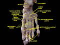
There are three cuneiform ("wedge-shaped") bones in the human foot:
- the first or medial cuneiform
- the second or intermediate cuneiform, also known as the middle cuneiform
- the third or lateral cuneiform
They are located between the navicular bone and the first, second and third metatarsal bones and are medial to the cuboid bone.[1]
Structure
[edit]There are three cuneiform bones:
- The medial cuneiform (also known as first cuneiform) is the largest of the cuneiforms. It is situated at the medial side of the foot, anterior to the navicular bone and posterior to the base of the first metatarsal. Lateral to it is the intermediate cuneiform. It articulates with four bones: the navicular, second cuneiform, and first and second metatarsals. The tibialis anterior and fibularis longus muscle inserts at the medial cuneiform bone.[2]
- The intermediate cuneiform (second cuneiform or middle cuneiform) is shaped like a wedge, the thin end pointing downwards. The intermediate cuneiform is situated between the other two cuneiform bones (the medial and lateral cuneiforms), and articulates with the navicular posteriorly, the second metatarsal anteriorly and with the other cuneiforms on either side.
- The lateral cuneiform (also known as third cuneiform or external cuneiform) intermediate in size between the other two cuneiform bones, is also wedge-shaped, the base being uppermost. It occupies the center of the front row of the tarsal bones, between the intermediate cuneiform medially, the cuboid laterally, the navicular posteriorly and the third metatarsal in front. The tibialis posterior inserts at the lateral cuneiform, while the flexor hallucis brevis originates from it.[2]
Muscle attachments
[edit]| Muscle | Direction | Attachment[2] |
| Tibialis anterior | Insertion | Medial cuneiform |
| Fibularis longus | Insertion | Medial cuneiform |
| Tibialis posterior | Insertion | Medial cuneiform |
| Flexor hallucis brevis | Origin | Lateral cuneiform |
Injuries
[edit]- Lisfranc fracture – in which one or all of the metatarsals are displaced from the tarsus[3]
- Cuneiform fracture - Due to the ligamentous support of the midfoot, isolated cuneiform fractures are rare [4]
Additional images
[edit]-
 Bones of the right foot. Dorsal surface.
 -
 Bones of the right foot. Plantar Surface.
 -
 Skeleton of foot. Medial aspect.
 -
 Skeleton of foot. Lateral aspect.
 -
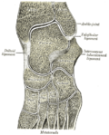 Oblique section of left intertarsal and tarsometatarsal articulations, showing the synovial cavities.
 -
 Bones of foot
 -
 Cuneiform. Superior view.
 -
 Cuneiform. Superior view.


Other animals
[edit]This section needs expansion. You can help by adding to it. (April 2015) |
See also
[edit]- Cuneiform, for writing by pressing a wedge-shaped reed into wet clay.
References
[edit]Wikimedia Commons has media related to Cuneiform bones.
- ^ Bojsen-Møller, Finn; Simonsen, Erik B.; Tranum-Jensen, Jørgen (2001). Bevægeapparatets anatomi [Anatomy of the Locomotive Apparatus] (in Danish) (12th ed.). p. 245. ISBN 978-87-628-0307-7.
- ^ a b c Bojsen-Møller, Finn; Simonsen, Erik B.; Tranum-Jensen, Jørgen (2001). Bevægeapparatets anatomi [Anatomy of the Locomotive Apparatus] (in Danish) (12th ed.). pp. 364–367. ISBN 978-87-628-0307-7.
- ^ TheFreeDictionary > Lisfranc's fracture Citing: Mosby's Medical Dictionary, 8th edition. 2009
- ^ Mabry LM, Patti TN, Ross MD, Bleakley CM, Gisselman AS (July 2021). "Isolated Medial Cuneiform Fractures: A Systematic Search and Qualitative Analysis of Case Studies". J Am Podiatr Med Assoc. 111 (4): 1–9. doi:10.7547/20-047. PMID 34478529. S2CID 225705519.
{{cite journal}}: CS1 maint: multiple names: authors list (link)
Cuneiform bones
View on Grokipediafrom Grokipedia
Anatomy
Overview and nomenclature
The cuneiform bones are a set of three wedge-shaped tarsal bones located in the midfoot of the human foot, consisting of the medial (first), intermediate (second), and lateral (third) cuneiforms.[2] These bones are positioned distally to the navicular bone and proximally to the bases of the first, second, and third metatarsals, contributing to the structure of the medial longitudinal and transverse arches of the foot.[6] The medial cuneiform is the largest and most prominent of the three, situated on the medial side of the foot, while the intermediate cuneiform is the smallest and the lateral cuneiform is intermediate in size.[2][6] The term "cuneiform" derives from the Latin words cuneus (wedge) and forma (shape), reflecting the bones' characteristic wedge-like morphology that aids in forming the foot's arches.[7][8] They are also referred to by their positional names—medial, intermediate, and lateral—to distinguish their anatomical roles and relationships within the tarsus.[6] The cuneiform bones were first described in ancient Greek and Roman anatomical texts, such as those by Galen in the 2nd century AD, with shape-based nomenclature for tarsal bones evolving through Renaissance anatomists like Andreas Vesalius in the 16th century.[8] Modern standardized terminology for these bones was established in the 19th century through efforts like the Nomina Anatomica, formalizing their designation in anatomical literature.[9]Osteology and articulations
The cuneiform bones, consisting of the medial, intermediate, and lateral cuneiforms, are three wedge-shaped tarsal bones located in the midfoot, positioned between the navicular proximally and the metatarsals distally.[10] Their osteology reflects their role in forming the transverse arch of the foot, with varying degrees of wedging that contribute to the medial longitudinal arch.[11] Each bone exhibits distinct surfaces adapted for articulation and soft tissue attachment, with the medial cuneiform being the largest, the intermediate the smallest, and the lateral intermediate in size.[12] The medial cuneiform is the largest of the three, featuring a rectangular base and a prominent tubercle on its plantar surface for attachment of the tibialis anterior tendon.[11] Its dorsal surface is narrow and convex, while the plantar surface is flat and broader, accommodating a slip of the tibialis posterior tendon.[10] The medial surface is rough and subcutaneous, presenting a vertical convexity with an impression for the tibialis anterior muscle; the lateral surface includes dorsal facets separated by ridges for articulation and a groove for the peroneus longus tendon.[11] Proximally, it has a pyriform (pear-shaped) facet, and distally, a reniform (kidney-shaped) surface.[11] The intermediate cuneiform is the smallest and most wedge-shaped, with a narrow plantar base that enhances its tapered form.[12] Its dorsal surface is rectangular and rough, the plantar surface narrow and receiving a slip of the tibialis posterior tendon, and the proximal surface features a concave triangular facet.[13] The distal surface is also triangular, while the medial and lateral surfaces are partly articular with appositional contact to the adjacent cuneiforms and include L-shaped facets medially.[13] A unique feature is its formation of a mortise-like structure that partially encases the base of the second metatarsal.[10] The lateral cuneiform is the most distinctly wedge-shaped, with a dorsal convexity and a rectangular rough dorsal surface.[12] Its plantar surface is narrow, giving origin to the flexor hallucis brevis muscle and attachment for the tibialis posterior tendon, while the medial surface articulates with the intermediate cuneiform via two small facets.[14] The lateral surface presents a facet for the cuboid, the proximal surface a triangular articulation, and the distal surface contacts the bases of the second and third metatarsals.[14] It lies with its edge downward, positioned between the intermediate cuneiform and cuboid.[14] All three cuneiform bones articulate proximally with the navicular bone via three separate concave facets, one on each bone, forming the cuneonavicular joints.[15] Distally, the medial cuneiform articulates with the base of the first metatarsal (and dorsomedially with the second), the intermediate with the second metatarsal, and the lateral with the bases of the second and third metatarsals, comprising the tarsometatarsal joints.[10] Intercuneiform joints exist between the medial and intermediate cuneiforms, and between the intermediate and lateral cuneiforms, with the lateral cuneiform additionally articulating laterally with the cuboid via the cuneocuboid joint.[15] These synovial joints are reinforced by interosseous ligaments.[13] Ossification of the cuneiform bones occurs postnatally, with primary centers appearing in the first few years of life: the lateral cuneiform at approximately 1 year, the medial at 3 years, and the intermediate at 3-4 years, often being the last tarsal to ossify.[16] Each bone typically develops from a single ossification center, though the medial may have two, and full ossification is generally complete by late childhood, around 10-12 years.[10]Ligaments and soft tissue relations
The cuneiform bones are stabilized by a network of ligaments that connect them to adjacent tarsal and metatarsal bones, ensuring midfoot integrity. The dorsal and plantar cuneonavicular ligaments reinforce the articulations between the navicular bone and the three cuneiforms, providing anterior-posterior stability while allowing limited flexion and extension. Interosseous ligaments bind the non-articular surfaces between the medial, intermediate, and lateral cuneiforms, limiting excessive lateral or medial translation during weight-bearing activities. Cuneometatarsal ligaments, including dorsal, plantar, and interosseous components, link the distal surfaces of the cuneiforms to the bases of the first through third metatarsals, distributing compressive forces across the transverse arch. Central to this ligamentous complex is the Lisfranc ligament, an oblique band arising from the lateral plantar surface of the medial cuneiform and inserting onto the medial base of the second metatarsal. This ligament, comprising dorsal, interosseous, and plantar components, acts as the primary restraint against midfoot diastasis. The surrounding soft tissues include the proximity of the tibialis anterior tendon along the dorsomedial aspect of the medial cuneiform and the peroneus longus tendon near its lateral surface, which contribute to dynamic stabilization without direct bony insertion in this region. Attachments to the plantar fascia occur via extensions that blend with the cuneiform bases, supporting the medial longitudinal arch. Fat pads and bursae in the midfoot, particularly beneath the cuneiforms, provide cushioning against shear forces during gait. Biomechanically, these ligaments prevent dorsal displacement of the metatarsals relative to the cuneiforms and maintain longitudinal arch alignment under load. The Lisfranc joint complex forms a "Roman arch" configuration, where the recessed intermediate cuneiform and second metatarsal base serve as the keystone, with the Lisfranc ligament anchoring the medial pillar to resist plantarflexion and abduction stresses. This setup enhances overall foot rigidity, particularly in propulsion phases of walking. Pathological ligament laxity, often seen in hypermobile feet, can compromise cuneiform stability, leading to excessive first ray motion and predisposition to midfoot deformities such as flatfoot or hallux valgus.Vascular and neural supply
The cuneiform bones receive their arterial supply from branches of the dorsalis pedis artery, which provides blood to the dorsal aspects via the medial and lateral tarsal arteries.[17] These arteries anastomose with branches from the medial plantar artery, which supplies the plantar surfaces, forming arcades around the tarsometatarsal joints that ensure comprehensive perfusion of the midfoot region.[17] For the medial cuneiform specifically, a middle pedicle branch from the dorsalis pedis contributes to the dorsal supply, while the medial and superficial medial plantar arteries nourish the plantar aspect, supported by a dense intraosseous capillary network with one major nutrient artery and several minor ones.[18] Venous drainage of the cuneiform bones parallels the arterial supply, occurring through the dorsal and plantar venous plexuses of the foot.[19] These plexuses ultimately drain into the great saphenous vein medially and the small saphenous vein laterally, facilitating return of deoxygenated blood to the lower limb venous system.[20] Sensory innervation to the cuneiform bones arises from the deep peroneal nerve on the dorsal side and the medial and lateral plantar nerves on the plantar side, providing periosteal sensation to these skeletal structures.[19] As non-muscular elements, the cuneiform bones receive no significant motor innervation.[10] Notably, the intermediate cuneiform is susceptible to avascular necrosis following fractures.[21]Function
Biomechanical role in the foot
The cuneiform bones play a pivotal role in forming the arches of the foot, which are essential for maintaining structural integrity and dynamic function. The medial and intermediate cuneiforms are key components of the medial longitudinal arch (MLA), forming its anterior pillar alongside the navicular bone and the first three metatarsals, while all three cuneiforms contribute to the transverse arch through their wedge-shaped morphology that elevates the central foot region.[22] This wedged configuration, with the base of each cuneiform oriented dorsally, creates a stable yet resilient framework that supports the foot's adaptability to varying loads.[6][23] During gait, the cuneiform bones facilitate efficient load transmission by transferring weight from the talus and navicular proximally to the metatarsals distally across the tarsometatarsal joints, which exhibit slight mobility to absorb impact forces. This mechanism allows the midfoot to act as a shock absorber, dissipating vertical ground reaction forces during the stance phase and enabling smooth propulsion in the push-off phase.[22] The interlocking articulations between the cuneiforms and adjacent bones, reinforced by ligamentous tension such as the Lisfranc ligament complex, establish a rigid platform that enhances overall foot stability while permitting controlled pronation and supination to adapt to terrain irregularities.[24][6] In pathomechanics, misalignment of the cuneiform bones, as seen in flat feet (pes planus), disrupts normal arch geometry, leading to altered stress distribution where loads are unevenly shifted from the cuneiforms to the navicular and other midfoot structures, potentially causing overload and compensatory strain on adjacent bones and soft tissues.[25] This redistribution increases the risk of midfoot instability and pain, particularly under dynamic loading like walking or running, as the reduced arch height diminishes the foot's natural shock-absorbing capacity.[26]Muscle and tendon attachments
The cuneiform bones serve as key attachment sites for several extrinsic and intrinsic foot muscles, facilitating movements such as dorsiflexion, inversion, eversion, and plantarflexion while contributing to the stability of the transverse and medial longitudinal arches of the foot. The medial cuneiform, in particular, receives the primary insertion of the tibialis anterior tendon on its medial and plantar surfaces, enabling dorsiflexion and inversion of the foot during the swing phase of gait.[2][3] A slip from the tibialis posterior tendon also inserts on the plantar surface of the medial cuneiform, supporting inversion and arch maintenance, while the peroneus longus tendon attaches to its base and that of the first metatarsal, promoting eversion and plantarflexion to balance medial forces.[6][2] The intermediate cuneiform has more limited muscular attachments, primarily receiving a second slip of the tibialis posterior tendon on its plantar surface, which reinforces the medial arch and aids in controlled inversion during weight-bearing activities.[2] In contrast, the lateral cuneiform features an origin site for the flexor hallucis brevis muscle on its plantar aspect, allowing this intrinsic muscle to flex the proximal phalanx of the hallux and stabilize the forefoot during toe-off in the gait cycle.[3][6] Tendon relations further integrate the cuneiform bones into foot dynamics. The dorsal surfaces of the cuneiforms provide supportive relations for tendons like those of the extensor digitorum brevis, though primary attachments remain on adjacent structures. These muscular interfaces enable fine-tuned adjustments, such as coordinated propulsion in gait, where imbalances in tibialis anterior or peroneus longus tension can alter forefoot alignment.[6] Anatomical variations in attachments are uncommon but include occasional accessory slips of the tibialis posterior to the intermediate or lateral cuneiforms, or rare sesamoid bones embedded near the peroneus longus insertion on the medial cuneiform, potentially affecting local biomechanics.[2]Clinical relevance
Injuries and disorders
The cuneiform bones are susceptible to various traumatic injuries, primarily fractures that occur due to direct trauma or repetitive stress. Avulsion fractures of the medial cuneiform tubercle are uncommon but can result from forceful contraction of the tibialis anterior tendon, pulling a bone fragment at its insertion site.[27] Stress fractures of the cuneiform bones are rare and can affect any of the three, often seen in runners or athletes subjected to high repetitive loading, leading to microtrabecular failure over time.[4] Fractures of the lateral cuneiform are rare in isolation and typically occur in conjunction with cuboid injuries, such as in high-energy midfoot trauma involving compression or nutcracker mechanisms.[28] Osteochondrosis of the cuneiform bones is a rare, self-limiting condition primarily affecting children, characterized by aseptic necrosis that can cause midfoot pain and a limp, typically resolving spontaneously within months.[29] Bipartite cuneiforms, most commonly of the medial cuneiform, are rare anatomical variants with a prevalence of approximately 0.8-2%, which may be asymptomatic but can lead to chronic midfoot pain or be mistaken for fractures due to non-fusion of ossification centers.[30] Lisfranc injuries represent a spectrum of midfoot trauma involving the tarsometatarsal joints, where the cuneiform bones articulate with the metatarsals, often resulting in sprains, fractures, or dislocations. These injuries disrupt the stability of the medial (first), intermediate (second), and lateral (third) cuneiform-metatarsal articulations, with the second metatarsal base serving as a keystone for alignment. The original classification by Quenu and Kuss categorizes these as homolateral (all metatarsals displaced in the same direction), isolated (single metatarsal affected), or divergent (first metatarsal medially displaced with others laterally).[31] Degenerative and congenital disorders also impact the cuneiform bones, altering midfoot biomechanics. Osteoarthritis commonly develops at the inter-cuneiform joints, characterized by cartilage loss and osteophyte formation, often secondary to prior trauma or chronic overload.[32] Tarsal coalition, a congenital fusion anomaly, infrequently involves the cuneiforms, with reported cases of intermediate-lateral cuneiform synostosis leading to restricted motion and pain during adolescence or early adulthood.[33] Stress reactions, precursors to full fractures, manifest in athletes as periosteal edema in the cuneiforms due to repetitive impact, particularly in the medial or intermediate bones. Risk factors for cuneiform injuries and disorders include participation in high-impact sports like running or basketball, which impose repetitive axial loading on the midfoot. Systemic conditions such as rheumatoid arthritis predispose to inflammatory arthropathy in the inter-cuneiform joints, accelerating degenerative changes. Biomechanical abnormalities, notably pes planus (flatfoot), increase vulnerability by altering load distribution across the cuneiforms and tarsometatarsal complex.[34][35][36]Diagnosis and management
Diagnosis of cuneiform bone-related issues begins with a thorough clinical evaluation, focusing on patient history and physical examination to identify midfoot pathology. Patients often present with localized midfoot pain exacerbated by weight-bearing, swelling, ecchymosis, and tenderness upon palpation over the cuneiforms or tarsometatarsal joints. For Lisfranc injuries involving the cuneiforms, specific tests include the pronation-abduction drawer test, which assesses instability by applying anterior-posterior and rotational stress to the midfoot, and the single-leg heel-rise test, where inability to rise on toes indicates disruption. Gait analysis may reveal biomechanical abnormalities contributing to stress on the medial column, such as excessive pronation or arch collapse.[37][38] Imaging modalities are essential for confirming diagnosis, as initial plain radiographs may miss subtle injuries. Weight-bearing anteroposterior and lateral X-rays are first-line, revealing key signs such as the fleck sign—an avulsion fracture from the medial cuneiform base indicating Lisfranc ligament disruption—or diastasis greater than 2 mm between the first and second metatarsal bases. If non-weight-bearing views are inconclusive, computed tomography (CT) provides detailed assessment of fracture extent, displacement, and joint involvement, particularly for isolated cuneiform fractures where occult lesions occur in up to 73% of initial radiographs. Magnetic resonance imaging (MRI) is preferred for evaluating soft tissue and ligament damage, such as intercuneiform or tarsometatarsal ligament tears, while ultrasound aids in detecting dynamic instability during stress maneuvers. For tarsal coalitions involving cuneiforms, CT delineates bony bridging, with MRI assessing associated cartilage or soft tissue changes. Diagnostic delays average 65 days for isolated medial cuneiform fractures due to subtle findings.[37][39][40] Management strategies depend on injury stability, displacement, and patient factors, prioritizing restoration of midfoot alignment and function. Conservative approaches are indicated for nondisplaced or stable injuries, including immobilization in a short-leg cast or walking boot with non-weight-bearing for 4-6 weeks, followed by progressive weight-bearing and orthotics to support the longitudinal arch. Stress fractures of the cuneiforms respond well to this regimen, often resolving within 4-6 weeks with protected activity. For tarsal coalitions causing pain, initial nonsurgical management involves orthotics, activity modification, and anti-inflammatory measures. Surgical intervention is required for displaced fractures, unstable Lisfranc injuries, or irreparable joint surfaces, typically involving open reduction and internal fixation (ORIF) with transarticular screws or dorsal plates to maintain medial column length and tarsometatarsal stability. In cases of severe comminution or coalitions with degenerative changes, arthrodesis of the affected joints is performed to achieve fusion and alleviate pain. Primary fusion may be considered for high-energy injuries with ligamentous incompetence.[41][39][42] Outcomes vary by injury type and timeliness of intervention, with conservative management yielding good results in reported cases of nondisplaced isolated cuneiform fractures, allowing return to full activity within 3-6 months without complications. Surgical ORIF for Lisfranc injuries achieves anatomical reduction in most cases, with favorable functional scores in stable postoperative patients, though risks include posttraumatic arthritis in 20-40% long-term. Rehabilitation protocols emphasize early mobilization post-immobilization, incorporating physical therapy for strengthening, proprioception, and arch-supportive orthotics to prevent recurrence. Delayed diagnosis increases complication rates, underscoring the need for vigilant evaluation.[39][38][43]Comparative anatomy
Structure in other mammals
In non-human mammals, the cuneiform bones—typically the three distal tarsals (medial, intermediate, and lateral)—exhibit significant morphological variations compared to the distinct, wedge-shaped structures seen in humans, often reflecting locomotor adaptations. In carnivores such as dogs and cats, the three cuneiform bones remain distinct and unfused, forming part of the seven tarsal bones that support agile, digitigrade locomotion.[44] These bones articulate proximally with the navicular and distally with the metatarsals, maintaining a configuration that allows for flexibility during predation and rapid directional changes. In contrast, many quadrupedal ungulates show reductions and fusions; for instance, in horses, the medial and intermediate cuneiforms are fused into a single mediointermediate bone, resulting in six tarsal bones overall, which enhances stability for high-speed galloping.[45] Ruminants like deer and sheep further reduce the count to five tarsal bones through fusion of the intermediate and lateral cuneiforms with other elements, integrating them into a more compact structure suited to cursorial lifestyles.[46] Articulations of the cuneiform bones also vary to accommodate species-specific demands. In felines, the lateral cuneiform articulates more extensively with the cuboid bone laterally, providing additional support for powerful leaps and pouncing, while the medial cuneiform maintains close contact with the navicular for precise foot placement.[44] Ungulates exhibit enhanced wedge-like shapes in their cuneiforms (or fused equivalents), with broader proximal surfaces for weight distribution across fewer digits, as seen in the elongated, robust forms in deer that facilitate efficient force transmission during sustained running.[47] Primates, particularly non-human ones, generally retain human-like separation of the three cuneiform bones, though with subtle differences; for example, in chimpanzees, the medial cuneiform displays a more convex distal facet, aiding in arboreal grasping and climbing.[48] Functional adaptations further distinguish cuneiform morphology across mammals. In cursorial species like deer, the cuneiforms are elongated and integrated into fused complexes that prioritize rigidity and shock absorption for speed over rough terrain, minimizing lateral movement during high-velocity pursuits.[47] Conversely, in brachiating primates such as gibbons, the cuneiforms are relatively reduced in size and contribute to a highly flexible, prehensile foot structure, with the medial cuneiform's articulation allowing greater dorsiflexion and opposition of the hallux for secure branch gripping during suspension.[49] A striking example of extreme reduction occurs in cetaceans, where distinct cuneiform bones are absent due to the evolutionary loss of hindlimbs and tarsal elements altogether, replaced by a streamlined flipper formed primarily from modified forelimb bones.[50] In equids, the central tarsal bone, homologous to the navicular, provides a pivotal role in the locked hock joint for propulsion.[44]Evolutionary aspects
The cuneiform bones of mammals are derived from the distal row of tarsal elements present in reptilian ancestors, where primitive reptiles possessed up to five distal tarsals that contributed to the foundational structure of the tarsus.[51] In early tetrapods, homologs of the cuneiforms formed part of a more extensive tarsal mosaic, with proximal elements fusing into the astragalus-calcaneus complex through ontogenetic repatterning of the tibiale, intermedium, and fibulare, as observed in taxa like Hylonomus lyelli, the earliest known amniote.[52] This configuration diverged in therapsids, the synapsid lineage leading to mammals, where progressive reduction and specialization of distal tarsals established the seven-bone mammalian tarsus, including three distinct cuneiforms, by the late Permian to early Triassic.[53] Bipedal evolution in hominids accentuated the wedge-shaped morphology of the cuneiforms to support longitudinal arch formation and efficient weight transfer during upright locomotion. In Australopithecus afarensis (ca. 3.0–3.4 million years ago), the medial cuneiform exhibits transitional features, such as a dorsally convex distal facet for the non-opposable hallux, indicating early adaptation for bipedal stability while retaining some arboreal capabilities.[54] Primates generally show loss of tarsal fusion among the cuneiforms, allowing greater midfoot flexibility for grasping and varied locomotion, in contrast to artiodactyls like ruminants, where fusion of the cuneiforms with the navicular and cuboid persists, enhancing rigidity for cursorial gaits.[50] Fossil evidence from Miocene apes, such as those from East African sites (ca. 20–14 million years ago), reveals elongated cuneiforms adapted for arboreal locomotion, with extended distal facets facilitating toe-off during suspension and climbing in forested environments.[55] Post-Australopithecus, biomechanical shifts in the Homo lineage (ca. 1.8 million years ago onward) further refined cuneiform geometry for endurance walking, as seen in elevated arches in Homo erectus footprints from Koobi Fora (1.5 million years ago), which optimized elastic energy return and reduced metabolic cost during long-distance travel.[56] In modern humans, vestigial features persist, such as the small size of the intermediate cuneiform, reflecting evolutionary reduction of the second ray relative to the robust medial and lateral rays, a legacy of diminished reliance on medial foot grasping in favor of bipedal propulsion.[54]References
- https://en.wikisource.org/wiki/The_Osteology_of_the_Reptiles/Chapter_5