Hubbry Logo
SubcloningSubcloningMain
Open search
Subcloning
Community hub
Subcloning
logo
8 pages, 0 posts
0 subscribers
Be the first to start a discussion here.
Contribute something
Subcloning
Subcloning
from Wikipedia
This image diagrams the procedure of subcloning as outlined to the left.

In molecular biology, subcloning is a technique used to move a particular DNA sequence from a parent vector to a destination vector.

Subcloning is not to be confused with molecular cloning, a related technique.

Procedure

[edit]

Restriction enzymes are used to excise the gene of interest (the insert) from the parent. The insert is purified in order to isolate it from other DNA molecules. A common purification method is gel isolation. The number of copies of the gene is then amplified using polymerase chain reaction (PCR).

Simultaneously, the same restriction enzymes are used to digest (cut) the destination. The idea behind using the same restriction enzymes is to create complementary sticky ends, which will facilitate ligation later on. A phosphatase, commonly calf-intestinal alkaline phosphatase (CIAP), is also added to prevent self-ligation of the destination vector. The digested destination vector is isolated/purified.

The insert and the destination vector are then mixed together with DNA ligase. A typical molar ratio of insert genes to destination vectors is 3:1;[1] by increasing the insert concentration, self-ligation is further decreased. After letting the reaction mixture sit for a set amount of time at a specific temperature (dependent upon the size of the strands being ligated; for more information see DNA ligase), the insert should become successfully incorporated into the destination plasmid.

Amplification of product plasmid

[edit]

The plasmid is often transformed into a bacterium like E. coli. Ideally when the bacterium divides the plasmid should also be replicated. In the best case scenario, each bacterial cell should have several copies of the plasmid. After a good number of bacterial colonies have grown, they can be miniprepped to harvest the plasmid DNA.

Selection

[edit]

In order to ensure growth of only transformed bacteria (which carry the desired plasmids to be harvested), a marker gene is used in the destination vector for selection. Typical marker genes are for antibiotic resistance or nutrient biosynthesis. So, for example, the "marker gene" could be for resistance to the antibiotic ampicillin. If the bacteria that were supposed to pick up the desired plasmid had picked up the desired gene then they would also contain the "marker gene". Now the bacteria that picked up the plasmid would be able to grow in ampicillin whereas the bacteria that did not pick up the desired plasmid would still be vulnerable to destruction by the ampicillin. Therefore, successfully transformed bacteria would be "selected."

Example case: bacterial plasmid subcloning
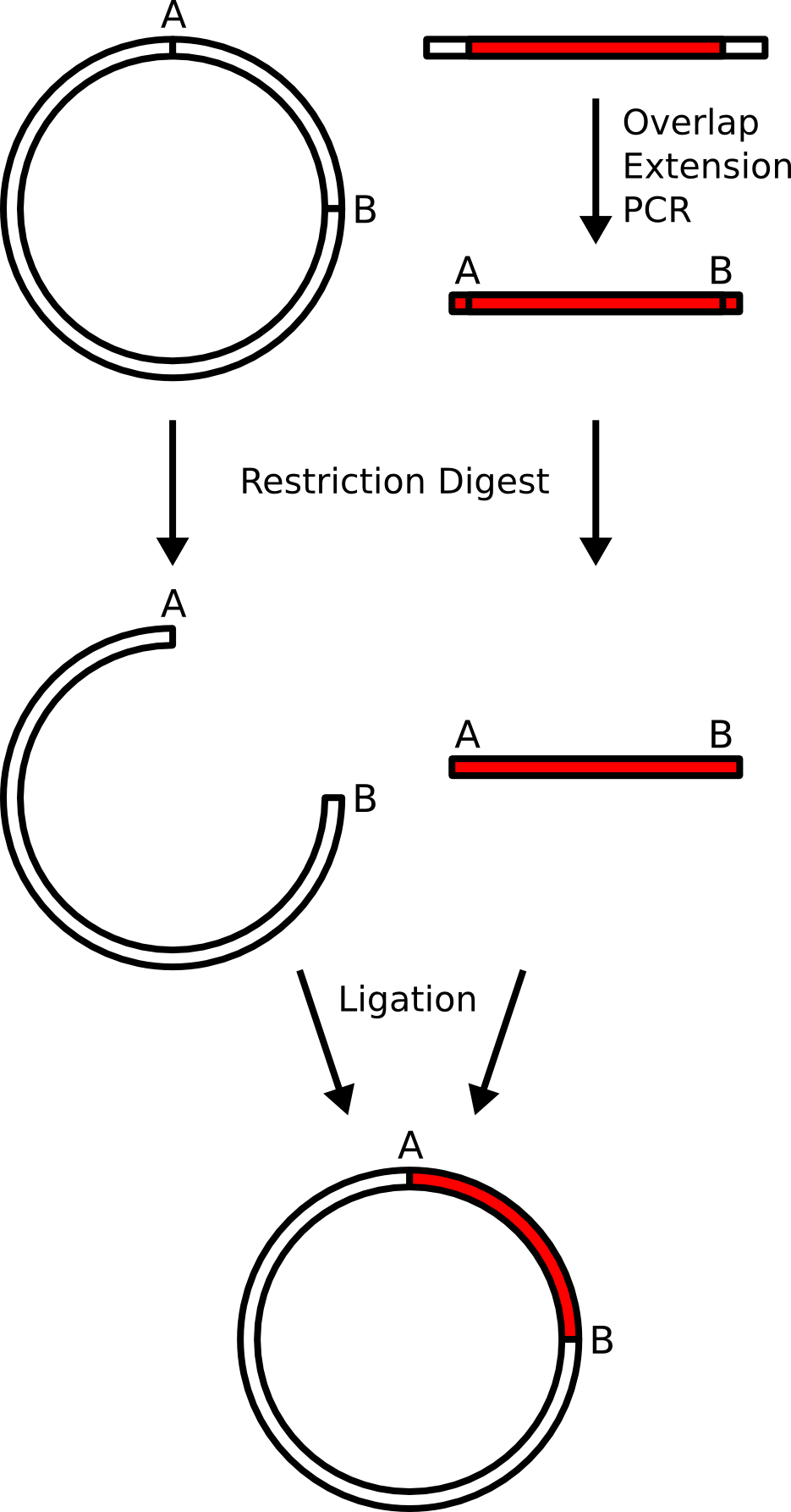
[edit]

In this example, a gene from mammalian gene library will be subcloned into a bacterial plasmid (destination platform). The bacterial plasmid is a piece of circular DNA which contains regulatory elements allowing for the bacteria to produce a gene product (gene expression) if it is placed in the correct place in the plasmid. The production site is flanked by two restriction enzyme cutting sites "A" and "B" with incompatible sticky ends.

The mammalian DNA does not come with these restriction sites, so they are built in by overlap extension PCR. The primers are designed to put the restriction sites carefully, so that the coding of the protein is in-frame, and a minimum of extra amino acids is implanted on either side of the protein.

Both the PCR product containing the mammalian gene with the new restriction sites and the destination plasmid are subjected to restriction digestion, and the digest products are purified by gel electrophoresis.

The digest products, now containing compatible sticky ends with each other (but incompatible sticky ends with themselves) are subjected to ligation, creating a new plasmid which contains the background elements of the original plasmid with a different insert.

The plasmid is transformed into bacteria and the identity of the insert is confirmed by DNA sequencing.

See also

[edit]

References

[edit]
Revisions and contributorsEdit on WikipediaRead on Wikipedia
from Grokipedia
Subcloning is a core technique in for transferring a specific DNA fragment, or insert, from a parent vector to a destination vector, enabling targeted , expression, or manipulation of the genetic material in appropriate host systems. This process is essential for adapting cloned DNA to new experimental contexts, such as changing expression hosts from to mammalian cells or incorporating regulatory elements like promoters and tags. The primary method of subcloning involves restriction enzyme digestion to excise the DNA insert from the parent vector and linearize the destination vector, followed by ligation using enzymes like T4 DNA ligase to join the compatible ends. Directional cloning is often achieved with two different restriction enzymes producing incompatible sticky ends, ensuring the insert orients correctly, while blunt-end ligation can be used for fragments without suitable restriction sites by employing polymerases such as T4 DNA polymerase. Alternative approaches, including recombination-based systems like Gateway cloning, utilize site-specific recombinases to facilitate seamless transfer without relying on restriction sites, which is particularly useful for large or complex inserts. Following ligation, the recombinant vector is introduced into competent host cells via transformation, typically using bacterial strains like E. coli JM109, and colonies are screened for successful insertion through methods such as colony PCR, restriction analysis, or sequencing. Subcloning's versatility supports diverse applications, from and to constructing gene fusions and targeting vectors for .

Overview

Definition and Purpose

Subcloning is a core technique in that entails isolating a specific DNA fragment, referred to as the insert, from a source vector and transferring it into a destination vector to enable improved functionality, such as increased copy number, specialized promoters, or compatibility with particular host systems. This process refines the genetic construct by repositioning the insert within a new backbone, facilitating more targeted experimentation or propagation. The primary purposes of subcloning include enhancing through integration into vectors with optimized regulatory elements, constructing expression libraries to screen gene functions on a large scale, preparing refined DNA fragments for high-throughput sequencing, and adapting inserts for diverse host organisms, for instance, moving bacterial-optimized sequences to mammalian expression systems. These applications support advanced studies in , , and cross-species . Subcloning relies on foundational concepts of , particularly the formation of , which involves the enzymatic joining of DNA segments from different origins to create hybrid molecules that can be replicated and expressed in host cells like . This technique was first described in the early 1970s as part of the pioneering work in technology, revolutionizing scalable DNA manipulation and laying the groundwork for modern .

Historical Context

Subcloning emerged in the mid-1970s alongside the pioneering experiments of Stanley Cohen and , who in 1973 successfully cloned antibiotic resistance genes into the low-copy-number plasmid pSC101 in , establishing the foundation for plasmid-based . Early vectors like pSC101, with only 5–10 copies per cell, yielded insufficient for downstream applications, necessitating subcloning techniques to transfer inserts into higher-copy-number plasmids for enhanced production and analysis. This approach addressed key limitations in yield and scalability, transforming initial proofs-of-concept into practical tools for manipulation. The 1980s saw further innovations with the development of , which allowed inserts to be propagated across species; notably, in 1978, Albert Hinnen, , and Gerald Fink introduced the first -E. coli shuttle vector, facilitating eukaryotic and cross-host transfers that expanded subcloning's utility beyond prokaryotes. By the 1990s, subcloning integrated with (PCR) technology—developed by in 1983 and commercialized in the mid-1980s—to generate precise inserts without dependence on natural restriction sites, streamlining workflows and reducing labor-intensive steps. This period marked a shift from purely restriction enzyme-reliant methods toward more versatile amplification-based strategies. The technique evolved further in the early 2000s with recombination systems, exemplified by Invitrogen's platform launched in 1999, which employed bacteriophage lambda for directional, scarless transfer of inserts between vectors, significantly improving efficiency, throughput, and fidelity over traditional ligation. Subcloning's advancements proved instrumental in the (1990–2003), where it enabled the fragmentation and transfer of large genomic inserts from bacterial artificial chromosomes into specialized sequencing vectors, supporting high-throughput assembly and analysis of the .

Molecular Principles

Role of Vectors and Inserts

In subcloning, vectors serve as the essential DNA backbone that enables the replication, maintenance, and selection of recombinant molecules within host cells, while inserts represent the specific genetic sequences of interest that are transferred into these vectors for further manipulation or expression. Destination vectors, such as high-copy plasmids from the pUC series, are widely used for efficient propagation of inserts in due to their mutated , which supports copy numbers exceeding 500 per cell, facilitating high-yield DNA production. These vectors include key features like a (MCS)—a polylinker region containing unique recognition sites for precise insert integration—an (ori) for autonomous propagation, and selectable markers such as the ampicillin resistance gene (bla) to identify transformed hosts. Expression vectors like the pET series, which incorporate the T7 promoter for inducible high-level in T7 polymerase-expressing strains, extend this functionality by providing regulatory elements for downstream applications in protein studies. Shuttle vectors, designed with multiple origins of replication (e.g., one for and another for or mammalian cells), allow seamless transfer and propagation of inserts across diverse host organisms, enhancing versatility in multi-host experiments. Inserts in subcloning are DNA fragments carrying the genetic cargo, such as genes, promoters, or regulatory elements, that are excised or amplified from source material like PCR products, genomic libraries, or existing recombinant clones, and then incorporated into the destination vector to confer new properties or enable functional analysis. These inserts typically range in size from 100 base pairs (bp) to 10 kilobases (kb), balancing ease of manipulation with the complexity of the sequence; smaller inserts (under 1 kb) are common for regulatory elements, while larger ones (up to 10 kb) suit full genes or operons, though efficiency decreases beyond this due to recombination risks in bacterial hosts. Sources of inserts include polymerase chain reaction (PCR) amplification for targeted sequences or restriction digestion of parental plasmids, ensuring the fragment ends are compatible with the vector's MCS. Compatibility between vectors and inserts is governed by principles that ensure efficient and directional ligation, primarily through matching cohesive or blunt ends generated by restriction enzymes at the MCS and insert termini. For instance, using enzymes like or creates compatible sticky ends that anneal precisely, while linkers or adapters can bridge incompatible sites by adding tailored restriction motifs. To prevent vector self-ligation, which would yield empty recombinants, treatment dephosphorylates the vector's 5' ends, blocking recircularization without affecting the insert's phosphorylated termini. Overall, vectors provide the structural and selective framework for stable propagation, whereas inserts deliver the functional payload, forming the core of subcloning's .

Key Enzymes and Reactions

Type II restriction endonucleases serve as the primary tools for preparing DNA inserts and vectors in subcloning by recognizing specific short DNA sequences and cleaving the phosphodiester backbone at precise locations. These enzymes, such as and , are classified as Type II, meaning they require only Mg²⁺ as a cofactor and cut within or near their 4- to 8-base-pair palindromic recognition sites, producing either sticky (cohesive) or blunt ends depending on the stagger of the cut. For instance, recognizes the sequence 5'-GAATTC-3' and cleaves between the G and A residues, generating 5' sticky ends with a 4-base AATT overhang; recognizes 5'-AAGCTT-3' and cleaves between the first A and A, also producing 5' sticky ends with an AGCT overhang. This cleavage specificity allows for the generation of compatible ends that facilitate precise fragment assembly without random cuts. DNA ligase, most commonly T4 DNA ligase from T4, is essential for joining the prepared DNA fragments by catalyzing the formation of phosphodiester bonds between the 5'-phosphate and adjacent 3'-hydroxyl groups on double-stranded DNA substrates. The reaction is ATP-dependent and occurs in three enzymatic steps: first, the ligase forms an enzyme-AMP intermediate by reacting with ATP; second, the AMP group is transferred to the 5'-phosphate terminus of the DNA; and third, the activated 5'-phosphoryl attacks the 3'-hydroxyl, sealing the nick and releasing AMP. Optimal conditions for this ligation include incubation at 16°C overnight to maximize efficiency with cohesive ends, though higher temperatures like 25°C can be used for shorter times with blunt ends. Additional enzymes support subcloning by modifying ends to prevent artifacts or enable alternative strategies. Alkaline phosphatase, such as calf intestinal alkaline phosphatase (CIP), dephosphorylates the 5'-ends of linearized vectors by hydrolyzing the phosphate group, thereby preventing recircularization during ligation since T4 DNA ligase cannot join dephosphorylated ends without a 5'-phosphate donor from the insert. For blunt-end subcloning, when sticky ends are absent or incompatible, polymerases like T4 DNA polymerase or the Klenow fragment (lacking 5'→3' exonuclease activity) of Escherichia coli DNA polymerase I are used; these fill in 5' overhangs via 5'→3' polymerase activity or excise 3' overhangs via 3'→5' exonuclease activity, creating flush ends for non-directional ligation. The core reaction principles in subcloning emphasize compatibility and stoichiometry to ensure efficient recombinant formation. Directional cloning exploits asymmetric sticky ends from enzymes like and at opposite insert termini, allowing compatible pairing with the vector in only one orientation and reducing inverted inserts. Blunt-end reactions, while less directional, rely on the same ligase mechanism but achieve specificity through . To promote insert incorporation over vector self-ligation, a molar ratio of insert to vector DNA of approximately 3:1 is standard, as it saturates vector ends while minimizing multimers.

Detailed Procedure

Insert and Vector Preparation

The preparation of the insert in subcloning begins with the isolation of the desired DNA fragment, typically through digestion of the parent vector, or alternatively through (PCR) amplification from a template such as a or existing when necessary to introduce restriction sites. For PCR-based preparation, primers are designed to incorporate recognition sites at their 5' ends, flanking the target sequence, to generate compatible overhangs for subsequent ligation; for example, sequences like AAGCTT () or GTCGAC (SalI) are added to enable precise directional . The PCR reaction employs high-fidelity to minimize errors, with typical conditions including an initial denaturation at 98°C for 2 minutes, followed by 18–30 cycles of denaturation (98°C, 10 seconds), annealing (55–65°C, 15–30 seconds), and extension (72°C, 30 seconds per kb), ending with a final extension at 72°C for 1–5 minutes. After amplification, the product is verified and purified via on a 0.8–1.5% gel stained with or safer alternatives like , allowing separation based on size (100 bp to 25 kb range) and excision of the specific band to isolate the insert from primer dimers or non-specific products. Gel purification of the insert involves dissolving the excised agarose slice in a chaotropic salt buffer (e.g., guanidine thiocyanate), binding the DNA to a silica column, washing away contaminants, and eluting in low-salt buffer (e.g., 10 mM Tris-HCl, pH 8.0) for recovery yields of 50–80%. Quantification follows using UV spectrophotometry, where the concentration is calculated as 50 μg/mL per absorbance unit at 260 nm (A260) for double-stranded DNA, with purity assessed by an A260/A280 ratio of approximately 1.8 indicating minimal protein contamination and an A260/A230 ratio near 2.0–2.2 showing low organic contaminants. Vector preparation entails digestion of the plasmid with one or two restriction enzymes to linearize it and create ends compatible with the insert, using manufacturer-recommended buffers and incubation at 37°C for 1–2 hours (or overnight for complete digestion). To prevent self-ligation, the linearized vector is treated with (e.g., calf intestinal phosphatase, CIP, at 0.01 U/pmol ends) for 30 minutes at 37°C, followed by heat inactivation or purification to remove the enzyme. For vectors with internal restriction sites, partial digestion may be employed by limiting enzyme amount or time to ensure only the is cleaved, preventing fragmentation. Post-digestion and dephosphorylation, the linearized vector is separated from uncut supercoiled plasmid via (1% gel), followed by extraction of the linear band using silica-based kits to achieve >90% purity and remove enzymes or buffer salts. Quality control for both insert and vector includes agarose gel electrophoresis to confirm fragment size against a DNA ladder (e.g., 100 bp to 10 kb markers) and assess purity by the absence of smearing or extraneous bands, ensuring integrity for ligation. Contaminants such as RNases, which can co-purify from cellular sources and degrade any residual RNA or affect downstream steps, are minimized by using RNase-free reagents, disposable gloves, and dedicated workspaces to prevent skin-derived enzyme introduction. To optimize ligation efficiency, yields are calculated based on molar ratios, typically using ~100 ng of vector (e.g., 3 kb plasmid, ~0.05 pmol) with ~50 ng of insert (e.g., 1 kb fragment, ~0.15 pmol) to achieve a 3:1 insert:vector molar (calculated as pmol insert / pmol vector, using approximate MW = bp × 660 g/mol), promoting directional insertion while avoiding excess that could favor concatemers.

Ligation and Transformation

The ligation process in subcloning begins with the assembly of the prepared insert and linearized vector DNA fragments in a reaction mixture containing T4 DNA ligase enzyme and 10x ligase buffer, which provides necessary cofactors such as ATP and Mg²⁺ for enzymatic activity. Typical reaction volumes range from 10-20 μL, with 50-200 ng of vector DNA and a calculated amount of insert DNA to achieve the desired molar ratio. To promote the formation of recombinant molecules through phosphodiester bond formation, the insert-to-vector molar ratio is optimized, commonly at 3:1, to favor insert incorporation over vector self-ligation or insert multimers, thereby increasing the yield of desired circular plasmids. For cohesive ends typically generated in restriction enzyme-based subcloning, incubation occurs at room temperature (20-25°C) for 10 minutes to 4 hours or at 16°C overnight, allowing efficient annealing and ligation while minimizing non-specific reactions. Vector-only ligation controls are routinely included to quantify background religation rates, which should be low (e.g., <1% of total transformants) for effective subcloning. Following ligation, the recombinant DNA is introduced into host cells through transformation, a critical step that delivers the ligated plasmids into competent Escherichia coli for propagation. Chemical transformation via heat shock is widely used, where 1-5 μL of ligation mixture is added to 50-100 μL of competent cells on ice, followed by a 30-90 second incubation at 42°C to facilitate DNA uptake. Alternatively, electroporation involves mixing 1-2 μL of DNA with 20-50 μL of electrocompetent cells and applying a high-voltage pulse (e.g., 1.8-2.5 kV in 0.1-0.2 cm cuvettes), offering higher throughput for large libraries. Transformation efficiencies vary by method and cell strain, typically ranging from 10⁶ to 10⁹ colony-forming units (CFU) per μg of DNA for heat shock and up to 10¹⁰ CFU/μg for electroporation, influenced by DNA quality and cell viability. Chemical competence in E. coli is achieved by treating log-phase cells with ice-cold 50-100 mM CaCl₂, which neutralizes negative charges on lipopolysaccharides in the outer membrane, promoting electrostatic binding of negatively charged DNA and altering membrane permeability to enable uptake during heat shock. After transformation, cells are immediately placed on ice to stabilize the process, then diluted into 250-1000 μL of pre-warmed SOC medium (a nutrient-rich broth containing tryptone, yeast extract, NaCl, KCl, MgCl₂, MgSO₄, and glucose) and incubated at 37°C with shaking (200-250 rpm) for 45-60 minutes. This recovery step allows expression of antibiotic resistance genes from the plasmid, enhances cell viability, and can double transformation efficiency compared to standard LB medium.

Amplification of Recombinant Plasmids

Following successful transformation, recombinant s are amplified by inoculating a small volume of transformed Escherichia coli cells into Luria-Bertani (LB) broth supplemented with the appropriate antibiotic to maintain selective pressure and ensure propagation of plasmid-bearing cells. The culture is incubated at 37°C with agitation (typically 200–250 rpm) to facilitate oxygen transfer and logarithmic growth, with optical density at 600 nm (OD600) monitored to assess cell density; growth is often continued to the late log phase (OD600 ≈ 0.4–0.6) if induction of expression is planned, or overnight to stationary phase for maximal biomass accumulation in routine subcloning. Plasmid yield per cell varies significantly based on the vector's , with high-copy-number plasmids such as achieving 500–700 copies per cell under standard conditions, compared to low-copy-number vectors like , which maintain only 15–20 copies per cell. This difference directly impacts the amount of recoverable, as higher copy numbers enable greater amplification efficiency during host . Following growth, plasmids are typically extracted using an miniprep method, which selectively denatures chromosomal DNA while preserving supercoiled plasmid integrity through neutralization and precipitation steps. For initial screening in subcloning workflows, amplification is performed on a small scale using 1–5 mL cultures, yielding sufficient DNA (typically 5–50 μg total) for downstream verification without specialized equipment. Larger-scale production scales to liter volumes in shake flasks or bioreactors/fermenters, where controlled parameters like pH, dissolved oxygen, and nutrient feeding optimize yields; for E. coli hosts, 37°C remains the standard temperature to balance growth rate and plasmid stability. Post-amplification purification via alkaline lysis or column-based methods routinely yields 5–50 μg of plasmid DNA per mL of culture, with higher outputs from high-copy vectors under optimized conditions.

Selection of Clones

Selection of clones in subcloning involves distinguishing recombinant plasmids containing the desired insert from non-recombinant vectors and untransformed cells, primarily through phenotypic markers integrated into the cloning vector. Antibiotic selection serves as the initial step to identify transformed bacterial cells, utilizing resistance genes such as the bla gene encoding β-lactamase for ampicillin resistance, which allows only cells harboring the plasmid to survive on media containing the antibiotic. This method, derived from early cloning vectors like pBR322, ensures high initial enrichment but does not differentiate between self-ligated vectors and true recombinants. To further select for recombinants, blue-white screening exploits the lacZα gene fragment in vectors such as , where insertion of foreign DNA into the disrupts α-complementation of β-galactosidase, preventing hydrolysis of substrate and resulting in white colonies, while non-recombinants form blue colonies on indicator plates. This technique, introduced with the pUC series, provides a rapid visual distinction, with recombinant frequencies often reaching 80-95% under optimized conditions. Insertional inactivation extends this principle to antibiotic resistance markers; for instance, cloning into the tetracycline resistance gene (tetR) of disrupts its function, rendering recombinants sensitive to while retaining ampicillin resistance from the intact bla gene. For vectors with dual markers, or patch tests enable sequential screening: colonies are transferred from plates to media, identifying sensitive patches as potential recombinants. Without such screening, recombinant yields typically range from 1-10% of total transformants due to prevalent vector religation. The reliability of these disruption-based methods increases with insert size, as fragments larger than 1 kb more effectively inactivate marker genes by causing significant frameshifts or steric hindrance.

Verification and Analysis

Screening Techniques

Screening techniques in subcloning serve as initial, low-resolution methods to verify the presence and correct orientation of the inserted DNA fragment in transformed bacterial clones, prior to more definitive . These approaches enable efficient filtering of potential recombinants from a pool of transformants, distinguishing those with the desired subcloned insert from empty vectors or religated backbones. By focusing on rapid, cost-effective assays, researchers can prioritize promising clones for further processing, such as extraction detailed in amplification protocols. Colony PCR is a widely adopted technique for directly assessing insert presence without requiring plasmid isolation. In this method, a small portion of a bacterial colony is resuspended and lysed, typically by brief boiling, to release the plasmid DNA, which is then used as a template in a polymerase chain reaction with insert-specific primers. The reaction amplifies a region spanning the insert or junction sites, and the products are analyzed by agarose gel electrophoresis to confirm the expected band size corresponding to the insert length. For inserts under 3 kb, this approach provides quick results within hours, making it suitable for screening dozens of colonies. Primers flanking the multiple cloning site on the vector can also be used, where the absence of an insert yields a smaller amplicon than one with the insert. This technique is particularly valuable in subcloning workflows due to its simplicity and ability to detect inserts as small as 100 bp. Restriction digest screening offers a complementary verification step by examining the physical structure of the recombinant . Following miniprep isolation of DNA from overnight cultures of selected colonies, the DNA is subjected to double with restriction enzymes that flank the insertion site or cut within the insert. The resulting fragments are separated by , revealing expected patterns such as the vector backbone and released insert if ligation was successful; for example, a 3 kb insert in a 5 kb vector might produce bands of 5 kb and 3 kb upon , contrasting with a single ~5 kb band for an empty vector (linearized, assuming negligible multiple cloning site fragment). This method confirms both insert presence and approximate size, though it requires more time than colony PCR due to the need for culture growth and purification. To determine insert orientation, particularly in blunt-end or non-directional ligations, PCR-based checks employ asymmetric primer pairs—one annealing to the vector backbone and the other to an internal within the insert. Amplification occurs only if the insert is in the correct orientation, producing a specific band size on gel analysis; for instance, a forward vector primer paired with a reverse insert primer yields a product solely for the desired direction. This approach, adaptable to colony lysates, rapidly distinguishes forward from reverse inserts without sequencing. It has been demonstrated as a reliable alternative to restriction mapping for orientation verification in various systems. These techniques support , often in 96-well formats for large clone libraries, allowing parallel processing of hundreds of candidates via automated PCR or setups. However, they are prone to false positives, such as amplification of residual ligation mixture or incomplete digests, necessitating orthogonal like sequencing for final validation.

Confirmation Methods

of subcloned constructs is essential to ensure the integrity, accuracy, and functionality of the transferred insert within the new vector, distinguishing this step from preliminary screening by providing high-fidelity validation of the full and assembly. These methods focus on verifying the precise composition, detecting any unintended alterations such as mutations introduced during PCR amplification or ligation errors, and confirming proper integration at the vector-insert junctions. While initial screens like restriction digests offer rapid insights, techniques deliver definitive proof of the construct's fidelity before proceeding to downstream applications. Sanger sequencing remains the gold standard for validating individual subcloned plasmids due to its high accuracy and ability to resolve sequences up to approximately 1,000 base pairs per read. This chain-termination method involves using vector-specific primers, such as those annealing to flanking regions like T7 or SP6 promoters, to initiate sequencing from known sites adjacent to the insert. Primer walking extends coverage across longer inserts by designing sequential primers based on initial read results, ensuring comprehensive analysis of the insert ends and ligation junctions where scars or mismatches may occur. For instance, primers targeting 20-50 base pairs into the vector backbone allow detection of insertions, deletions, or point mutations arising from enzymatic steps like PCR, with error rates typically around 10^{-6} per base pair when using high-fidelity polymerases, minimizing the introduction of mutations during amplification. This approach confirms the exact sequence of the subcloned fragment, identifying any discrepancies from the reference design. For high-throughput scenarios involving clone libraries or multiple variants, next-generation sequencing (NGS) technologies provide scalable confirmation by generating millions of short reads that can be assembled into full contigs. Platforms like Illumina or Oxford Nanopore enable parallel sequencing of numerous constructs, with long-read options from the latter achieving complete resolution in a single run without the need for extensive primer design. Post-sequencing, de novo assembly software aligns reads to reference vectors, verifying the insert's full length and orientation while quantifying coverage depth—typically aiming for 50-100x to minimize errors. This method is particularly useful for detecting rare variants in diverse subcloning pools, offering greater efficiency than Sanger for projects exceeding dozens of clones. Functional confirmation through assays complements sequence-based validation by assessing the subcloned construct's ability to produce the intended protein or , thereby verifying molecular integrity beyond accuracy. In these assays, the is transfected into appropriate host cells (e.g., HEK293 for mammalian systems), followed by evaluation of expression via , , or activity, confirming that the insert is not only correctly sequenced but also transcriptionally active without regulatory disruptions at junctions. Such tests are applied judiciously to subcloning workflows, focusing on constructs destined for expression studies, and typically require 24-48 hours for results. Standard criteria for successful confirmation include achieving 100% sequence identity to the expected reference across the entire insert and vector junctions, with no evidence of frameshifts, stop codons, or ligation artifacts like extra . Junction analysis specifically examines the 10-20 base pairs at each insert-vector interface to ensure seamless integration, often using targeted primers for bidirectional sequencing. These benchmarks ensure the subcloned construct is reliable for subsequent experiments, with any deviations prompting re-cloning.

Applications and Variations

In Bacterial Systems

Subcloning in bacterial systems, particularly using as a host, leverages the prokaryotic cell's simplicity for efficient DNA propagation and expression. These systems offer advantages such as rapid bacterial growth rates, which allow for quick amplification of recombinant plasmids, straightforward transformation protocols via or heat shock, and cost-effectiveness, making them ideal for large-scale library screening and . A typical involves subcloning a of interest from a into an like pET for E. coli expression. The process begins by isolating the target insert from the library through digestion or PCR amplification, followed by ligation into the of the pET vector under the control of the T7 promoter. The recombinant is then transformed into competent E. coli BL21(DE3) cells, selected using antibiotic markers such as resistance, and induced with (IPTG) to drive high-level protein production via the lac operator-regulated T7 RNA polymerase. This approach enables scalable expression, often yielding milligrams of purified protein per liter of culture. An early demonstration of bacterial subcloning involved transferring resistance genes between plasmids, as shown in foundational experiments where restriction endonuclease-generated fragments from separate plasmids—one carrying kanamycin resistance and another tetracycline resistance—were joined to form functional recombinant molecules capable of conferring dual resistance upon transformation into E. coli. This technique highlighted the potential for constructing novel plasmids with combined traits. For handling larger DNA fragments, variations employ bacterial artificial chromosomes (), which are low-copy F-factor-based vectors that stably propagate inserts exceeding 100 kb, up to 300 kb, without rearrangements, facilitating subcloning of complex genomic regions for functional studies.

In Eukaryotic Expression Systems

Subcloning into eukaryotic expression systems enables the production of recombinant proteins that require post-translational modifications, such as , which are often absent in bacterial hosts. These systems utilize specialized vectors designed for propagation in both bacterial and eukaryotic cells, facilitating the transfer of inserts from prokaryotic plasmids to eukaryotic hosts like , , or mammalian cells. Key adaptations include promoters responsive to factors, selection markers compatible with eukaryotic , and elements ensuring proper mRNA processing, such as signals. In systems, shuttle vectors like pYES2 are commonly employed for subcloning into . These vectors contain a bacterial for initial amplification in E. coli, alongside yeast-specific elements including the GAL1 promoter for inducible expression and the auxotrophic marker for selection in uracil-deficient strains. The process involves subcloning the of interest into the of pYES2, followed by transformation into yeast cells, where induction activates transcription; this system has been foundational for efficient DNA manipulation in yeast since its development in the late 1980s. For mammalian expression, vectors such as pcDNA3.1 are widely used to subclone cDNAs under the control of the (CMV) immediate-early promoter, which drives high-level transcription in a broad range of cell lines. Subcloning requires consideration of eukaryotic mRNA , including the incorporation of introns for enhanced splicing efficiency in some constructs and the bovine growth hormone (BGH) polyA signal to ensure proper 3' end formation and mRNA stability. These features support transient or stable expression in cells like HEK293, enabling the study of proteins in a native-like cellular environment. In cell systems, subcloning often utilizes baculovirus vectors like pFastBac for expression in or cells. The gene of interest is subcloned into pFastBac, which is then transformed into DH10Bac E. coli cells containing a bacmid; site-specific transposition generates a recombinant bacmid, which is subsequently transfected into cells to produce virus particles for protein expression. This recombination-based approach leverages the polyhedrin promoter for high-yield production, particularly suited for secreted or membrane proteins requiring complex . A critical aspect of subcloning for eukaryotic systems is codon optimization, which adjusts the gene sequence to match the host's tRNA usage bias, thereby enhancing translation efficiency and protein yield. For instance, optimizing for or mammalian codon preferences can increase expression levels by several fold compared to native sequences, as rare codons may stall ribosomes in hosts. This step is typically performed prior to subcloning and is essential for therapeutic protein production.

Advantages and Challenges

Benefits Over Direct Cloning

Subcloning provides significant efficiency gains by allowing researchers to transfer a verified DNA insert from an initial to an optimized destination vector, such as those designed for enhanced stability, higher copy number, or improved expression levels, without requiring re-amplification or re-extraction from the original genomic or PCR source material. This approach minimizes the introduction of errors that can occur during repeated PCR amplification and leverages pre-existing, sequence-verified constructs to streamline downstream applications. The technique offers substantial flexibility, enabling easy host switching—such as from bacterial strains sensitive to (e.g., using ⁻/dcm⁻ hosts like JM110) to those optimized for mammalian expression—and the addition of regulatory elements like promoters or tags without disrupting the insert integrity. By first purifying the insert via isolation or PCR cleanup, subcloning reduces background noise in libraries, as it eliminates contaminating vector DNA or non-specific fragments that plague direct attempts from complex mixtures. In terms of cost and time savings, subcloning reuses confirmed inserts, avoiding the labor-intensive and expensive steps of repeated genomic DNA extractions or PCR optimizations, while compatible restriction ends typically yield high success rates, with blunt-end ligations achieving over 90% insert-containing transformants when using optimized buffers. These efficiencies are particularly evident in streamlined protocols, such as rapid 5–15 minute ligations, which can complete the entire process in under an hour. Furthermore, subcloning facilitates modular cloning strategies by allowing inserts to be excised and integrated into type IIS restriction enzyme-based systems like assembly, where hierarchical constructs can be built iteratively using standardized parts and fusion sites for seamless, one-pot multi-fragment assemblies. This integration supports scalable applications, such as constructing genetic circuits or multi-gene pathways, by treating subcloned products as reusable modules compatible across vector standards like MoClo or GoldenBraid.

Common Limitations and Troubleshooting

Subcloning procedures can encounter several limitations that impact efficiency and reliability. One primary challenge is the low ligation efficiency associated with blunt-end ligation, which is typically 10-fold less efficient than cohesive-end ligation due to the lack of complementary overhangs guiding the reaction. Another limitation arises from insert instability, particularly when genes that produce toxic proteins to the host cell, leading to deletions, rearrangements, or failed propagation as the host selects against harmful sequences. Host restrictions further complicate subcloning; for instance, standard E. coli ⁺ strains methylate DNA at GATC sites, rendering certain restriction enzymes inactive and necessitating the use of ⁻ strains, which may introduce other propagation issues like reduced stability. Troubleshooting these issues requires targeted adjustments to the . For poor transformation efficiency, often resulting in few or no colonies, employing freshly prepared competent cells is essential, as repeated freeze-thaw cycles can significantly diminish competency. If no recombinant clones are obtained despite successful ligation—as confirmed by control reactions—increasing the insert-to-vector molar ratio to 3:1 or higher can favor productive joins, or switching to recombinase-based systems like In-Fusion cloning can bypass ligation altogether for seamless assembly. , such as activity degrading DNA, can be mitigated through rigorous sterile techniques, including working in a dedicated clean area and treating reagents with DNase to eliminate extraneous nucleic acids. When issues persist, alternatives like PCR-free methods (e.g., ) or commercial kits such as offer faster workflows by reducing reliance on restriction-ligation steps and minimizing error-prone handling. Additionally, for inserts generated via PCR, using high-fidelity polymerases is critical to minimize mutations, as standard has an error rate of approximately 1 error per 10,000 bases (10⁻⁴ per bp), while high-fidelity polymerases achieve rates as low as 10⁻⁷ per bp. As of 2025, emerging techniques such as CRISPR-assisted cloning and optimized commercial kits continue to address these limitations by improving precision and reducing error rates in complex assemblies.

References

Add your contribution
Related Hubs
Contribute something
User Avatar
No comments yet.