Recent from talks
TGF alpha
Knowledge base stats:
Talk channels stats:
Members stats:
TGF alpha
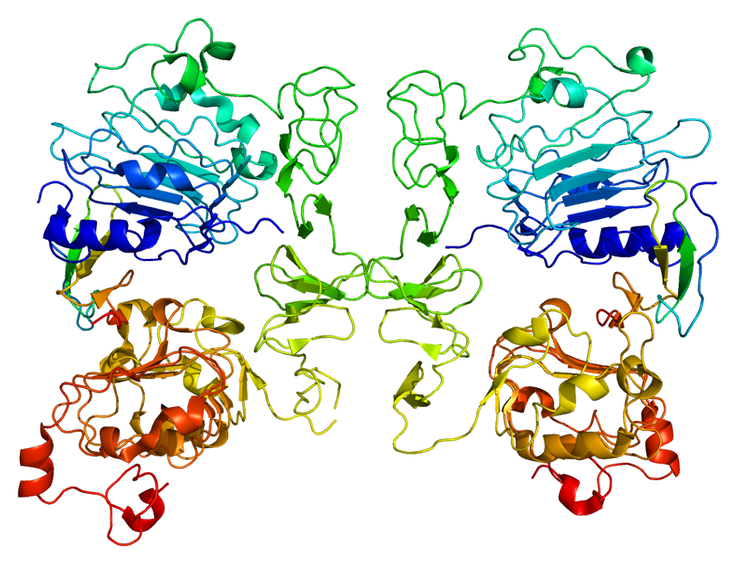
Transforming growth factor alpha (TGF-α) is a protein that in humans is encoded by the TGFA gene. As a member of the epidermal growth factor (EGF) family, TGF-α is a mitogenic polypeptide. The protein becomes activated when binding to receptors capable of protein kinase activity for cellular signaling.
TGF-α is a transforming growth factor that is a ligand for the epidermal growth factor receptor, which activates a signaling pathway for cell proliferation, differentiation and development. This protein may act as either a transmembrane-bound ligand or a soluble ligand. This gene has been associated with many types of cancers, and it may also be involved in some cases of cleft lip/palate.
TGF-α is synthesized internally as part of a 160 (human) or 159 (rat) amino acid transmembrane precursor. The precursor is composed of an extracellular domain containing a hydrophobic transmembrane domain, 50 amino acids of TGF-α, and a 35-residue-long cytoplasmic domain. In its smallest form, TGF-α has six cysteines linked together via three disulfide bridges. Collectively, all members of the EGF/TGF-α family share this structure. The protein, however, is not directly related to TGF-β.
Limited success has resulted from attempts to synthesize of a reductant molecule to TGF-α that displays a similar biological profile.
In the stomach, TGF-α is manufactured within the normal gastric mucosa. TGF-α has been shown to inhibit gastric acid secretion.
TGF-α can be produced in macrophages, brain cells, and keratinocytes. TGF-α induces epithelial development. Considering that TGF-α is a member of the EGF family, the biological actions of TGF-α and EGF are similar. For instance, TGF-α and EGF bind to the same receptor. When TGF-α binds to EGFR it can initiate multiple cell proliferation events. Cell proliferation events that involve TGF-α bound to EGFR include wound healing and embryogenesis. TGF-α is also involved in tumerogenesis and believed to promote angiogenesis.
TGF-α has also been shown to stimulate neural cell proliferation in the adult injured brain.
A 170-kDa glycosylated protein known as the EGF receptor binds to TGF-α allowing the polypeptide to function in various signaling pathways. The EGF receptor is characterized by having an extracellular domain that has numerous amino acid motifs. EGFR is essential for a single transmembrane domain, an intracellular domain (containing tyrosine kinase activity), and ligand recognition. As a membrane anchored-growth factor, TGF-α can be cleaved from an integral membrane glycoprotein via a protease. Soluble forms of TGF-α resulting from the cleavage have the capacity to activate EGFR. EGFR can be activated from a membrane-anchored growth factor as well.
Hub AI
TGF alpha AI simulator
(@TGF alpha_simulator)
TGF alpha
Transforming growth factor alpha (TGF-α) is a protein that in humans is encoded by the TGFA gene. As a member of the epidermal growth factor (EGF) family, TGF-α is a mitogenic polypeptide. The protein becomes activated when binding to receptors capable of protein kinase activity for cellular signaling.
TGF-α is a transforming growth factor that is a ligand for the epidermal growth factor receptor, which activates a signaling pathway for cell proliferation, differentiation and development. This protein may act as either a transmembrane-bound ligand or a soluble ligand. This gene has been associated with many types of cancers, and it may also be involved in some cases of cleft lip/palate.
TGF-α is synthesized internally as part of a 160 (human) or 159 (rat) amino acid transmembrane precursor. The precursor is composed of an extracellular domain containing a hydrophobic transmembrane domain, 50 amino acids of TGF-α, and a 35-residue-long cytoplasmic domain. In its smallest form, TGF-α has six cysteines linked together via three disulfide bridges. Collectively, all members of the EGF/TGF-α family share this structure. The protein, however, is not directly related to TGF-β.
Limited success has resulted from attempts to synthesize of a reductant molecule to TGF-α that displays a similar biological profile.
In the stomach, TGF-α is manufactured within the normal gastric mucosa. TGF-α has been shown to inhibit gastric acid secretion.
TGF-α can be produced in macrophages, brain cells, and keratinocytes. TGF-α induces epithelial development. Considering that TGF-α is a member of the EGF family, the biological actions of TGF-α and EGF are similar. For instance, TGF-α and EGF bind to the same receptor. When TGF-α binds to EGFR it can initiate multiple cell proliferation events. Cell proliferation events that involve TGF-α bound to EGFR include wound healing and embryogenesis. TGF-α is also involved in tumerogenesis and believed to promote angiogenesis.
TGF-α has also been shown to stimulate neural cell proliferation in the adult injured brain.
A 170-kDa glycosylated protein known as the EGF receptor binds to TGF-α allowing the polypeptide to function in various signaling pathways. The EGF receptor is characterized by having an extracellular domain that has numerous amino acid motifs. EGFR is essential for a single transmembrane domain, an intracellular domain (containing tyrosine kinase activity), and ligand recognition. As a membrane anchored-growth factor, TGF-α can be cleaved from an integral membrane glycoprotein via a protease. Soluble forms of TGF-α resulting from the cleavage have the capacity to activate EGFR. EGFR can be activated from a membrane-anchored growth factor as well.
Recent media