Hubbry Logo
ThelaziasisThelaziasisMain
Open search
Thelaziasis
Community hub
Thelaziasis
logo
8 pages, 0 posts
0 subscribers
Be the first to start a discussion here.
Thelaziasis
Thelaziasis
from Wikipedia
Thelaziasis
Other namesThelaziosis
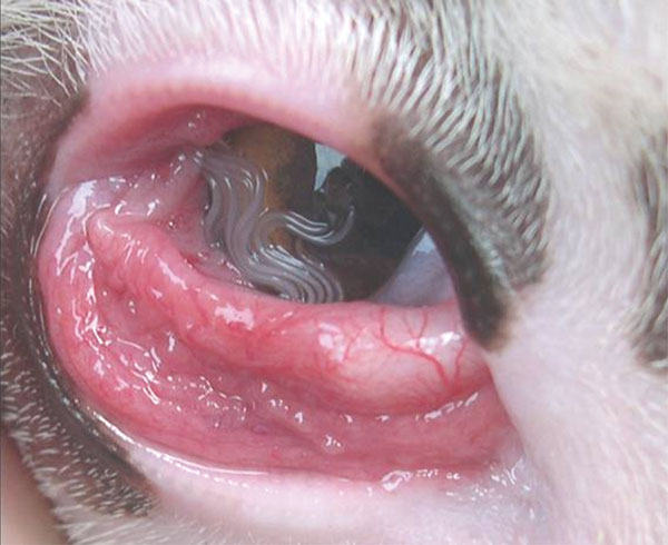
Thelazia callipaeda infestation in a dog[1]
SpecialtyInfectious disease

Thelaziasis is the term for infestation with parasitic nematodes of the genus Thelazia. The adults of all Thelazia species discovered so far inhabit the eyes and associated tissues (such as eyelids, tear ducts, etc.) of various mammal and bird hosts, including humans.[2] Thelazia nematodes are often referred to as "eyeworms".

Signs and symptoms

[edit]

In animal and human hosts, infestation by Thelazia may be asymptomatic, though it frequently causes watery eyes (epiphora), conjunctivitis, corneal opacity, or corneal ulcers (ulcerative keratitis).[1] Infested humans have also reported "foreign body sensation" – the feeling that something is in the eye.[3][4]

Cause

[edit]

Life cycle

[edit]

In the uterus of the adult female, the embryos develop into first-stage larvae (L1), which remain in the eggshell (sheath).[5] The female deposits these sheathed larvae in the tears of the mammal or bird definitive host, and the larvae are ingested by tear-feeding flies. In the fly, the larvae "hatch" (exsheath), penetrate the gut wall, and migrate to either the fat body, testes or egg follicles (depending on the species). There they develop into third-stage larvae (L3), which migrate to the head of the fly. The infective L3 larvae wiggle out of the straw-like feeding apparatus of the fly when it feeds on the tears of another mammal or bird host. The L3 larvae develop into adults in the eye or surrounding tissues of the host, where they may live for over one year.

In the definitive host, Thelazia have been found in various tissues of the orbit (or socket) of the eye, including within the eyelids, in the tear glands, tear ducts, or the so-called "third eyelid" (nictitating membrane) or in the eyeball itself.[6]

Hosts and geographic range

[edit]

While a few dozen species of Thelazia have been described in the literature, only three have been reported to infest humans, and only seven are commonly reported in veterinary contexts. The remaining species are occasionally found in birds or wild mammals.

In humans, dogs and cats, thelaziasis cases due to Thelazia callipaeda (Asia, Europe), and occasionally T. californiensis and T. gulosa (western North America), have been reported.[7]

Horses are infested by T. lacrymalis (worldwide) and, less frequently, by T. rhodesii (Africa, Asia, Europe).

In cattle, T. gulosa (Asia, Europe, North America), T. rhodesii (Africa, Asia, Europe) and T. skrjabini (Europe, North America) are the primary species of concern.

In camels, T. leesei infestations have been reported from the Post-Soviet states and India.

The intermediate hosts of several Thelazia species are known, and in each case they are tear-feeding flies of the genera Musca (family Muscidae), Phortica (family Drosophilidae), or Fannia (family Fanniidae).

Diagnosis

[edit]

Diagnosis involves simply examining the eyes and nearby tissues for the worms. Adult Thelazia are very active; one author described T. californiensis as a "short lively piece of nylon fishing line about 10 mm long."[8]

Treatment

[edit]

Because they live so close to the outside of the body, Thelazia is one of the few nematode infections which can be treated topically.

Topical treatment of livestock,[9] dogs and cats[10] with organophosphates (such as ecothiopate iodide or isofluorophate) and systemic treatment with anthelmintics (such as ivermectin, levamisole, and doramectin) are recommended by the Merck Veterinary Manual. Other sources have reported positive results treating dogs with moxidectin, imidacloprid, or milbemycin oxime.

For the treatment of human cases, removal of the worm is suggested. Topical treatment with cocaine or thiabendazole have also been reported to kill the worms in human cases.[8] Because most, if not all, species of Thelazia are spread by flies, sanitary practices which reduce the presence of flies will also reduce the spread of thelaziasis.

In canines, prevention against canine thelaziosis by monthly administrations of a combination of milbemycin oxime and afoxolaner (NexGard Spectra) has been found effective.[11]

Prevalence

[edit]

By one author's count, 257 human cases of Thelazia callipaeda had been reported worldwide by the year 2000,[12] though thelaziasis is still considered to be a rare disease.

Various livestock and wildlife surveys suggest that thelaziasis is quite common among animals.

  • A 1978 slaughterhouse survey in Guelph, Ontario, Canada found that about one-third (32%) of cattle over an eight-month period were infested with eyeworms.[13]
  • A survey of horses in Kentucky revealed a 42% rate of infestation with Thelazia lacrymalis.[14]
  • In Wyoming and Utah, a survey of hunter-harvested mule deer found 15% to be infested by Thelazia californiensis.[15]
  • A survey of various sites in Italy found 23-60% of dogs, 5% of foxes and 4 out of 4 cats to be infested with Thelazia callipaeda.[16]
  • In a study of dogs living in western Spain, 39.9% of the dogs were found to have Thelazia callipaeda worms living in their eyes.[17]

References

[edit]
[edit]
Revisions and contributorsEdit on WikipediaRead on Wikipedia
from Grokipedia
Thelaziasis is a zoonotic ocular caused by spirurid nematodes of the genus Thelazia, which primarily infest the conjunctival sac, tear ducts, and associated glands of mammals, including incidental hosts. These parasites, transmitted mechanically by lacrimophagous flies that feed on ocular secretions, lead to symptoms such as sensation, lacrimation, conjunctival inflammation, and in severe cases, corneal ulceration or . The primary causative species include Thelazia callipaeda (the Oriental eyeworm), T. californiensis, and T. gulosa, with T. callipaeda accounting for the majority of human cases (approximately 89%). Endemic in and parts of for T. callipaeda, the infection has a broader distribution for other species, including and , often linked to animal reservoirs like dogs, cats, , and . Transmission occurs when flies such as Phortica variegata (for T. callipaeda), Fannia spp. (for T. californiensis), or Musca autumnalis (for T. gulosa) ingest first-stage larvae from infected hosts and subsequently deposit infective third-stage larvae onto the eyes of new hosts during feeding. In humans, the disease is rare and typically unilateral, with over 130 reported cases globally as of recent systematic reviews, predominantly in rural Asian settings (e.g., , , ) where close contact with infected animals and poor fly control heighten risk. Clinically, infections may be asymptomatic or present with epiphora (excessive tearing, 34% of cases), conjunctival hyperemia (40%), , or , though intraocular involvement is uncommon. relies on direct visualization of the slender, worms (up to 2 cm in length) in the eye, confirmed morphologically or via molecular methods like PCR. Treatment involves manual extraction of worms under , often supplemented by topical antiparasitics such as or imidacloprid/moxidectin in veterinary contexts, though human efficacy data are limited. Prevention focuses on through fly repellents, animal , and environmental management in endemic areas, as human cases are considered emerging neglected zoonoses with increasing reports post-2000.

Etiology and Pathogen

Genus and Species

The Thelazia belongs to the family Thelaziidae within the order Spirurida of the phylum Nematoda. This places it among spirurid nematodes, distinguished from other eyeworm genera such as Oxyspirura (which primarily affects birds) by its exclusive parasitism in the ocular tissues of mammals and reliance on dipteran flies as vectors. The was established by Bosc in , encompassing vector-borne parasites that inhabit the conjunctival sacs, tear ducts, and associated structures of their hosts. Several species within the genus Thelazia are recognized as causative agents of thelaziasis, with Thelazia callipaeda being the most widespread and zoonotically significant. T. callipaeda primarily infects carnivores such as dogs and cats, but also humans as accidental hosts, and is endemic to , established in parts of , and emerging in . Another key species, Thelazia gulosa, predominates in and affects mainly , though it has been implicated in rare human cases. Thelazia skrjabini is chiefly found in ruminants like across , , and parts of , with occasional detections in wildlife such as . Less common species include Thelazia rhodesii, which has been reported in and wild ungulates in and . Other notable but rarer species encompass T. lacrymalis in horses and T. californiensis in western North American wildlife. The historical foundation of the genus traces to the description of T. callipaeda by Railliet and Henry in 1910 from specimens collected in , marking the first formal identification of a thelazial eyeworm in canines.

Morphology and Biology

Thelazia nematodes are slender, white, thread-like parasites adapted to the ocular environment of their hosts. worms exhibit , with females typically measuring 10–20 mm in length and 0.3–0.4 mm in width, while males are smaller at 7–13 mm long and 0.2–0.3 mm wide. The body is covered in fine cuticular striations, and the anterior end features a small, chitinous oral capsule without prominent , surrounded by four submedian papillae and amphids for sensory functions. In males, the posterior end is characterized by a ventrally curved bearing multiple pairs of caudal papillae (typically 8 precloacal and 3–5 postcloacal), two unequal spicules for copulation, and occasionally a small guiding the spicules, though this varies by species such as its presence in T. californiensis. Females have a straight with the positioned near mid-body and the subterminal, facilitating egg retention in the . Larval stages of Thelazia spp., particularly T. callipaeda, progress through three molts before reaching maturity. First-stage larvae (L1) are ensheathed, measuring 100–400 μm in length and 5–13.5 μm in width, with a blunt rounded anterior and pointed tail; they are released directly into the host's ocular secretions. Second-stage larvae (L2) grow to 0.46–3.2 mm long and 55–70 μm wide, developing more defined internal structures during migration in intermediate hosts. Third-stage larvae (L3), the infective form, reach approximately 2–3 mm in length, featuring a more robust body and migration to vector mouthparts for transmission to definitive hosts. Reproductively, Thelazia females are viviparous, retaining developing embryos in paired uteri and releasing motile L1 larvae into the conjunctival sac rather than depositing eggs externally. This strategy ensures direct dissemination via host tears, with achieved 1–2 months post-infection in the ocular tissues. Species like T. callipaeda show slight variations in adult size, with females often 11–15 mm and males 6–10 mm. These nematodes are specialized for surface-dwelling in the eye, inhabiting the conjunctival sacs, nictitating membranes, and lacrimal ducts while feeding on tears and glandular secretions, which contributes to their relatively low pathogenicity compared to tissue-invasive parasites. Their translucent and minimal mechanical damage allow prolonged residence without deep penetration, though heavy infestations can provoke .

Life Cycle and Transmission

Developmental Stages

The life cycle of Thelazia species exhibits direct development without free-living environmental stages, with all developmental phases occurring within definitive hosts (such as mammals) or intermediate hosts (primarily muscoid or drosophilid ). Gravid female nematodes, residing in the conjunctival sacs or lacrimal ducts of the definitive host, produce first-stage larvae (L1) that are released into ocular secretions. These L1 larvae are then ingested by suitable fly vectors during feeding on the host's , initiating further development within the intermediate host. Within the fly's digestive tract, the L1 larvae undergo two successive molts to progress through the second-stage larvae (L2) and reach the infective third-stage larvae (L3). The L3 larvae migrate to the fly's , positioning themselves for transmission. Upon the fly feeding on the ocular secretions of a new definitive host, the L3 larvae are deposited onto the eye surface and penetrate the to establish infection. In the definitive host, these L3 larvae further develop through additional molts into sexually mature adults, which migrate to the conjunctival fornix, , or lacrimal glands. Adult worms typically reach maturity and begin reproduction within 1 to 2 months post-infection, with females becoming gravid shortly thereafter. Larval development in the intermediate host generally spans 10 to 21 days, depending on species and conditions, with the shortest cycles observed around 14 days. For instance, in Phortica okadai flies infected with T. callipaeda L1 larvae, third-stage larvae emerge by day 18 and persist up to day 30. Adult nematodes have a lifespan of up to 1 year in the definitive host, during which they continue to shed L1 larvae into ocular secretions to perpetuate the cycle. Flies serve as vectors in this process, facilitating transmission without undergoing significant alteration themselves beyond hosting the larval stages. Development rates are influenced by environmental factors, particularly and , which accelerate or inhibit larval progression in the vector. Optimal conditions for T. callipaeda larval development occur at 23.4–29.7°C, with maintenance at 28 ± 2°C and 75 ± 10% relative yielding successful cycles in 14 days. Higher temperatures within this range shorten the time to infective L3 formation, while deviations can prolong development or reduce viability. levels support survival and thus vector competence, indirectly affecting overall transmission efficiency.

Vectors and Intermediate Hosts

The primary vectors of Thelaziasis are lacrimophagous dipteran flies that serve as intermediate hosts for different Thelazia species, facilitating mechanical and biological transmission of the nematode larvae. For Thelazia callipaeda, the main vector is the fruit fly Phortica variegata (Drosophilidae) in Europe and P. okadai in Asia, with males exhibiting zoophilic behavior that promotes uptake of first-stage larvae (L1) from host ocular secretions. In North America, Thelazia gulosa is primarily transmitted by the face fly Musca autumnalis (Muscidae), while Thelazia californiensis relies on latrine flies of the genus Fannia, including F. canicularis and F. benjamini. The transmission process begins when adult female nematodes release L1 larvae into the host's conjunctival sac and tear secretions; these larvae are ingested by flies during feeding on the ocular fluids. Within the fly's digestive tract and tissues, the L1 molt twice over approximately 2–3 weeks, developing into infective third-stage larvae (L3), which then migrate to the fly's mouthparts. of a new definitive host occurs when the fly deposits L3 onto the eye surface while feeding on secretions, allowing the larvae to penetrate the . This cycle integrates with the fly's life history, as the parasite's development aligns with the insect's adult stage longevity, typically spanning days to weeks under suitable temperatures. Vector biology emphasizes the flies' dependence on warm, humid conditions for peak activity, with M. autumnalis emerging in spring (March–April) and remaining active through summer and autumn until in cooler months, concentrating transmission during these periods. Similarly, P. variegata exhibits heightened lachryphagous activity in summer and early autumn at temperatures of 20–25°C, correlating with increased fly densities and parasite prevalence in endemic areas. Experimental evidence confirming vector competence dates from the early , with foundational studies in the demonstrating M. autumnalis as a competent host for T. gulosa, where L1 led to L3 development and larviposition after 10–14 days. Later in the quantified survival rates and sex ratios of developing larvae in face flies, supporting biological transmission efficiency. For T. callipaeda, 2000s studies verified P. variegata's role under natural and conditions, including male-mediated transmission and L3 infectivity after 20 days of development at 25°C. These findings, spanning field observations to modern molecular confirmations, underscore the flies' essential role without direct host-to-host transfer.

Epidemiology

Primary and Accidental Hosts

Thelazia parasites, belonging to the genus Thelazia (Nematoda: Spirurida), primarily infect the ocular tissues of various mammals, with host specificity varying by species. Primary hosts include both domestic and wild animals that serve as natural reservoirs for the parasite's life cycle. Domestic primary hosts encompass dogs, cats, , and sheep, while wildlife reservoirs include foxes, wolves, deer, and rabbits. Host specificity is evident among Thelazia species; for instance, T. callipaeda predominantly infects canids (such as dogs and foxes) and felids (such as cats), as well as lagomorphs like rabbits, whereas T. gulosa primarily affects ungulates including , sheep, and deer. Infection occurs through ocular exposure when vectors, such as drosophilid or face flies, deposit infective larvae on the host's eyes while feeding on lacrimal secretions. Humans serve as accidental or dead-end hosts for Thelazia species, with infections being rare and zoonotic in nature, typically resulting from close contact with infected pets like dogs or cats. Documented cases, often involving T. callipaeda or T. gulosa, highlight the potential for incidental transmission, though humans do not contribute to sustained parasite propagation. Other mammals, such as pigs, may act as accidental hosts for certain Thelazia species in specific contexts, but infections remain uncommon and non-reservoir roles.

Geographic Distribution

Thelaziasis, caused primarily by Thelazia callipaeda, is endemic in several Asian countries, including , , , , and , where it has been reported in both animals and humans for decades. In these regions, the parasite is particularly prevalent among dogs, which serve as primary reservoirs, facilitating its maintenance in rural and agricultural settings. In , T. callipaeda has emerged as an expanding since the early , initially reported in and subsequently spreading to , , and during the . The parasite's distribution has further extended into southern and , including , , , and , often through wildlife such as foxes and mustelids acting as dispersers. In , thelaziasis is less common and primarily involves T. gulosa in and T. californiensis in various hosts across the , with rare human cases documented in . The historical introduction of T. callipaeda to is thought to have occurred via the movement of infected animals through trade or migration from , with ongoing monitoring through veterinary surveys in wildlife and domestic populations. Distribution patterns are influenced by climatic conditions suitable for fruit fly vectors, as well as underreporting in rural areas where human-animal contact is frequent.

Prevalence and Incidence

Thelaziasis exhibits varying among animal hosts, particularly dogs and , in endemic regions. In , where the disease is longstanding, prevalence in dogs can reach up to 84.6% in high-risk rural areas of , though urban and overall rates are lower, around 3-4% in . In , canine prevalence ranges from 1% to over 40% in foci such as and , with rates up to 41.8% reported in specific Italian regions; in , infections are widespread but typically lower, at 0.5-10%. For , primarily affected by Thelazia gulosa and related species in , prevalence is estimated at 5-25%, with studies in showing 21.5% in beef herds and 25.7% in . Human thelaziasis remains rare globally, with a systematic review documenting 134 cases across 18 countries as of 2023, predominantly caused by Thelazia callipaeda. In , reports the highest burden, with 32 cases documented in the review (from 1949 to 2023), though local reports suggest up to 658 cases from 1917 to 2020. In , human cases are emerging but sporadic, with 17 confirmed infections across countries like , , and since the first autochthonous case in in 2006. Epidemiological trends indicate an increasing incidence in , driven by the expansion of fruit fly vectors like Phortica variegata into new territories, facilitating zoonotic spillover from and pets. Infections peak seasonally in late summer and autumn, aligning with vector activity from spring to fall. Key risk factors include rural residence, pet ownership—especially of outdoor dogs—and underdiagnosis in veterinary settings, where clinical signs are often mistaken for other ocular conditions, leading to only about 16% of cases being correctly identified at initial referral. A 2024 case in urban suggests possible emergence in non-rural settings.

Clinical Features

Signs and Symptoms in Animals

Thelaziasis in animals primarily manifests through ocular caused by the mechanical movement of adult nematodes in the conjunctival sac, , or lacrimal ducts. Common clinical signs include epiphora (excessive tearing), ocular pruritus, , , and visible worms in the eye, often leading to mild to moderate discomfort. Additional symptoms such as , ocular discharge, and conjunctival hyperemia are frequently reported, particularly in cases with multiple worms. In dogs and cats, infections are typically unilateral and cause mild , with signs like excessive lacrimation and conjunctival inflammation predominating; or slow-healing ulcers may occur but are uncommon. In cattle, symptoms include lacrimation, , and , with flies often clustering around affected eyes; corneal ulcers remain rare even in heavier infestations. Wildlife species, such as deer or foxes, frequently serve as carriers, though mild bilateral has been noted in some cases. If untreated, thelaziasis can lead to complications such as secondary bacterial infections, resulting in purulent discharge, or with potential corneal ulceration. Chronic infections may cause corneal pigmentation and persistent . Infections are more prevalent in young and outdoor animals due to increased exposure to vectors like flies, with adult dogs and in extensive management systems showing higher rates; breed predispositions vary, with local or large breeds in some regions at greater risk.

Signs and Symptoms in Humans

Thelaziasis in humans is a rare zoonotic ocular infection caused by nematodes of the genus Thelazia, primarily T. callipaeda, transmitted from animal hosts via vectors. In affected individuals, adult worms or larvae inhabit the l sac, tear ducts, or lacrimal glands, leading to a range of mild to moderate symptoms. The condition is often unilateral, affecting 90.3% of cases, with a median of 3 parasites per eye (range 1–32), and worms are typically visible on the or in tears. The most common symptoms prompting medical consultation include a sensation in 53% of cases, conjunctival hyperemia in 39.6%, and epiphora in 33.6%. Other frequent manifestations are ocular pruritus (27.6%), direct visualization of parasites by the patient (26.9%), and red eye (21.6%). Lacrimation and may also occur, particularly in heavier infestations, alongside varying degrees of . Approximately 5.2% of cases are and discovered incidentally. Complications are uncommon but can include corneal ulcers (1.5%), corneal abrasions, keratitis, secondary bacterial infections such as preseptal cellulitis, and rarely, vision impairment. Reinfection occurs in about 6% of cases. Human cases predominantly occur in rural areas of Asia, accounting for 82.8% of reported infections in a 2025 systematic review of 134 global cases, though Chinese literature reports over 650 cases in China up to 2018 alone, indicating potential underreporting in international databases. Affected individuals range in age from children to adults (mean 40.5 years, interquartile range 23–60.5 years) and show a slight male predominance (59.7%).

Pathogenesis

Thelazia nematodes, primarily species such as Thelazia callipaeda and Thelazia gulosa, induce ocular through a combination of mechanical and biochemical mechanisms localized to the eye's surface structures. Adult worms and larvae inhabit the conjunctival sac, lacrimal ducts, and , where their active movement causes direct abrasion of the conjunctival and via the worms' serrated cuticular surface. This mechanical disrupts the ocular surface integrity, leading to localized trauma and secondary bacterial in heavier infestations. Additionally, the parasites release enzymatic secretions and excretory-secretory products that exhibit toxic effects, promoting and further exacerbating tissue damage. Larval migration within the tear ducts and along the ocular surface contributes to persistent , as first- and third-stage larvae navigate these confined spaces during development. The host's immune response to Thelazia infestation is predominantly local and confined to the ocular region, characterized by an eosinophilic conjunctivitis driven by type 2 immune pathways. Eosinophil infiltration and IgE-mediated hypersensitivity occur in response to the parasites' antigens, resulting in follicular hyperplasia and exudative inflammation of the conjunctiva. These reactions manifest as chemosis, hyperemia, and increased lacrimal secretion, but systemic involvement remains minimal due to the superficial, non-invasive nature of the infection, with rare reports of broader allergic responses. The overall immune activation is self-limiting in many cases, potentially leading to parasite expulsion through heightened tear production and ocular flushing. Tissue-level effects of thelaziasis include progressive epithelial damage, where repeated mechanical and enzymatic insults lead to corneal erosions, opacity, and ulceration in severe cases. Histological changes often involve in the conjunctival , enhancing production as a protective response to . While the parasites typically remain ectoparasitic on the ocular surface, rare instances of deeper invasion into subconjunctival tissues can occur, particularly with high larval burdens, potentially forming cysts or proliferative lesions. Severity of thelaziasis is influenced by several host-parasite interaction factors, including worm burden, which correlates with the intensity of mechanical damage and inflammatory response—higher loads (e.g., >5 worms per eye) often result in more pronounced clinical signs. Host immunity plays a key role, with immunocompromised individuals or animals (e.g., due to or stress) exhibiting increased susceptibility and prolonged infestations, while robust local defenses may promote self-resolution. The parasites demonstrate low overall , with most infections remaining subclinical or mild, rarely progressing to vision-threatening complications unless untreated.

Diagnosis

Clinical Detection

Clinical detection of thelaziasis relies on direct visualization of the nematodes during ophthalmologic or veterinary examination, often prompted by symptoms such as sensation or ocular irritation. Key examination techniques include slit-lamp biomicroscopy to identify worms on the ocular surface, sometimes aided by topical to facilitate visualization without discomfort. Flushing the conjunctival sac with sterile saline solution can also dislodge and reveal hidden parasites, allowing for their observation in the tear film or fornix. Diagnostic indicators typically involve spotting translucent or semihyaline adult worms, measuring 1-2 cm in length, crawling on the , , or within the lacrimal ducts; unilateral eye involvement is common. In veterinary practice, routine eye examinations using these methods are recommended for dogs and cats in endemic regions to detect subclinical infections early. In human ophthalmology, detection is usually opportunistic, occurring during evaluations for unrelated ocular complaints in areas where the parasite is prevalent. Challenges in clinical detection include asymptomatic presentations where worms cause no noticeable signs, leading to overlooked infections, and frequent misdiagnosis as a simple due to the worms' mobile and thread-like appearance.

Laboratory Confirmation

Laboratory confirmation of thelaziasis involves the collection and analysis of biological samples to identify Thelazia definitively, distinguishing them from other ocular parasites or conditions. Samples are typically obtained by manual removal of adult worms from the conjunctival sac using non-toothed under or slit-lamp guidance, ensuring minimal trauma to the ocular surface. Lacrimal secretions can also be collected via smears or flushing with saline to detect first-stage larvae, which are then examined microscopically. Microscopic examination of extracted worms remains the cornerstone of identification, relying on morphological characteristics such as body length, cuticular striations, and reproductive structures. Thelazia worms measure 5–20 mm in length, with males generally shorter (5–12 mm) than females (10–19 mm). Key diagnostic features include the number and arrangement of caudal papillae—typically 7 pairs preanal and 2 pairs postanal in males—and spicule length, which varies by species (e.g., 0.7–1.31 mm in T. callipaeda males). Larvae in tear fluid are identified by their elongated, sheathed form and size (approximately 0.3–0.4 mm). These morphological traits provide high specificity for species differentiation when performed by trained parasitologists. Molecular methods, particularly (PCR) targeting the mitochondrial subunit 1 (cox1) , offer confirmatory identification, especially in cases where morphology is ambiguous due to damaged specimens or larval stages. PCR amplification followed by sequencing of the cox1 (e.g., producing 658–689 bp fragments) enables , which has been used to verify T. callipaeda and other like T. skrjabini with phylogenetic accuracy. Serological tests are limited and not routinely employed for due to lack of validated assays specific to Thelazia antigens. Morphological microscopy yields high specificity (>95% in expert settings) for adult worm identification but may be less reliable for larvae or mixed infections, where molecular tools enhance precision through genetic markers. Emerging approaches are increasingly integrated in research to resolve cryptic species diversity and support epidemiological tracking.

Management and Prevention

Treatment Options

The primary treatment for thelaziasis involves mechanical removal of adult Thelazia worms from the conjunctival sac, typically performed under using fine , cotton swabs, or saline to flush out visible parasites. This method is considered definitive for cases and achieves near-complete efficacy (close to 100%) against accessible adult worms, though it may miss larval stages or embedded parasites. In veterinary practice, mechanical extraction is similarly the first-line approach for dogs and cats, often combined with gentle flushing to minimize ocular trauma. Pharmacological interventions target both adult and larval stages, particularly in animals. administered subcutaneously at 0.2 mg/kg or, off-label, as topical , has demonstrated high (up to 87-100%) in eliminating Thelazia infections in and dogs by paralyzing and killing the nematodes, though topical use should be approached cautiously due to potential adverse reactions. Oral milbemycin oxime, at a minimum dose of 0.5 mg/kg monthly, effectively clears infections in companion animals like dogs, with rates exceeding 90% when used systemically. In humans, pharmacological options are limited, and mechanical removal remains the standard, though adjunctive has been explored in select cases without established routine use. Additionally, topical (5 mg/mL applied every 30 minutes for four doses) has shown in treating T. callipaeda infections without significant , as reported in a 2025 study. Supportive care addresses secondary complications such as bacterial infections or . Topical antibiotic drops, including tobramycin or , are commonly prescribed to prevent or treat and resulting from worm migration or removal procedures. agents, such as dexamethasone or pranoprofen eye drops, help reduce ocular swelling, redness, and discomfort, particularly in cases with significant conjunctival irritation. These measures are essential for symptom management in both human and animal patients. Treatment outcomes are generally favorable, with cure rates approaching 98% following mechanical removal and supportive therapy, and low recurrence (under 3%) in monitored cases. However, reinfection can occur if vector flies or intermediate hosts remain untreated in the environment, necessitating follow-up examinations. In animals, protocols incorporating monthly administration post-treatment enhance long-term resolution.

Preventive Measures

Preventing Thelaziasis involves integrated strategies targeting its primary vectors, lacryphagous flies such as Phortica variegata in and Phortica okadai in , which transmit the nematodes by feeding on ocular secretions. Vector control measures include the deployment of baited traps on farms and in endemic areas; for instance, transparent traps with 14 small holes, filled with a 50:50 blend of and vinegar, effectively capture male Phortica flies when suspended 1.6–1.8 meters high under tree canopies during peak activity periods. Insecticides applied to and companion animals, such as pour-on formulations, reduce fly populations landing on hosts, while environmental management practices like prompt removal of organic waste and minimize breeding sites for drosophilid flies in rural and agricultural settings. Repellents like , released at approximately 5 mg per day from baited traps, have shown significant reduction in fly captures compared to untreated controls. Host management focuses on domestic and wild animals as reservoirs, with regular prophylactic recommended during fly seasons. Monthly oral administration of moxidectin-based products, such as sarolaner/moxidectin/ at doses of 24 µg/kg moxidectin, achieves 100% efficacy in preventing Thelazia callipaeda infections in dogs over six months in endemic and . Similarly, milbemycin oxime at a minimum dose of 0.5 mg/kg monthly eliminates infections and prevents reinfection in dogs and cats, offering a practical option for year-round prophylaxis in . For imported animals, veterinary screening and potential protocols are advised to curb cross-border spread, particularly in emerging European foci, alongside monitoring of populations like foxes and deer through to detect and manage subclinical infections. Human prevention emphasizes reducing direct exposure to infected flies, especially in rural or farming communities. Wearing protective eyewear and using insect repellents on the face during outdoor activities in endemic areas significantly lowers the risk of larval deposition in the eyes. Regular screening and of household pets, combined with on zoonotic transmission risks, further mitigates household-level spread; for example, campaigns promoting bed nets at night protect sleeping individuals from fly contact. Maintaining personal hygiene, such as avoiding eye rubbing after fly encounters, is also advised. Public health initiatives include ongoing surveillance programs in emerging regions like , where autochthonous cases in humans and animals are rising, to track prevalence and guide interventions through a framework integrating veterinary, medical, and environmental efforts. No vaccines are currently available for Thelaziasis prevention in humans or animals, underscoring the reliance on vector and host controls.

References

Add your contribution
Related Hubs
User Avatar
No comments yet.