Hubbry Logo
KeratitisKeratitisMain
Open search
Keratitis
Community hub
Keratitis
logo
8 pages, 0 posts
0 subscribers
Be the first to start a discussion here.
Contribute something
Keratitis
Keratitis
from Wikipedia
Keratitis
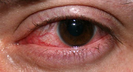
An eye with non-ulcerative sterile keratitis.
SpecialtyOphthalmology

Keratitis is a condition in which the eye's cornea, the clear dome on the front surface of the eye, becomes inflamed.[1] The condition is often marked by moderate to intense pain and usually involves any of the following symptoms: pain, impaired eyesight, photophobia (light sensitivity), red eye and a 'gritty' sensation.[2] Diagnosis of infectious keratitis is usually made clinically based on the signs and symptoms as well as eye examination, but corneal scrapings may be obtained and evaluated using microbiological culture or other testing to identify the causative pathogen.[3]

Classification (by chronicity)

[edit]

Classification (infective)

[edit]

Viral

[edit]
Dendritic corneal ulcer after fluorescein staining under cobalt blue illumination
Adenoviral keratitis of a 24-year-old woman

The most common causes of viral keratitis include herpes simplex virus (HSV) and varicella zoster virus (VZV), which cause herpes simplex keratitis and herpes zoster keratitis (a subtype of herpes zoster ophthalmicus) respectively.[3] Herpes simplex keratitis occurs due to latent HSV reactivation in the ophthalmic nerve (the V1 branch of the trigeminal nerve).[3] Herpes keratitis is classically associated with a branching (dendritic) infiltrate pattern of inflammation in the corneal epithelium and may cause clouding of the cornea.[3]

Approximately 8-20% of cases of shingles (due to VZV reactivation) involve the eyes as herpes zoster ophthalmicus.[4][3] And VZV keratitis occurs in 13-76% of cases of herpes zoster ophthalmicus, usually 1 month after onset of symptoms.[3] Herpes zoster ophthalmicus is also associated with reactivation of ZVZ in the V1 branch (the ophthalmic nerve) of the trigeminal nerve.[4] VZV keratitis presents as a dendriform epithelial keratitis pattern early in the course of the infection.[4] ZVZ keratitis may cause clouding of the cornea, with 50% of cases involving inflammatory infiltrates in the stromal layer of the cornea, corneal scarring is a possible complication of VZV keratitis.[3] Vaccination with the zoster vaccine is highly effective in preventing shingles, as well as herpes zoster ophthalmicus and herpes zoster keratitis.[3]

Bacterial

[edit]
  • Bacterial keratitis. Bacterial infection of the cornea can follow from an injury or from wearing contact lenses. The bacteria involved are Staphylococcus aureus and for contact lens wearers, Pseudomonas aeruginosa. Pseudomonas aeruginosa produces enzymes that can digest the cornea.[5]
  • In those who wear contact lenses, bacteria are the most common causative agent of keratitis, with 90% of cases being due to a bacterial pathogen. Of those 90% of cases, Pseudomonas aeruginosa is responsible for 40%.[3] Staph aureus and streptococci are other common bacterial pathogens responsible for infectious keratitis in contact lens wearers.[3] Lens cases, used to store contact lenses, may form a biofilm leading to colonization of the contact lenses by bacteria, this is especially common with poor contact lens hygiene or improper storage.[3]

Fungal

[edit]

Amoebic

[edit]
Acanthamoeba keratitis

Parasitic

[edit]

Microbial keratitis (due to bacterial, fungal, or parasitic pathogens), as opposed to viral keratitis, is more commonly associated with the formation of corneal ulcers. Other risk factors for corneal ulcer formation include contact lens use, keratitis in the setting of eye trauma, underlying corneal disease or ocular surface diseases (such as severe chronic dry eye).[3] Infectious keratitis sometimes presents as corneal edema, or with a hypopyon (a collection of inflammatory cells in the anterior chamber of the eye).[3]

Classification (by stage of disease)

[edit]

Classification (by environmental aetiology)

[edit]

Treatment

[edit]

Treatment depends on the cause of the keratitis. Infectious keratitis can progress rapidly, and generally requires urgent antibacterial, antifungal, or antiviral therapy to eliminate the pathogen. Antibacterial solutions include levofloxacin, gatifloxacin, moxifloxacin, ofloxacin. It is unclear if steroid eye drops are useful.[11]

In addition, contact lens wearers are typically advised to discontinue contact lens wear and replace contaminated contact lenses and contact lens cases. (Contaminated lenses and cases should not be discarded as cultures from these can be used to identify the pathogen).

Topical ganciclovir or oral valacyclovir, famciclovir or acyclovir are used for HSV keratitis.[3] Steroids should be avoided as application of steroids to a dendritic ulcer caused by HSV may result in rapid and significant worsening of the ulcer to form an 'amoeboid' or 'geographic' ulcer, so named because of the ulcer's map like shape.[12]

Prevention

[edit]

In those who wear contact lenses, good lens hygiene and storage practices reduce the risk of keratitis. Specific lens care practices which may lead to infectious keratitis include wearing contact lenses overnight or in the shower, not replacing contact lens cases, storing lenses in tap water rather than contact lens solution and topping off lens solution rather than replacing it regularly.[3] Improper lens storage may lead to bacterial biofilm formation in the contact lens case and subsequent colonization of the lenses by bacteria.[3] Exposure of the lens to tap water through improper storage or use may lead to acanthamoeba infection, as the amoeba is commonly found in tap water.[3]

Acyclovir prophylaxis has been found to reduce the risk of additional episodes of herpes simplex viral eye diseases (as well as oral or facial herpes) including a 50% reduction in the incidence of HSV keratitis. There was no rebound effect, or increased rate of HSV related eye disease upon stopping acyclovir prophylaxis.[13]

Prognosis

[edit]

Some infections may scar the cornea, thereby limiting vision. Others may result in perforation of the cornea, endophthalmitis (an infection inside the eye), or even loss of the eye. With proper medical attention, infections can usually be successfully treated without long-term visual loss.[citation needed]

Acanthamoebic and fungal keratitis are difficult to treat and are associated with a poor prognosis.[3]

In non-humans

[edit]
  • Feline eosinophilic keratitis — affecting cats and horses; possibly initiated by feline herpesvirus 1 or other viral infection.[14]

See also

[edit]

References

[edit]
[edit]
Revisions and contributorsEdit on WikipediaRead on Wikipedia
from Grokipedia
Keratitis is the of the , the clear, dome-shaped tissue covering the front of the eye that protects the iris and . It can result from infectious agents such as , viruses, fungi, or parasites, or from noninfectious causes like , autoimmune conditions, or exposure to irritants. If untreated, keratitis may lead to corneal scarring, ulceration, or permanent vision loss, making it a significant cause of ocular morbidity worldwide. Infectious keratitis, the most common form, accounts for a substantial portion of cases and is often associated with risk factors including contact lens wear, ocular trauma, and compromised ocular surface integrity. Bacterial keratitis, frequently caused by pathogens like Pseudomonas aeruginosa or Staphylococcus aureus, is prevalent among contact lens users, with an incidence rate up to 130 cases per 100,000 person-years in this group compared to 14 per 100,000 in non-wearers (as of 2010). Viral keratitis, particularly from herpes simplex virus (HSV), affects the corneal epithelium and stroma, leading to recurrent episodes and dendritic ulcers. Fungal and parasitic forms, such as those caused by Aspergillus species or Acanthamoeba, are more common in agricultural settings or with exposure to contaminated water, and they pose challenges due to delayed diagnosis. Noninfectious keratitis may stem from systemic diseases like rheumatoid arthritis or nutritional deficiencies such as vitamin A shortage, resulting in stromal inflammation without microbial involvement. Emerging antimicrobial resistance in bacterial pathogens has complicated treatment in recent years. Epidemiologically, keratitis contributes to corneal blindness, affecting an estimated 1.3 million individuals globally due to infectious keratitis as of 2015, and ranks as the fifth leading cause of blindness, particularly in developing regions like where incidence can reach 799 per 100,000 in high-risk areas (as of 2007). More recent estimates as of 2020 indicate that corneal opacities, largely from keratitis, cause blindness or moderate/severe in approximately 5.5 million people bilaterally and 6.2 million unilaterally worldwide. , it prompted over 1 million annual visits to eye care professionals as of 2010. Common symptoms include eye pain, redness, , excessive tearing, , and a sensation of a , which can vary in severity based on the underlying . Diagnosis typically involves clinical examination, corneal scrapings for and sensitivity, (PCR) testing, or to identify the causative agent. Treatment strategies are tailored to the type and severity of keratitis, emphasizing prompt intervention to prevent complications. Bacterial cases are managed with topical fortified antibiotics like or fluoroquinolones, while viral infections require antivirals such as acyclovir. Fungal keratitis responds to topical or , and parasitic infections like necessitate biguanides such as polyhexamethylene . For noninfectious forms, corticosteroids or immunosuppressive agents may be used cautiously to reduce , alongside addressing underlying conditions; recent advances include therapies for neurotrophic cases. depends on early detection, with scarring occurring in many cases but vision preservation possible through timely therapy or surgical options like in advanced scenarios.

Clinical Presentation

Symptoms

Patients with keratitis typically present with a range of subjective ocular symptoms, primarily involving and due to of the . Ocular is a hallmark , often described as ranging from mild irritation or a gritty foreign body sensation to severe, sharp discomfort that worsens with blinking or eye movement. This may be accompanied by significant , making exposure to light intensely uncomfortable and prompting patients to keep their eyes closed. Visual disturbances are central to the experience of keratitis, with blurred or hazy vision being the most common report, resulting from corneal edema and irregularity that scatters light entering the eye. Reduced , particularly in the affected eye, can occur rapidly, and some patients notice halos around lights, especially at night, due to corneal swelling. These symptoms are specific to corneal involvement and may lead to difficulty with daily activities like reading or driving. Associated discomforts include excessive tearing, a sensation of eye redness (though subjective), and discharge that can feel sticky or watery. Symptom severity and onset vary by underlying cause; for instance, bacterial keratitis often presents with acute, intense pain and purulent discharge, whereas viral keratitis, such as , tends to have a more gradual onset with milder discomfort and watery discharge. Fungal keratitis may initially cause subtler symptoms, with less pronounced pain compared to bacterial forms. In untreated cases, symptoms typically progress rapidly, with pain and visual blurring intensifying within hours to days, potentially leading to profound vision loss if the spreads or causes corneal thinning. This progression correlates briefly with observable signs like , underscoring the urgency of symptom recognition.

Signs

Clinical signs of keratitis are primarily identified through slit-lamp biomicroscopy, which reveals characteristic corneal abnormalities and associated ocular changes. Epithelial defects appear as punctate erosions or ulcers that stain with fluorescein, often overlying stromal infiltrates that manifest as focal opacities ranging from small, gray-white lesions to larger, denser areas of . Stromal involvement may include and cellular infiltration, while anterior chamber reaction typically presents as cells and flare, indicating inflammatory response in the aqueous humor. Specific slit-lamp findings vary by keratitis type. In (HSV) keratitis, dendritic ulcers are hallmark features, appearing as branching, linear epithelial lesions with terminal bulbs that enhance under light after fluorescein staining. often shows a ring-shaped stromal infiltrate in advanced stages, accompanied by radial keratoneuritis—linear opacities extending from the limbus—and epitheliopathy resembling pseudodendrites. Bacterial keratitis typically exhibits an epithelial defect overlying a suppurative stromal infiltrate with irregular, undermined edges, while fungal keratitis displays dry, elevated lesions with feathery margins and satellite infiltrates. Severity of corneal involvement is assessed using standardized grading systems. The Oxford grading scheme evaluates the extent of corneal staining post-fluorescein application on a 0–5 scale, where grade 0 indicates no staining and grade 5 represents confluent staining across the entire corneal surface. Ulcer depth is measured via slit-lamp pachymetry or , often categorized as superficial (limited to ), stromal (involving mid-layers), or deep (approaching ), with depths exceeding 50% of corneal thickness signaling high risk for . Associated ocular signs include conjunctival injection, which may be focal (ciliary flush around the limbus) or diffuse, reflecting vascular response to corneal inflammation. Lid edema contributes to periorbital swelling, particularly in herpetic cases, while —a layered collection of and debris in the anterior chamber—involves up to 20–30% of cases, most commonly in bacterial or fungal infections, and appears as a dependent, yellowish level on upright slit-lamp view.

Etiology and Risk Factors

Infectious Causes

Infectious keratitis arises from a variety of microbial pathogens that invade the corneal epithelium, leading to inflammation and potential vision loss. These infections typically occur following corneal trauma, contact lens overuse, or exposure to contaminated environments, allowing opportunistic microbes to breach the ocular surface barrier. Bacterial keratitis is predominantly caused by gram-positive and gram-negative organisms, with Staphylococcus aureus and Pseudomonas aeruginosa being among the most common pathogens. S. aureus often colonizes the ocular surface in cases of chronic blepharitis or after surgical interventions, while P. aeruginosa is frequently implicated in contact lens-related infections due to its ability to thrive in moist environments and form biofilms on lens surfaces, which protect it from host defenses and disinfectants. These biofilms contribute to outbreaks, as seen in increased incidence among extended-wear contact lens users exposed to contaminated solutions. Other notable bacteria include Staphylococcus epidermidis and Streptococcus pneumoniae, which exploit epithelial defects to initiate infection. Viral keratitis is primarily driven by herpesviruses, with type 1 (HSV-1) accounting for the majority of cases through recurrent epithelial and stromal infections. HSV-1 establishes latency in trigeminal ganglia and reactivates due to triggers like stress or , leading to dendritic ulcers and neurotrophic effects that impair corneal sensation and healing. Varicella-zoster virus (VZV), reactivating as herpes zoster ophthalmicus, similarly damages corneal nerves, causing neurotrophic keratitis with pseudodendrites and stromal involvement. Adenovirus contributes to epidemic keratoconjunctivitis, featuring superficial punctate keratitis that can progress to subepithelial infiltrates. These viruses evade immunity by inhibiting responses and persisting in neuronal tissues. Fungal keratitis involves opportunistic pathogens such as Candida species and species, often following ocular trauma with vegetative matter like plant material, which introduces spores into the corneal stroma. and species predominate in agricultural settings, where hyphal forms invade stromal layers, eliciting a robust inflammatory response. Candida typically affects immunocompromised individuals or those with prior steroid use, penetrating via endothelial routes in severe cases. The hyphal invasion by filamentous fungi like disrupts architecture and promotes through enzymatic degradation. Parasitic and protozoal causes include species and , both associated with exposure to contaminated sources. * arises from trophozoites or resilient cysts entering via contact lens storage in or during , with cysts exhibiting exceptional durability—surviving for years and resisting standard multipurpose solutions due to their double-walled structure. This leads to ring-shaped infiltrates and radial . , spore-forming , cause punctate epitheliopathy after ocular exposure to or , embedding spores in corneal cells and evading . Water-related risks are heightened in wearers, where poor hygiene facilitates protozoal adhesion. The general of infectious keratitis involves microbial adhesion to the via adhesins or pili, followed by evasion of innate immunity through capsule formation or toxin-mediated suppression of . Pathogens like P. aeruginosa and S. aureus produce exotoxins (e.g., alpha-toxin) and proteases that degrade epithelial tight junctions and , causing ulceration and influx that amplifies tissue destruction. Fungi and employ hyphal or penetration to invade deeper layers, while viruses induce cell and immune dysregulation. This cascade results in if unchecked.

Non-Infectious Causes

Non-infectious keratitis encompasses a range of conditions where corneal arises from immune-mediated, mechanical, chemical, or neurological disruptions rather than microbial invasion. These etiologies often result in sterile corneal infiltrates characterized by epithelial defects, stromal haze, and ulceration without suppuration, distinguishing them from infectious forms that typically exhibit purulent discharge and rapid progression. Autoimmune and inflammatory processes play a significant role in non-infectious keratitis, particularly through mechanisms involving immune complex deposition and reactions. In , peripheral ulcerative keratitis (PUK) develops when immune complexes activate the , recruiting neutrophils and macrophages that release collagenase and proteases, leading to stromal degradation and crescent-shaped limbal ulcers. Similarly, Sjögren's syndrome contributes via lymphocytic infiltration of lacrimal glands, causing aqueous tear deficiency and keratoconjunctivitis sicca, which manifests as filamentary keratitis due to disrupted tear film stability and elevated proinflammatory cytokines like IL-6 and TNF-α. These conditions highlight immune-driven sterile inflammation, often requiring systemic evaluation to address underlying collagen vascular diseases. Nutritional deficiencies, particularly , can lead to and , resulting in corneal drying, melting, and ulceration due to impaired epithelial integrity and increased susceptibility to secondary infections. Traumatic and chemical injuries represent common external triggers for non-infectious keratitis, initiating sterile inflammatory responses through direct corneal damage. Abrasive injuries from foreign bodies, scratches, or trichiasis cause epithelial denudation and subsequent stromal infiltration without initial microbial involvement, though secondary infection risk exists. Chemical burns and (UV) exposure, as in or "snow blindness," damage the via photochemical reactions, leading to punctate erosions and transient inflammation that resolves without antibiotics if uncomplicated. These mechanisms emphasize the cornea's vulnerability to environmental insults, resulting in self-limited sterile keratitis unless exacerbated by ongoing exposure. Neurotrophic and exposure-related keratitis stem from impaired corneal innervation or inadequate eyelid closure, promoting epithelial breakdown through reduced protective reflexes. damage, such as post-herpetic neuralgia following herpes zoster ophthalmicus, diminishes corneal sensation and trophic support, fostering persistent epithelial defects and neurotrophic ulcers via halted and healing. , often from or proptosis, causes incomplete eyelid closure, leading to exposure keratopathy where desiccated corneal surfaces develop superficial punctate keratitis and potential ulceration from chronic drying. These forms underscore the role of neural and mechanical factors in maintaining ocular surface integrity, yielding sterile changes that mimic infection but lack microbial elements. Iatrogenic causes arise from medical interventions that compromise corneal health, often through overuse or procedural trauma. Postoperative complications following or other refractive surgeries can induce diffuse lamellar keratitis via epithelial disruption or inflammatory debris under the flap, presenting as sterile central infiltrates. Prolonged use of topical s, such as in abuse cases, toxic to the , results in anesthetic keratopathy with ring-shaped infiltrates and delayed healing due to inhibited cellular proliferation. These iatrogenic insults produce non-suppurative , contrasting with infectious keratitis by the absence of pathogens and reliance on resolution through intervention cessation. In summary, non-infectious keratitis features sterile infiltrates driven by immune, traumatic, or neurotrophic mechanisms, differing from infectious suppuration by the lack of microbial proliferation and purulent response, though clinical overlap may necessitate diagnostic differentiation.

Classification

By Chronicity

Keratitis is classified by chronicity into acute, chronic, and recurrent forms based on the duration and pattern of corneal . Acute keratitis is typically defined as an episode lasting less than 3 months, characterized by rapid onset and progression that can lead to severe complications such as corneal thinning and if not addressed promptly. This form is exemplified by bacterial corneal ulcers, where aggressive inflammatory responses cause swift tissue destruction, often within days to weeks. Chronic keratitis, in contrast, persists for more than 3 months, involving ongoing that may result in corneal scarring and vascularization over time. Examples include herpetic stromal keratitis, an immune-mediated condition following initial viral infection, and marginal keratitis associated with prolonged , both of which maintain low-grade but sustained corneal involvement. These cases often stem from unresolved acute episodes or underlying immune dysregulation, leading to protracted healing. Recurrent keratitis manifests as episodic flares separated by periods of quiescence, with new episodes occurring at least 3 months after resolution of the prior one or within 3 months of healing. This pattern is common in viral infections like or autoimmune-related cases, triggered by factors such as , fever, hormonal changes, or ultraviolet light exposure. Without timely intervention, acute keratitis can transition to chronic forms through persistent stromal and inadequate epithelial repair, particularly in cases involving viral agents that establish latency and provoke repeated immune responses. Bacterial agents predominate in acute presentations, while viral pathogens are more typical in chronic and recurrent types.

By Infective Agent

Infectious keratitis is classified by the causative microbial agent, with viral, bacterial, fungal, protozoal, and parasitic subtypes each presenting distinct clinical features and epidemiological s. Viral keratitis encompasses several subtypes, primarily herpetic caused by (HSV), adenoviral from human adenovirus (HAdV), and (CMV) infections, particularly in immunocompromised individuals. Herpetic keratitis, the most common viral form, is subdivided into epithelial, stromal, neurotrophic, and endothelial types based on the affected corneal layer; the epithelial subtype characteristically exhibits a dendritic of branching ulcers visible on fluorescein staining, resulting from direct viral replication in corneal epithelial cells. Adenoviral keratitis, often manifesting as epidemic keratoconjunctivitis (EKC), is typically caused by HAdV serotypes 8, 19, and 37, leading to subepithelial infiltrates that can persist and cause chronic ; it spreads rapidly in community settings like schools or healthcare facilities. CMV keratitis, though rarer, predominantly affects immunocompromised patients such as those with AIDS or post-transplant , presenting as corneal endotheliitis with endothelial cell loss, occasionally associated with iris atrophy in cases involving anterior , often confirmed via on aqueous humor. Bacterial keratitis is differentiated by Gram staining characteristics, with Gram-positive and Gram-negative organisms requiring culture-based identification for . , such as coagulase-negative staphylococci and , predominate in cases linked to ocular surface colonization or overuse, forming central ulcers with stromal infiltrates and ; these are identified through growth on blood or , showing positive Gram staining and positivity. In contrast, like are more aggressive, often associated with wear or trauma in humid environments, producing rapid corneal thinning and ring abscesses due to production; cultures on reveal lactose-nonfermenting colonies, with Gram-negative rods confirming the diagnosis. This Gram-based differentiation guides initial empiric antibiotics, as Gram-negatives like exhibit higher resistance to certain agents. Fungal keratitis is categorized into filamentous and yeast forms, with regional variations influenced by environmental exposure. Filamentous fungi, including Aspergillus and Fusarium species, are prevalent in tropical and agricultural regions, frequently following trauma with vegetative matter like plant material, which introduces spores into the corneal stroma; these cause feathery-edged infiltrates with satellite lesions, diagnosed via potassium hydroxide smears showing septate hyphae. Yeast-like fungi, such as Candida species, are more common in temperate climates and immunocompromised hosts, often presenting as fluffy white plaques in the stroma without a history of trauma; they grow as budding yeasts on Sabouraud dextrose agar and are associated with endogenous spread from systemic candidiasis. Agricultural workers in developing countries face elevated risk for filamentous infections due to frequent ocular exposure to soil and plant debris during farming activities. Protozoal and parasitic keratitis involves free-living amoebae and helminths, each tied to specific environmental or vector-borne exposures. , caused by the free-living protozoan species, arises from contaminated water sources or improper hygiene, progressing through pseudodendrites, radial keratoneuritis, and ring infiltrates due to trophozoite invasion; it is diagnosed by revealing double-walled cysts. Helminthic keratitis, exemplified by from , contributes to river blindness in endemic African and Latin American regions, where microfilariae carried by blackfly bites induce sclerosing keratitis with formation and ; this chronic process leads to blindness in untreated cases. Post-2020, emerging infectious agents in keratitis include -associated cases and drug-resistant bacterial strains, reflecting pandemic-related shifts. has been linked to secondary bacterial or fungal superinfections exacerbating keratitis in hospitalized patients, potentially via direct viral conjunctival invasion or immune dysregulation, though primary viral keratitis remains uncommon. Drug-resistant strains, such as extensively drug-resistant , have surged in outbreaks tied to contaminated products like , causing severe necrotizing keratitis with poor response to standard antibiotics; genomic sequencing identifies multi-locus sequence types like ST244 in these resistant isolates. These trends underscore the need for vigilant surveillance in high-risk settings.

By Disease Stage

Keratitis progresses through distinct stages characterized by increasing depth of corneal involvement and inflammatory response, typically beginning with superficial epithelial changes and potentially advancing to deep stromal damage if untreated. In the early stage, the disease primarily affects the , manifesting as superficial punctate keratitis with small, scattered areas of epithelial erosion and minimal underlying stromal alteration, often accompanied by mild and inflammatory cell infiltration. This phase is marked by symptoms such as ocular discomfort and , but structural integrity remains largely preserved. As the condition advances to the intermediate stage, stromal infiltration becomes prominent, leading to localized suppuration, ulceration of the epithelial and anterior stromal layers, and the initial onset of neovascularization as blood vessels begin to invade the peripheral to support . Ulceration in this stage involves tissue and sloughing, with surrounding stromal and possible anterior chamber reaction, increasing the risk of deeper extension. Neovascularization emerges as fine, looping vessels at the ulcer margin, aiding in the delivery of inflammatory cells but potentially contributing to persistent opacity. In the advanced stage, progressive stromal thinning occurs due to ongoing enzymatic degradation and inflammation, heightening the risk of corneal perforation and forming a descemetocele, where the thin bulges forward through the weakened stroma. This phase features severe corneal haze, significant , and potential , necessitating urgent intervention to prevent rupture. During the resolution stage, active subsides, leading to re-epithelialization and stromal remodeling, often resulting in corneal scarring, persistent superficial vascularization, and long-term visual sequelae such as irregular or central opacity. may contract over time, further altering corneal curvature, while residual vessels can regress partially but contribute to chronic haze. Staging systems for keratitis severity aid in assessing progression and guiding ; for instance, the modified Jones grading for bacterial keratitis categorizes cases as mild (superficial with minimal stromal involvement), moderate (deeper stromal changes with moderate ), or severe (extensive ation and ). These systems emphasize epithelial defects, infiltrate depth, and ulcer size to track evolution from early epithelial disruption to advanced stromal compromise.

By Environmental Exposure

Keratitis can arise from various environmental exposures that compromise the corneal surface or introduce pathogens. wear is a prominent , where poor practices, such as inadequate cleaning or storage, allow bacterial , leading to microbial keratitis; overwearing lenses reduces oxygen supply to the , increasing susceptibility to infections like . Solution further exacerbates this, with multipurpose solutions sometimes failing to eradicate , resulting in severe cases. Occupational exposures heighten vulnerability in specific professions. Agricultural workers face elevated risks of fungal keratitis due to corneal trauma from materials, such as vegetable matter harboring species, which penetrate minor abrasions during fieldwork. Similarly, swimmers and those using for lens care are prone to , as the thrives in chlorinated and biofilms on storage cases. Geographic and climatic factors influence keratitis incidence by altering environmental stressors. In arid regions, dry winds and low humidity contribute to exposure keratitis by desiccating the ocular surface, often seen in desert climates where ultraviolet radiation intensifies epithelial damage. Tropical areas, with their warm, humid conditions, foster parasitic infections like microsporidial keratitis from soil or exposure during daily activities. Iatrogenic exposures occur in healthcare settings, where contaminated instruments during ocular procedures can transmit , leading to postoperative keratitis. Immunosuppression following corneal transplants or systemic therapies heightens risk from environmental opportunists like airborne fungi. Post-2023 research highlights how amplifies these risks, with rising temperatures and altered precipitation patterns increasing UV exposure and trauma events, potentially leading to increased incidence of infectious keratitis in vulnerable regions, with projections estimating up to a 36.5% rise in ocular disease prevalence as of 2025. Such exposures often favor infectious agents like and fungi, as detailed in classifications by infective agent.

Diagnosis

Clinical Evaluation

Clinical evaluation of keratitis begins with a detailed to identify potential risk factors and guide suspicion toward specific etiologies. Patients are queried about the onset and progression of symptoms, such as acute pain, , or , which may suggest infectious causes like bacterial or viral keratitis. A of ocular trauma, including foreign body injury or chemical exposure, is crucial, as it predisposes to bacterial, fungal, or atypical mycobacterial infections. Contact lens use is a major risk factor, particularly for Pseudomonas or Acanthamoeba keratitis, with details on wear duration, overnight use, hygiene practices, and activities like swimming in lenses being essential. Travel to regions with soil or water exposure may indicate fungal or parasitic risks, while queries about immunosuppression—such as HIV, diabetes, or corticosteroid use—raise concern for opportunistic infections like herpes simplex virus or microsporidial keratitis. Visual acuity assessment forms the cornerstone of the initial evaluation, providing a baseline measure of functional impact. Standard testing is performed, often supplemented by pinhole occlusion to differentiate refractive errors from corneal ; significant reduction in acuity correlates with central or deep stromal involvement. This testing helps quantify vision loss, which is a common presenting complaint in moderate to severe cases. The relies on basic bedside tools to detect corneal abnormalities. Penlight inspection reveals ciliary injection, lid edema, , and purulent discharge, indicating anterior segment confined to the . Fluorescein under light highlights epithelial defects, revealing patterns such as linear abrasions from trauma, dendritic figures in herpetic keratitis, or punctate erosions in viral cases; the uptake delineates the infiltrate's size, depth, and location, aiding in severity assessment. Differential diagnosis during evaluation emphasizes corneal exclusivity to distinguish keratitis from mimicking conditions. Unlike , which features diffuse conjunctival hyperemia without stromal involvement, keratitis presents with focal corneal infiltrates, , or ulceration. In contrast to , characterized by anterior chamber cells and flare but intact , keratitis shows primary epithelial or stromal changes with possible secondary chamber reaction like . These clues, linked to the hallmark symptoms of pain and , direct further suspicion toward keratitis when corneal signs predominate.

Laboratory and Imaging Tests

Laboratory tests for keratitis primarily involve obtaining corneal scrapings to identify infectious agents through staining and culture techniques. Corneal scrapings are collected under topical using a sterile or from the base and edges, followed by Gram staining to detect bacterial morphology and arrangement, which aids in presumptive identification of pathogens like Gram-positive cocci or rods. is particularly useful for visualizing intracellular organisms, , or viral inclusions, enhancing detection in cases of suspected or viral keratitis. Cultures from these scrapings are inoculated onto media such as blood agar for , Sabouraud agar for fungi, and for fastidious organisms, with subsequent sensitivity testing via disk diffusion or to guide selection; however, culture sensitivity varies, achieving positive yields in 60-80% of bacterial cases but lower for fungi at around 40-50%. Molecular diagnostics, such as (PCR), provide rapid and sensitive detection of viral pathogens in keratitis. Real-time PCR assays target viral DNA or , including (HSV) types 1 and 2, and varicella-zoster virus (VZV), with multiplex panels allowing simultaneous detection of multiple agents from corneal scrapings or tear samples; these tests exhibit sensitivities exceeding 90% and specificities near 100% for HSV in epithelial keratitis. For instance, multiplex PCR panels like those for HSV and VZV enable quantification of viral load, correlating with disease severity and treatment response. These tests are particularly valuable when clinical suspicion guides sampling, as in dendritic ulcers suggestive of herpetic involvement. In vivo confocal microscopy offers non-invasive imaging of corneal microstructures to identify specific pathogens without tissue removal. This technique uses a laser scanning confocal to visualize cysts, appearing as double-walled, ovoid structures measuring 10-25 μm in the epithelium and stroma, facilitating early with sensitivity up to 80% in suspected cases. For fungal keratitis, it detects branching hyphae as highly reflective, linear elements, often 5-10 μm wide, outperforming traditional stains in speed and specificity for non-responsive ulcers. Anterior segment (AS-OCT) provides high-resolution cross-sectional imaging to assess keratitis structural changes. It quantifies stromal depth involvement, revealing hyporeflective areas in infectious infiltrates and measuring thinning, where central corneal thickness may reduce to below 300 μm in severe cases, aiding in risk assessment. AS-OCT delineates epithelial defects, stromal , and hyperreflective debris, with resolution down to 5-10 μm, supporting monitoring of progression without contact. Recent advancements in 2024-2025 have integrated (AI) with for accelerated identification in keratitis. models applied to images achieve over 90% accuracy in classifying fungal and by analyzing hyphal branching patterns and cyst morphologies. Systems like DeepIK, developed in 2024, use convolutional neural networks on slit-lamp data to differentiate infectious types in real-time, reducing diagnostic time from days to minutes and improving in resource-limited settings. These AI tools, validated on diverse datasets, enhance specificity to 95% for rapid .

Management

Pharmacological Treatment

Pharmacological treatment of keratitis is etiology-specific, aiming to eradicate the infectious agent, reduce , and support corneal healing while minimizing complications such as scarring or . For infectious cases, anti-infective agents form the cornerstone, with adjunctive therapies addressing and secondary . Treatment selection relies on clinical suspicion, results, and sensitivity testing to combat rising . In bacterial keratitis, broad-spectrum topical antibiotics are initiated empirically, with fortified preparations preferred for severe or central ulcers. Common regimens include alternating hourly drops of fortified tobramycin (14 mg/mL) and (50 mg/mL), or monotherapy with fluoroquinolones such as 0.5% or 0.3% every hour while awake. For (MRSA) infections, fortified (25-50 mg/mL) is recommended, often combined with tobramycin. Dosing typically starts hourly and tapers based on clinical improvement, with cultures guiding de-escalation after 48-72 hours; monitoring involves daily slit-lamp exams for infiltrate resolution and avoidance of overtreatment to prevent . A Cochrane review found no significant differences among common topical antibiotics, emphasizing broad coverage initially. Viral keratitis, particularly (HSV), is managed with antivirals to inhibit replication. For epithelial HSV keratitis, topical acyclovir 3% ointment five times daily or oral acyclovir 400 mg five times daily for 7-10 days is standard, with oral valacyclovir 500 mg twice daily as an alternative. Stromal involvement requires concomitant oral antivirals and cautious topical corticosteroids (e.g., 1% four times daily) after epithelial healing to reduce , per Herpetic Eye Disease Study (HEDS) results showing faster resolution without increased perforation risk. Monitoring includes weekly follow-up for recurrence, with prophylactic oral acyclovir 400 mg twice daily reducing stromal keratitis relapse by 50% over one year. Fungal keratitis treatment centers on topical antifungals, with 5% suspension as first-line for filamentous fungi like , applied hourly initially then tapered over weeks. For yeast infections such as Candida, 0.15% is preferred, while 1% serves as an adjunct for deeper penetration in non-responsive cases, though the Mycotic Ulcer Treatment Trial (MUTT I) showed it inferior to natamycin for smear-positive ulcers. Oral (200 mg twice daily) may be added for scleral extension, but MUTT II reported higher adverse events without superior outcomes. Therapeutic monitoring entails or cultures to assess response, with poor stromal penetration necessitating adjunctive . Acanthamoeba keratitis requires prolonged anti-parasitic therapy targeting both trophozoites and cysts. Dual topical therapy with polyhexamethylene biguanide (PHMB) 0.02% and propamidine isethionate 0.1% (or dibromopropamidine 0.15%) is initiated hourly day and night for the first 3-5 days, then every 2 hours while awake, continuing for 6-12 months with slow tapering. 0.02% may substitute for PHMB in dual regimens, and 1% is added for refractory pain or . Multi-month regimens are essential due to cyst persistence, with monitoring via confocal microscopy for cyst clearance and clinical signs of resolution; pain often precedes visible improvement. Anti-inflammatory agents like topical corticosteroids (e.g., 1%) are used cautiously in infectious keratitis after 24-48 hours of effective anti-infective and organism identification, to mitigate corneal scarring. The Steroids for Corneal Ulcers (SCUT) demonstrated no overall vision or healing benefit but improved in certain subgroups such as those with low baseline vision or central ulcers, with worsening in cases; risks of worsening also apply to fungal infections. In non-infectious keratitis, such as autoimmune or exposure-related, corticosteroids are primary, starting at low doses and tapering per response. Adjunctive therapies include cycloplegics like atropine 1% or two to four times daily to relieve ciliary and , particularly in anterior chamber involvement. For neurotrophic keratitis, preservative-free lubricants (e.g., or carbomer gels) are applied every 1-2 hours to protect the and promote healing. Resistance patterns, such as fluoroquinolone resistance in 80% of MRSA isolates, necessitate vigilance and culture-directed adjustments. Overall, close ophthalmologic monitoring ensures therapeutic drug levels and prevents complications like resistance or .

Surgical Interventions

Surgical interventions are reserved for severe cases of keratitis where medical therapy has failed, particularly when there is significant stromal involvement, impending or actual , or intraocular complications such as . These procedures aim to preserve ocular integrity, promote healing, and restore visual function, with timing critical to prevent irreversible damage—often indicated when the involves more than 50% of stromal depth or progresses despite treatment. Preoperative preparation typically involves optimizing pharmacological control of to minimize intraoperative risks. Therapeutic penetrating keratoplasty (TPK) is the gold standard for managing perforated s or deep stromal ulcers in infectious keratitis, involving full-thickness replacement of the diseased with a donor graft to infected tissue and restore structural stability. It is particularly indicated for bacterial, fungal, or with descemetocele formation or , where medical therapy alone cannot eradicate the . Graft survival rates for TPK in infectious keratitis vary, with studies reporting approximately 70-80% anatomical success at one year, though functional outcomes depend on the underlying and timely intervention. Amniotic membrane transplantation (AMT) serves as an adjunctive or primary surgical option for non-perforated with persistent epithelial defects, leveraging the membrane's anti-inflammatory, anti-scarring, and growth-promoting properties to facilitate re-epithelialization and reduce stromal melting. It is commonly used in bacterial or fungal unresponsive to topical agents, applied as a graft or patch over the bed to enhance healing without full-thickness excision. Clinical outcomes demonstrate improved and epithelial healing in over 80% of cases, with lower rates of progression to compared to conservative management alone. Photoactivated (PACK-CXL) is an emerging adjunctive procedure for severe infectious keratitis, involving application and UV-A irradiation to strengthen the and enhance effects, halting melting and preventing in nearly all treated cases as of 2024 reviews. Conjunctival flap procedures or provide protective coverage for chronic or exposure-related keratitis, where ongoing ulceration threatens corneal integrity due to inadequate lid closure or neurotrophic changes. A conjunctival flap involves advancing bulbar over the to shield it from desiccation and promote vascularized healing, indicated in recalcitrant infectious or bullous keratopathy cases. , by partially suturing the eyelids, reduces exposure in severe keratitis with , achieving healing success in about 90% of non-healing epithelial defects. Common complications across these interventions include graft rejection in TPK (up to 20% in infectious cases), recurrence of infection particularly in fungal keratitis (10-30% post-transplant), and flap-related issues such as retraction or melt in conjunctival procedures (1-5%). Other risks encompass elevation, formation, and endothelial failure, necessitating close postoperative monitoring.

Supportive Care

Supportive care in keratitis management focuses on alleviating symptoms, promoting corneal healing, and preventing complications through non-specific interventions that complement primary treatments. is a key component, as keratitis often causes significant discomfort due to corneal and exposure. Oral analgesics, such as non-steroidal drugs (NSAIDs) like ibuprofen or acetaminophen combinations, are commonly recommended to control without interfering with healing. Cold compresses applied to the closed for 10-15 minutes several times a day can reduce swelling and provide symptomatic relief by constricting blood vessels and numbing the area. Long-term use of topical anesthetics, such as proparacaine, must be avoided, as they can delay epithelial repair, increase risk, and lead to toxic keratopathy. Protective strategies aim to shield the , maintain moisture, and facilitate epithelial regeneration during the acute phase. patching or pressure patches may be used short-term to immobilize the , reduce on the healing , and promote re-epithelialization, particularly in cases of persistent epithelial defects; however, patching is contraindicated in active infectious keratitis to avoid trapping pathogens. Moisture chambers, such as specialized or spectacles that create a humid environment around the eye, help prevent and support corneal repair by retaining natural tears and reducing , especially beneficial in exposure-related or neurotrophic forms. These measures are typically combined with frequent but should be monitored to ensure they do not exacerbate underlying issues. Nutritional support is particularly relevant in regions where contributes to and secondary keratitis, common in developing countries with . Supplementation with high-dose (e.g., 200,000 IU orally for adults) can reverse early corneal changes, restore epithelial integrity, and prevent progression to ulceration or perforation by supporting function and production. In deficiency-related cases, dietary improvements incorporating -rich foods like liver, carrots, and leafy greens are advised alongside supplements to sustain ocular surface . This approach is most effective when initiated promptly upon of deficiency. Follow-up protocols are essential to track healing, adjust care, and educate patients on to minimize recurrence risks. Patients typically require daily or every-other-day clinic visits during the initial week to assess epithelial progress, pain levels, and response to therapy, with intervals extending to weekly thereafter until resolution. Education emphasizes hand , avoiding eye rubbing, proper care if applicable (e.g., daily cleaning and no overnight wear), and recognizing signs of worsening such as increased redness or discharge to prompt immediate return. These measures empower patients to maintain ocular and adhere to routines. Holistic aspects address the broader impact of keratitis, including the psychological burden of potential vision impairment. Vision loss from scarring or complications can lead to anxiety, depression, and reduced , with studies showing higher rates of emotional distress in affected individuals. Referrals to vision rehabilitation services, such as low-vision aids training or counseling, are recommended for patients with persistent visual deficits to improve , , and mental through strategies like devices and support. Integrating these elements ensures comprehensive care beyond physical symptoms.

Prevention

General Measures

Maintaining proper eye is a cornerstone of preventing keratitis, particularly for individuals who wear es or are prone to corneal irritation. Regular handwashing with and water before handling contact lenses significantly reduces the risk of introducing to the ocular surface, as unclean hands can transfer pathogens directly to the . Avoiding rubbing the eyes prevents mechanical trauma to the , which can create entry points for infectious agents and exacerbate vulnerability to . For contact lens users, safe handling practices are essential; this includes never rinsing lenses or cases with tap water, which may contain , a rare but severe cause of keratitis, and always using fresh, sterile solution for disinfection. Protective plays a vital role in shielding the eyes from environmental and occupational hazards that could lead to keratitis. with 100% UVA and UVB are recommended to prevent , a painful corneal triggered by exposure during outdoor activities like sunbathing or . In such as or , polycarbonate-framed protective reduces the risk of corneal trauma by up to 90%, minimizing opportunities for secondary infections. For occupational risks, such as or chemical handling, safety glasses compliant with ANSI Z87.1 standards provide impact and splash resistance, preventing corneal abrasions that predispose to keratitis. Addressing general health conditions that compromise corneal integrity further supports keratitis prevention. Individuals with should use preservative-free multiple times daily to maintain ocular surface lubrication and reduce epithelial breakdown, which heightens infection susceptibility. For those with , rigorous blood glucose management—targeting an A1C below 7%—helps mitigate diabetic keratopathy, a condition involving delayed corneal healing and increased keratitis risk due to neuropathy and poor wound repair. Public health education campaigns emphasize compliance with these practices to curb keratitis incidence, especially among contact lens wearers. Initiatives like the CDC's Contact Lens Health Week highlight that approximately 99% of wearers engage in at least one risky behavior, such as topping off solution or sleeping in lenses, contributing to nearly one million annual U.S. visits for keratitis or contact lens complications. Non-compliance with lens care regimens is associated with increased risk of contact lens-related keratitis, underscoring the need for ongoing awareness efforts.

Specific Prophylaxis

Specific prophylaxis for keratitis targets high-risk subtypes and populations through tailored interventions to mitigate risks. For contact lens users, who face elevated chances of bacterial keratitis due to microbial contamination, recommendations emphasize the use of daily disposable lenses to minimize accumulation of pathogens and reduce disease severity compared to reusable lenses. Additionally, multipurpose contact lens solutions containing agents, such as polyhexamethylene or , are advised for effective disinfection during cleaning, thereby providing prophylactic protection against bacterial adhesion. These measures build on general practices by addressing subtype-specific vulnerabilities in lens wearers. In cases of recurrent herpetic keratitis caused by , oral antiviral prophylaxis with acyclovir at 400 mg twice daily for up to 12 months significantly reduces the recurrence rate of epithelial and stromal disease by approximately 50% in at-risk patients. This long-term suppressive therapy is particularly recommended for individuals with a history of multiple episodes, as it prevents viral reactivation and associated corneal damage. For acanthamoebic keratitis, a subtype linked to exposure to contaminated water, preventive strategies for swimmers and wearers include avoiding lens wear during water activities and using approved disinfecting solutions for lens care to eliminate amoebic cysts. Although is primarily employed in therapeutic regimens, its antimicrobial properties support its consideration in enhanced disinfection protocols for high-risk exposures, such as rinsing storage cases in contaminated environments, though standard prevention prioritizes water avoidance. Vaccinations play a role where applicable, notably the administered to children at 12-15 months and 4-6 years to prevent primary varicella-zoster virus infection, thereby reducing the lifetime risk of herpes zoster ophthalmicus, which can lead to keratitis in 50-70% of cases. This immunization is especially critical for preventing zoster-related ocular complications in pediatric populations. Among immunosuppressed groups, such as those with or on systemic corticosteroids, routine ophthalmic screening through regular eye examinations is essential to detect early signs of keratitis, given their heightened susceptibility to opportunistic infections. Post-ocular surgery, povidone-iodine prophylaxis at 5% concentration applied preoperatively reduces the incidence of postoperative infections, including keratitis, by broadly targeting microbial flora in these vulnerable patients.

Prognosis and Complications

Short-Term Outcomes

Short-term outcomes in keratitis primarily revolve around the resolution of acute epithelial defects and restoration of visual function following initial treatment. In mild cases, particularly bacterial keratitis, complete epithelial closure typically occurs within 7-14 days with prompt initiation of topical antimicrobial therapy, as early intervention minimizes stromal invasion and promotes rapid re-epithelialization. Factors such as the organism's and host further modulate this timeline; for instance, superficial ulcers respond faster than those with deeper involvement. Visual recovery in the immediate post-treatment period is often substantial, with many patients achieving best-corrected of 20/40 or better in bacterial keratitis when antibiotics are started within 24-48 hours of symptom onset. Randomized prospective studies demonstrate an average improvement of 2.9 logMAR lines in from presentation to three weeks post-treatment, reflecting the efficacy of fortified antibiotics in halting progression and allowing corneal repair. This early gain is most pronounced in central ulcers, where timely scraping and culture-guided therapy prevent scarring in the visual axis. Treatment failure is indicated by persistence of the epithelial defect beyond two weeks, often signaling , inadequate penetration, or underlying misdiagnosis such as fungal etiology in presumed bacterial cases. In such scenarios, re-evaluation including repeat cultures is essential, as non-resolution rates in uncontrolled studies exceed 15% without adjustment. Recent time-to-resolution metrics from randomized controlled trials up to 2025 underscore these patterns; for example, a 2022 trial comparing photoactivated cross-linking (PACK-CXL) to standard antimicrobials reported maximum epithelial of 28 days in the CXL group versus 50 days in controls for moderate infectious keratitis, highlighting adjunctive options for faster resolution in select cases. Similarly, a 2025 of amniotic membrane transplantation in corneal ulcers found consistently shorter times (reductions of several days, such as 2-3 days in some studies) compared to medical alone, with rates ranging from 73% to 93%, often above 85% in select studies in non-perforated cases. These data emphasize that while mild episodes resolve predictably, moderate-to-severe keratitis may require multimodal approaches to achieve short-term endpoints within 4-6 weeks.

Long-Term Effects

One of the primary long-term effects of keratitis is corneal scarring and opacity, which can distort the corneal surface and lead to irregular , resulting in permanent vision loss in approximately 14-20% of cases depending on the severity and . These scars reduce corneal transparency, impairing light transmission to the and often necessitating interventions like to restore function. Recurrence poses another significant risk, particularly in herpetic keratitis, where rates can reach up to 50% within five years, leading to cumulative corneal damage and progressive visual decline with each episode. Untreated or recurrent exacerbates , further compromising over time. Severe cases may also develop secondary complications such as , formation, or due to corneal perforation, each contributing to additional elevation, lens opacification, or intraocular that can cause irreversible vision impairment. These issues arise from prolonged or structural compromise, with secondary occurring in up to 38% of advanced infectious cases. The functional impacts of these long-term effects are profound, including driving restrictions due to reduced or field defects that fail to meet legal standards (e.g., 20/40 or better in many jurisdictions), alongside psychological burdens like anxiety and depression from loss. Patients often require ongoing rehabilitation, such as low-vision therapy or adaptive aids, to manage daily activities and maintain . Recent longitudinal data from tropical regions highlight persistent vision impairment in post-fungal keratitis cases, with over 50% of patients experiencing blindness and central axis involvement affecting 54% at presentation, underscoring the need for enhanced follow-up in high-risk areas.

Epidemiology

Global Incidence

Keratitis imposes a substantial challenge, with infectious forms predominating in many regions. Fungal keratitis has a worldwide incidence of 23.6 cases per 100,000 people, equating to approximately 1.2 million cases annually, with the highest burdens in and . Overall infectious keratitis is estimated to cause around 1.5-2 million cases of unilateral blindness annually, though total incidence may be higher due to underreporting. In developing countries, infectious causes comprise approximately 70-90% of all keratitis cases, contrasting with higher rates of non-infectious forms in developed settings. According to (WHO) data as of 2020, corneal opacities—often resulting from untreated keratitis—affect about 5.5 million people with bilateral blindness and 6.2 million with unilateral blindness globally. Recent trends indicate a rise in contact lens-related keratitis in urban areas, driven by increased lens usage and associated hygiene challenges, particularly in developed and rapidly urbanizing regions. Conversely, fungal keratitis remains prevalent in agriculture-heavy regions, where ocular trauma from plant matter facilitates infection by filamentous fungi. Meta-analyses highlight these patterns, with variations in etiology by region. The led to fewer reported cases and potential underreporting due to healthcare disruptions. Underreporting significantly skews incidence figures, especially in low-resource settings where limited access to labs and ophthalmologic services leads to misdiagnosis or overlooked cases. Projections suggest will drive increases in parasitic forms, such as and microsporidial keratitis, through expanded vector habitats, warmer temperatures, and more frequent events that heighten exposure risks. These trends underscore the need for enhanced surveillance in vulnerable areas.

Risk Group Distribution

Keratitis affects various demographic and clinical groups disproportionately, with risk profiles varying by age, geography, comorbidities, and . Among age groups, young adults aged 18-40 are particularly vulnerable to infectious keratitis due to wear, which accounts for up to 32% of cases in this demographic in developed countries. In contrast, elderly individuals over 65 face heightened risks from non-infectious forms such as exposure keratitis, often linked to or reduced blink rate, with studies showing increased incidence in this group due to age-related ocular surface changes. Children under 12, especially those under 2 years, are more prone to traumatic keratitis, where ocular trauma is a common , comprising 20-60% of pediatric cases in various cohorts. Geographically, infectious keratitis shows elevated rates in and , particularly among agricultural workers exposed to matter and , leading to fungal and bacterial etiologies that account for a significant portion of cases in these regions. For instance, in rural and , trauma-related infections predominate due to occupational hazards in farming, contrasting with higher non-infectious keratitis prevalence in arid climates like the , where dry eye exacerbates exposure risks. In Western countries, contact lens-associated cases are more common, reflecting lifestyle differences. Comorbidities substantially elevate keratitis risk across groups. Patients with diabetes mellitus experience approximately 1.3 times the risk of corneal ulcers and neurotrophic keratitis compared to non-diabetics (adjusted HR 1.31), attributed to impaired corneal innervation and delayed wound healing. Individuals with face opportunistic s, including recurrences and rare bacterial or fungal forms, affecting about 5% of cases and often compounded by or drug abuse. Post-transplant patients, particularly those on immunosuppressive therapy following organ or , show increased susceptibility to infectious keratitis, with graft infection rates reaching 4-6% due to epithelial defects and steroid use. Socioeconomic factors further stratify risk, with low-income populations exhibiting 3-5 times higher incidence rates of infectious keratitis, driven by poor , limited access to protective , and delayed care. Migrant agricultural workers face elevated risks of trauma-related eye infections from occupational exposures without adequate safety measures, particularly in regions like and . Socioeconomic disparities in access to care exacerbate outcomes in underserved areas.

Veterinary Aspects

In Domestic Animals

Keratitis in domestic animals manifests differently across species, often sharing etiological overlaps with human cases such as bacterial and viral infections, though adapted to veterinary contexts. In dogs and cats, ulcerative keratitis is a common presentation, frequently triggered by corneal trauma from scratches or foreign bodies, leading to epithelial defects that predispose to secondary bacterial infections. In cats specifically, feline herpesvirus-1 (FHV-1) serves as a primary cause, inducing dendritic ulcers and persistent epithelial erosions, particularly in young or stressed individuals. Treatment for these cases typically involves topical antibiotics such as oxytetracycline or triple antibiotic ointments to address secondary infections, alongside antivirals like famciclovir (40 mg/kg orally three times daily) for FHV-1, and supportive measures including oral L-lysine to reduce viral shedding. In horses, keratitis often arises in association with equine recurrent (ERU), an immune-mediated condition where repeated episodes of anterior uveal lead to secondary corneal , ulceration, and opacification, potentially causing vision impairment if unmanaged. Fungal keratitis, or keratomycosis, is another prevalent form, commonly resulting from corneal abrasion by plant material such as hay or bedding, allowing opportunistic fungi like spp. to invade the stroma. Management includes topical antifungal agents (e.g., or ), systemic non-steroidal anti-inflammatories for pain and control, and subconjunctival or intrastromal injections in severe cases to promote and prevent . Cattle and other livestock are particularly susceptible to infectious bovine keratoconjunctivitis (IBK), commonly known as pinkeye, a highly contagious condition primarily caused by Moraxella bovis bacteria, exacerbated by face flies and ultraviolet light exposure during summer months. This leads to rapid herd outbreaks, with clinical signs including corneal ulceration, purulent discharge, and blepharospasm, potentially resulting in temporary blindness and reduced productivity. Effective herd management strategies encompass prompt isolation of affected animals, fly control with insecticides, and vaccination against M. bovis strains, alongside topical oxytetracycline ointments or long-acting tetracyclines for treatment to minimize transmission and economic losses. Veterinary diagnostics for keratitis across species employ slit-lamp biomicroscopy to visualize corneal defects, depth of ulcers, and anterior segment inflammation, enabling precise assessment similar to human ophthalmology but tailored to animal restraint challenges. Species-specific pathogens require targeted identification; for instance, in , conjunctivae is a key etiologic agent in infectious keratoconjunctivitis, diagnosed via PCR on conjunctival swabs or culture on special media to confirm its role in outbreaks. Zoonotic transmission of keratitis-related pathogens is rare but documented, such as Chlamydia felis from infected cats causing chronic follicular in humans through direct ocular contact with secretions.

In Wildlife

In wild , such as free-ranging rhesus macaques, traumatic injuries to the represent a significant cause of keratitis, often resulting from environmental hazards like urban encroachment that increase encounters with sharp objects or aggressive interactions. These injuries can lead to and impaired vision, compromising foraging efficiency and predator evasion essential for survival in natural habitats. Parasitic infections also contribute to keratitis in , notably through eyeworms of the Thelazia. In wild birds, Thelazia species, particularly those in the Thelaziella, infest orbital cavities and conjunctival sacs, inducing that may progress to keratitis with symptoms including lacrimation and corneal ulceration. Similarly, in cervids like deer, Thelazia gulosa and related species cause ocular infestations leading to epiphora, pruritus, and keratitis, with hindering critical for detecting threats in forested environments. Among marine mammals, environmental factors exacerbate keratitis risks in wild populations. Seals and sea lions experience keratopathies linked to (UV) radiation exposure from environmental factors, causing and superficial damage that affects underwater navigation and hunting. Bacterial keratitis in these species can arise from pollution-related stressors, such as contaminated coastal waters harboring opportunistic pathogens like spp., leading to corneal ulcers that impair vision in foraging dives. In reptiles, fungal keratitis occurs sporadically in wild individuals, as documented in a gopher tortoise (Gopherus polyphemus) infected with an unidentified fungus, resulting in severe corneal ulceration and vision loss potentially tied to habitat degradation that weakens immune responses. For amphibians, while direct fungal keratitis cases are rare, habitat loss increases susceptibility to fungal pathogens like Batrachochytrium dendrobatidis, contributing to overall health decline in fragmented wetlands. Field studies on corneal health in endangered species highlight the prevalence of infectious keratitis. In wild koalas (Phascolarctos cinereus), chlamydial infections by Chlamydia pecorum cause keratoconjunctivitis in up to 47% of examined individuals, with ocular disease severity correlating directly with bacterial load in conjunctival swabs, leading to corneal scarring that reduces eucalyptus detection and mobility. These investigations, conducted in Queensland habitats, underscore how vision impairment exacerbates vulnerability in declining populations. In 2025, a vaccine against Chlamydia pecorum was approved for rollout in Australia, offering hope for reducing infection rates and aiding conservation efforts. From a conservation perspective, keratitis contributes to wildlife population declines by amplifying mortality in visually dependent . In koalas, chlamydial keratitis is a key driver of the ' endangered status, with infections facilitating broader disruptions through reduced and heightened predation risk. Emerging 2025 monitoring technologies, such as AI-enhanced camera traps and drone-based thermal imaging, enable non-invasive assessment of populations and general health in wild cohorts, informing habitat restoration and targeted interventions.

References

Add your contribution
Related Hubs
Contribute something
User Avatar
No comments yet.