Hubbry Logo
Ventricular flutterVentricular flutterMain
Open search
Ventricular flutter
Community hub
Ventricular flutter
logo
8 pages, 0 posts
0 subscribers
Be the first to start a discussion here.
Ventricular flutter
Ventricular flutter
from Wikipedia
Ventricular flutter
SpecialtyCardiology Edit this on Wikidata

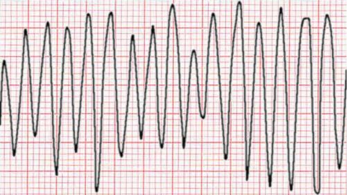
Ventricular flutter is an arrhythmia, more specifically a tachycardia affecting the ventricles with a rate over 250-350 beats/min, and one of the most indiscernible. It is characterized on the ECG by a sinusoidal waveform without clear definition of the QRS and T waves. It has been considered as a possible transition stage between ventricular tachycardia and fibrillation, and is a critically unstable arrhythmia that can result in sudden cardiac death.[citation needed][1]

It can occur in infancy,[2] youth,[3] or as an adult.

It can be induced by programmed electrical stimulation.[4][5]

References

[edit]
[edit]
Revisions and contributorsEdit on WikipediaRead on Wikipedia
from Grokipedia
Ventricular flutter is a potentially lethal cardiac characterized by an extremely rapid, hemodynamically unstable with rates typically exceeding 200 beats per minute, often 250 to 350 beats per minute. It manifests on the electrocardiogram (ECG) as a continuous sinusoidal without discernible QRS complexes, ST segments, or T waves, often resembling a that appears identical when inverted. This rhythm represents an extreme form of and serves as an unstable transitional state between organized and chaotic , frequently degenerating into the latter without prompt intervention. Ventricular flutter commonly arises in patients with underlying structural heart disease, such as , , or prior , where reentrant circuits in the abnormal myocardium propagate the rapid electrical activity. Additional risk factors include electrolyte imbalances (e.g., or hypomagnesemia), drug toxicities (e.g., from antiarrhythmics or stimulants), and ischemia, which exacerbate ventricular irritability. Pathophysiologically, it involves disrupted impulse conduction due to reentrant circuits in the ventricles, resulting in ineffective and swift hemodynamic collapse, often within minutes. Clinically, ventricular flutter presents with acute symptoms including , , , , and syncope, rapidly progressing to loss of consciousness and if untreated. relies on ECG identification of the characteristic monomorphic at rates exceeding 200 beats per minute, with no P waves or isoelectric baselines; supportive tests may include blood work for electrolytes and imaging to assess structural abnormalities. Management demands immediate and unsynchronized (120-200 J biphasic), followed by intravenous antiarrhythmics such as lidocaine or to stabilize the rhythm, alongside correction of precipitating factors. For survivors at risk of recurrence, implantation of an (ICD) is often recommended to prevent sudden death.

Definition and characteristics

Definition

Ventricular flutter is a lethal ventricular characterized by rapid, regular contractions of the ventricles at a rate typically ranging from 200 to 300 beats per minute, which results in ineffective mechanical ventricular activity and hemodynamic collapse. This represents a transitional state on the continuum between and , frequently progressing to the more disorganized and fatal within seconds to minutes if untreated.

Electrocardiographic features

Ventricular flutter manifests on the electrocardiogram (ECG) as a continuous sinusoidal or sine-wave pattern, featuring regular, undulating waves without identifiable isoelectric intervals. This morphology arises from rapid, organized ventricular activation that produces large, smooth oscillations resembling a continuous sine wave pattern that appears identical when inverted. The ECG lacks distinct P waves, QRS complexes, ST segments, or T waves, as the rapid cycling fuses the QRS and T-wave components into seamless oscillations. This absence of definable complexes differentiates it from slower ventricular tachycardias, where monomorphic QRS patterns remain discernible. The ventricular rate typically ranges from 200 to 300 beats per minute, often approximating 300 beats per minute, though it may vary slightly across leads. In precordial leads such as V1, the of the sine waves may appear more prominent compared to limb leads, where the pattern remains consistent but potentially lower in voltage.

Pathophysiology

Underlying mechanisms

Ventricular flutter primarily arises from macro-re-entrant circuits within the ventricular myocardium, where a self-sustaining of propagates in a circular manner around a fixed or functional pathway, leading to rapid, organized activation at rates typically exceeding 250 beats per minute. These circuits form in structurally altered tissue, such as post-infarction scars, allowing the impulse to recirculate without interruption once established. The re-entrant process requires specific electrophysiological conditions: two adjacent pathways with differing conduction velocities and refractory periods, a unidirectional block in one pathway to direct the impulse, and sufficiently slow conduction in the alternative pathway to permit recovery of excitability in the blocked tissue ahead of the . Heterogeneous conduction plays a central role in facilitating these circuits, often resulting from ischemia or fibrosis that creates areas of slowed conduction and unidirectional block. Ischemic regions exhibit variable durations and conduction velocities due to remodeling and disruption, promoting the formation of anatomical barriers or functional gradients that support re-entry. Fibrotic scars, common in cardiomyopathies, act as fixed obstacles around which the circulates, with surviving myocardial bundles providing the slow-conducting limbs essential for circuit maintenance. This heterogeneity ensures that the circulating impulse does not extinguish prematurely, sustaining the high-rate characteristic of ventricular flutter. Initiation of ventricular flutter typically involves premature ventricular complexes (PVCs) that exploit conduction heterogeneities to establish unidirectional block and trigger re-entry, with these PVCs often stemming from enhanced automaticity or triggered activity. Enhanced automaticity in Purkinje fibers or ventricular myocytes can generate spontaneous PVCs, particularly under adrenergic stimulation, which then propagate into vulnerable tissue to initiate the circuit. Triggered activity, mediated by early or delayed afterdepolarizations due to calcium overload or ion channel dysfunction, similarly produces initiating beats that align temporally—such as during the vulnerable R-on-T phase—to block conduction in one direction while allowing slow recovery in the other. The re-entry theory in ventricular flutter aligns with adaptations of the leading circle concept, originally described for atrial arrhythmias, where the circuit represents the smallest excitable pathway around an unexcitable core defined by the tissue's (conduction velocity multiplied by period). In ventricular tissue, this functional re-entry occurs when shortening—due to accelerated conduction or abbreviated refractoriness in ischemic or fibrotic zones—allows the wavefront to sustain itself by continuously invading partially tissue ahead. However, ventricular circuits more commonly incorporate anatomical elements, such as figure-of-8 re-entry around borders, where two opposing wavefronts collide centrally, adapting the leading circle dynamics to heterogeneous substrates. This interplay ensures persistent circular , distinguishing ventricular flutter from less organized arrhythmias.

Hemodynamic consequences

Ventricular flutter induces severe hemodynamic instability primarily through the disruption of coordinated atrial and ventricular activity. As a ventricular-origin , it eliminates effective atrioventricular synchrony, abolishing the atrial contribution to ventricular preload known as the atrial kick. This results in negligible , as the ventricles fail to fill adequately before contraction, leading to a sharp decline in and systemic perfusion. The hallmark rapid ventricular rate of ventricular flutter, typically ranging from 250 to 350 beats per minute, exacerbates these effects by markedly reducing diastolic filling time. With insufficient duration for ventricular relaxation and expansion, diminishes, further impairing ejection and overall cardiac performance. Concurrently, the abbreviated limits coronary artery , which relies heavily on diastolic gradients, thereby promoting myocardial oxygen supply-demand mismatch and worsening ischemia in already compromised tissue. Distinct from the chaotic disorganization of , the regular rhythm of ventricular flutter may transiently preserve minimal arterial pressure via limited, quasi-systolic mechanical activity before inevitable degeneration occurs. However, this episode is brief, as the commonly progresses to within seconds, culminating in total cessation of effective , profound , and circulatory arrest unless promptly intervened upon.

Etiology

Risk factors

Ventricular flutter, a life-threatening , is influenced by several non-modifiable factors that elevate susceptibility. Advanced age serves as an independent predictor, with the incidence of ventricular arrhythmias, including flutter, rising significantly in individuals over 65 due to cumulative structural and electrical changes in the myocardium. Male gender is associated with a higher , attributed to greater rates of underlying ischemic heart disease among men. A family history of sudden cardiac further increases , often linked to inherited channelopathies or structural cardiomyopathies that predispose to ventricular tachyarrhythmias. Modifiable risk factors play a critical role in precipitating ventricular flutter, particularly through disruptions in . Electrolyte imbalances, such as and hypomagnesemia, are common triggers; , for instance, can facilitate re-entrant circuits leading to flutter. Similarly, hypomagnesemia prolongs the and heightens vulnerability, especially when coexisting with . Drug toxicities also contribute substantially, with overdose inducing bidirectional ventricular tachycardia in severe cases. Certain antiarrhythmic agents, paradoxically, can provoke proarrhythmic effects, including rapid ventricular responses mimicking flutter. , such as cocaine or stimulants, and conditions causing high sympathetic tone (e.g., or ) are additional triggers. In specific clinical contexts, the risk is notably elevated; for example, patients in the acute phase following face a 5-10% incidence of ventricular arrhythmias, including flutter, often within the first 48 hours post-event.

Associated conditions

Ventricular flutter is frequently associated with underlying ischemic heart disease, particularly in the setting of acute , where it may arise as a complication of coronary artery occlusion leading to regional ischemia. Cardiomyopathies, including and , also predispose individuals to ventricular flutter due to structural and electrical remodeling of the ventricular myocardium. contributes to ventricular arrhythmias through and , creating a substrate for reentrant rhythms. Non-cardiac conditions commonly linked to ventricular flutter include severe disturbances, such as or hypomagnesemia, which alter myocardial excitability and conduction. In critical illness, hypoxia and further exacerbate the risk by impairing cellular metabolism and promoting arrhythmogenic triggers. Rare associations involve inherited channelopathies, including variants of , particularly in cases triggered by fever, leading to ventricular flutter as a precursor to more chaotic arrhythmias.

Clinical manifestations

Symptoms

Ventricular flutter most commonly manifests as sudden syncope or loss of , caused by cerebral hypoperfusion from severely compromised during the rapid . In instances of shorter episodes, patients may report prodromal sensations including , , , or prior to hemodynamic collapse. This rapid deterioration underscores the underlying hemodynamic instability that limits effective to vital organs.

Physical examination findings

Patients with ventricular flutter often exhibit profound hemodynamic instability on physical examination, characterized by absent or weak peripheral pulses due to severely reduced from the extremely rapid ventricular rate. is a hallmark finding, frequently accompanied by signs of such as cool, clammy extremities and if the episode persists. In instances where a peripheral or apical pulse remains palpable, it is typically rapid (often exceeding 250 beats per minute) and may feel regular, though the high rate often renders it faint or imperceptible. Neck vein examination may reveal prominent , resulting from atrioventricular dissociation where atrial contraction occurs against a closed . Conscious patients commonly display and diaphoresis, reflecting compensatory sympathetic activation and tissue hypoperfusion, alongside altered mental status ranging from to obtundation. These findings underscore the critical nature of the and indicate imminent cardiovascular collapse.

Diagnosis

Diagnostic criteria

The diagnosis of ventricular flutter is primarily established through electrocardiographic (ECG) demonstration of a regular, monomorphic ventricular rhythm at a rate exceeding 250 beats per minute, often around 300 beats per minute, characterized by a continuous sine-wave-like morphology without an isoelectric baseline or distinct QRS and T waves. This pattern reflects a cycle length of about 200 milliseconds, with the ECG tracing appearing identical when inverted, distinguishing it from other tachyarrhythmias. Supportive diagnostic elements include the clinical context of hemodynamic instability, such as , syncope, or , which typically accompanies the due to its rapid rate impairing effective . Artifacts must be excluded by confirming the rhythm across multiple ECG leads and correlating with clinical symptoms, as motion or equipment interference can mimic the sine-wave pattern. In non-acute settings, such as risk stratification post-myocardial infarction, an electrophysiology study may support the diagnosis if very fast (cycle lengths of 200-230 ms) or (cycle length <200 ms) is inducible by programmed ventricular stimulation, though inducibility alone does not always predict spontaneous recurrence.

Ventricular flutter must be differentiated from other ventricular arrhythmias and mimics that present with rapid, wide-complex rhythms on electrocardiography (ECG). Ventricular tachycardia (VT) typically exhibits a slower rate of less than 250 beats per minute and features distinct, recognizable QRS complexes, in contrast to the continuous pattern of ventricular flutter without identifiable QRS components. Ventricular fibrillation (VF) appears as a chaotic, irregular rhythm with fibrillatory waves of varying amplitude and no organized activity, distinguishing it from the regular, monomorphic of ventricular flutter. Supraventricular tachycardia (SVT) with aberrancy can mimic wide-complex tachycardias but lacks atrioventricular (AV) dissociation and often shows organized P waves or retrograde conduction, unlike the absence of such features in ventricular flutter. Artifacts, such as those caused by muscle tremors or loose ECG leads, may produce pseudo-sine wave patterns that resemble ventricular flutter but can be identified by their lack of clinical correlation and resolution upon artifact removal or patient stabilization. Rare mimics include hyperkalemia-induced rhythms, where severe electrolyte imbalance leads to a sine-wave ECG pattern from QRS-T wave fusion, potentially progressing to ventricular flutter-like activity but reversible with correction. , a polymorphic VT, features twisting QRS complexes with varying morphology and is often associated with prolonged QT intervals, differing from the uniform, regular waveform of ventricular flutter.

Treatment

Acute management

The acute management of ventricular flutter prioritizes immediate termination of the through electrical , as it is a life-threatening often associated with hemodynamic collapse. The first-line intervention is unsynchronized using a biphasic waveform at an initial energy level of 200 J, particularly in cases of pulseless ventricular flutter, which is treated similarly to due to its rapid rate (typically 240-300 beats per minute) and sine-wave morphology lacking identifiable QRS complexes. If the patient is pulseless, high-quality (CPR) must be initiated immediately and continued concurrently with attempts to maintain circulation and improve outcomes, following advanced cardiovascular (ACLS) protocols that emphasize minimal interruptions in chest compressions. Subsequent shocks should use equal or higher energy levels if the initial attempt fails, with CPR resumed after each shock until (ROSC) is achieved. For defibrillation-refractory ventricular flutter, pharmacologic therapy is administered to facilitate termination and suppress recurrence. For pulseless cases, intravenous 300 mg IV/IO push after the third defibrillation, with an additional 150 mg if needed; for hemodynamically stable monomorphic VT, 150 mg over 10 minutes, improving short-term survival and conversion rates compared to . Alternatively, lidocaine may be used at an initial dose of 1-1.5 mg/kg IV if is unavailable or ineffective, though it is less efficacious for shock-refractory polymorphic rhythms. Supportive care focuses on stabilizing the patient post-defibrillation, including securing the airway through endotracheal if ROSC occurs but respiratory compromise persists, and addressing post-shock with intravenous vasopressors such as norepinephrine to maintain above 65 mmHg. imbalances, such as or hypomagnesemia, should be corrected promptly to prevent recurrence, as they can exacerbate ventricular instability.

Long-term management

Following successful acute resuscitation from ventricular flutter, long-term management focuses on secondary prevention to mitigate the risk of recurrence and sudden cardiac death in survivors. Implantable cardioverter-defibrillator (ICD) placement is strongly recommended as a Class I indication for patients with structural heart disease who have survived ventricular flutter or hemodynamically unstable ventricular arrhythmias, demonstrating a 27% relative risk reduction in mortality at two years. This therapy is also indicated for secondary prevention in cases of sudden cardiac arrest due to ventricular flutter, particularly when reversible causes have been addressed. Antiarrhythmic serves as an adjunct to ICD to suppress recurrent episodes. Beta-blockers are recommended as first-line agents (Class I, Level of Evidence A) for reducing ventricular arrhythmia burden in survivors with underlying or post-myocardial infarction, as they effectively suppress recurrent ventricular flutter and improve one-year survival. may be considered (Class IIa, Level B) for symptomatic control in patients with frequent ICD shocks or when beta-blockers are insufficient, though it does not confer a survival benefit and is reserved due to potential adverse effects like dysfunction. For refractory monomorphic ventricular flutter involving re-entrant circuits, is indicated (Class I, Level of Evidence B-NR), with VT recurrence approximately 35% in ischemic and success exceeding 90% in idiopathic cases. Correcting underlying etiologies is integral to preventing recurrence. In patients with ischemia-related ventricular flutter, coronary revascularization is recommended (Class I, Level B) if viable myocardium is present, as it addresses the precipitating substrate and stabilizes risk. imbalances, such as , must be proactively managed by maintaining serum levels between 3.5 and 4.5 mmol/L (Class I recommendation), which prevents arrhythmogenic triggers in reversible cases like those associated with variants.

Prognosis and complications

Survival rates

Ventricular flutter, a life-threatening characterized by rapid, organized ventricular activity, rapidly progresses to hemodynamic collapse and if untreated, resulting in near 100% mortality within 3 to 5 minutes due to inadequate and cerebral hypoperfusion. In out-of-hospital settings, prompt significantly improves outcomes, with to hospital discharge rates ranging from 20% to 40% depending on response times and initial persistence. For instance, studies on shockable rhythms like —often a direct consequence or mimic of ventricular flutter—report an average 31.4% to discharge, rising to approximately 39% with within 2 minutes but falling to 22% if delayed beyond that threshold. Key factors influencing survival include whether the arrest is witnessed, the provision of bystander (CPR), and the interval to . Witnessed events and immediate bystander CPR more than double the likelihood of survival compared to unwitnessed or untreated collapses, while interventions within 5 minutes can substantially enhance odds by minimizing ischemic damage.

Potential outcomes

Ventricular flutter is a life-threatening characterized by rapid, regular ventricular contractions that often result in hemodynamic collapse due to inadequate . Without immediate intervention, such as , it frequently degenerates into , leading to sudden cardiac death. Survival rates plummet by 5-10% per minute without , underscoring the critical need for prompt treatment. In patients undergoing electrophysiologic studies, inducible ventricular flutter carries a prognosis comparable to sustained monomorphic ventricular tachycardia, with mortality rates of approximately 34% over a mean follow-up of 30 months. These patients exhibit a high risk of recurrent ventricular tachycardia or fibrillation, reported at 34% during 5-year follow-up post-myocardial infarction, intermediate between non-inducible cases (14%) and those with inducible ventricular fibrillation (17%). Outcomes are influenced significantly by underlying cardiac function; reduced left ventricular (LVEF <40%) is associated with higher cardiac mortality, reaching 31% in patients with syncope, primarily due to heart failure progression rather than arrhythmic events alone. In contrast, patients with preserved LVEF (>40%) face lower mortality rates of 3-5%, regardless of symptoms like syncope. Inducible ventricular flutter thus signals increased arrhythmic risk but does not independently predict sudden death when LVEF is adequate. For out-of-hospital cardiac arrests involving —often a sequela of untreated —survival to hospital discharge stands at about 31%, though many survivors experience neurological impairments from cerebral anoxia. Long-term management with implantable cardioverter-defibrillators can mitigate recurrence, but overall remains guarded in those with structural heart disease.

References

Add your contribution
Related Hubs
User Avatar
No comments yet.