

Community hub
Recent from talks
Contribute something
Nothing was collected or created yet.
Whiskers
View on WikipediaThis article may be too technical for most readers to understand. (April 2021) |


Whiskers, also known as vibrissae (/vəˈbrɪsi/; sg. vibrissa; /vəˈbrɪsə/) are a type of stiff, functional hair used by most therian mammals to sense their environment.[1] These hairs are finely specialised for this purpose, whereas other types of hair are coarser as tactile sensors. Although whiskers are specifically those found around the face, vibrissae are known to grow in clusters at various places around the body. Most mammals have them, including all non-human primates[2], marsupials [3][4], and especially nocturnal mammals. Monotremes, however, lack them.[5]
Whiskers are sensitive tactile hairs that aid navigation, locomotion, exploration, hunting, social touch and perform other functions.[6]
This article is primarily about the specialised sensing hairs of mammals, but some birds, fish, insects, crustaceans and other arthropods are known to have similar structures also used to sense the environment.
Etymology
[edit]Vibrissae (from Latin vibrāre 'to vibrate') from the characteristic motion seen in a small rodent that is otherwise sitting still. In medicine, the term also refers to the thick hairs found inside human nostrils.[7]
Evolution
[edit]The last common ancestor of all extant mammals had vibrissae.[8] All other extant mammal species besides great apes retain the same ancestral layout of the whiskers along with the special facial muscles that move them.[6]
Anatomy
[edit]Vibrissae are anatomically distinguished from other hair. They are easily visually identified since they are longer, stiffer, significantly larger in diameter, and stand above the surrounding fur by a considerable amount. In addition, they have well-innervated follicles, and an identifiable representation in the somatosensory cortex of the brain.[9] The largest number and the longest are found among the small, social, arboreal, and nocturnal mammals. Whiskers of aquatic mammals are the most sensitive. During foraging in complex, dark habitats, whiskers are rapidly moved in a cyclic way, tracing small circles at their tips. This motion, called "whisking" can occur at speeds of 25 Hz in mice, which is one of the fastest movements that mammals can make. Small animals use whisking to position their front paws during locomotion.[6]
Vibrissal groups
[edit]
Vibrissae typically grow in clusters. These groups vary somewhat in form and function, but they are relatively consistent among land mammals. Between land and marine mammals, there is less consistency (though commonalities are certainly present).
Many land mammals, like rats[10] and hamsters,[11] have four typical whisker groups on their heads (called cranial vibrissae), which might vary among animals due to different lifestyles. These cranial groups include:[12]
- above the eyes (supraorbital)
- on the cheeks (genal)
- where a moustache would be (mystacial)
- under the snout (mandibular).

The mystacial whiskers can be roughly identified as macrovibrissae (long whiskers for feeling the space around the head) and microvibrissae (small, down-pointing whiskers for identifying objects).[13] Not only are these two types hard to distinguish on an animal's face (see for example the image of a rat here), there are similarly weak distinctions in how they are used, though the distinction is nonetheless referred to ubiquitously in scientific literature and is considered useful in analysis.
Many land mammals, including domestic cats, also have vibrissae on the underside of the leg just above the paws (called carpal vibrissae).[14] Whilst these five major groups are often reported in studies of land mammals, several other groups have been reported more occasionally; for instance nasal, angular, and submental whiskers.[15]


Marine mammals can have substantially different arrangements of their vibrissae. For instance, whales and dolphins have lost their snout whiskers and gained vibrissae around their blowholes,[16] whereas every single one of the body hairs of the Florida manatee may be a vibrissa (see image).[17] Other marine mammals, like seals and sea-lions, have head vibrissae just like those on land mammals (see image), although these groups function quite differently.
Vibrissal follicles have evolved other functions in dolphins, such as electroreception.
Vibrissae
[edit]The vibrissal hair is usually thicker and stiffer than other types of (pelagic) hair[18] but, like other hairs, the shaft consists of an inert material (keratin) and contains no nerves.[18] However, vibrissae are different from other hair structures because they grow from a special hair follicle incorporating a capsule of blood called a blood sinus which is heavily innervated by sensory nerves.[19][20] Vibrissae are symmetrically arranged in groups on the face and supply the trigeminal nerve.[21]
The mystacial macrovibrissae are shared by a large group of land and marine mammals (see images), and it is this group that has received by far the most scientific study. The arrangement of these whiskers is not random: they form an ordered grid of arcs (columns) and rows, with shorter whiskers at the front and longer whiskers at the rear (see images).[13] In the mouse, gerbil, hamster, rat, guinea pig, rabbit, and cat, each individual follicle is innervated by 100–200 primary afferent nerve cells.[19] These cells serve an even larger number of mechanoreceptors of at least eight distinct types.[20] Accordingly, even small deflections of the vibrissal hair can evoke a sensory response in the animal.[22] Rats and mice typically have approximately 30 macrovibrissae on each side of the face, with whisker lengths up to around 50 mm in (laboratory) rats, 30 mm in (laboratory) mice, and a slightly larger number of microvibrissae.[13] Thus, an estimate for the total number of sensory nerve cells serving the mystacial vibrissal array on the face of a rat or mouse might be 25,000. Natural shapes of rat's mystacial pad vibrissae are well approximated by pieces of the Euler spiral. When all these pieces for a single rat are assembled together, they span an interval extending from one coiled domain of the Euler spiral to the other.[23]
Marine mammals may make even greater investment in their vibrissal sensory system than rats and mice. Seal whiskers, which are similarly arrayed across the mystacial region, are each served by around 10 times as many nerve fibres as those in rats and mice, so that the total number of nerve cells innervating the mystacial vibrissae of a seal has been estimated to be in excess of 300,000.[24] Manatees, remarkably, have around 600 vibrissae on or around their lips.[16][full citation needed]
Whiskers can be very long in some species; the length of a chinchilla's whiskers can be more than a third of its body length (see image).[25] Even in species with shorter whiskers, they can be very prominent appendages (see images). Thus, whilst whiskers certainly could be described as "proximal sensors" in contrast to, say, eyes, they offer a tactile sense with a sensing range that is functionally very significant.
Operation
[edit]Movement
[edit]
The follicles of some groups of vibrissae in some species are motile. Generally, the supraorbital, genal and macrovibrissae are motile,[11] whereas the microvibrissae are not. This is reflected in anatomical reports that have identified musculature associated with the macrovibrissae that is absent for the microvibrissae.[26] A small muscle 'sling' is attached to each macrovibrissa and can move it more-or-less independently of the others, whilst larger muscles in the surrounding tissue move many or all of the macrovibrissae together.[26][27]
Amongst those species with motile macrovibrissae, some (rats, mice, flying squirrels, gerbils, chinchillas, hamsters, shrews, porcupines, opossums) move them back and forth periodically in a movement known as whisking,[28] while other species (cats, dogs, raccoons, pandas) do not appear to.[9] The distribution of mechanoreceptor types in the whisker follicle differs between rats and cats, which may correspond to this difference in the way they are used.[20] Whisking movements are amongst the fastest produced by mammals.[29] In all whisking animals in which it has so far been measured, these whisking movements are rapidly controlled in response to behavioural and environmental conditions.[9] The whisking movements occur in bouts of variable duration, and at rates between 3 and 25 whisks/second. Movements of the whiskers are closely coordinated with those of the head and body.[9]
Function
[edit]Generally, vibrissae are considered to mediate a tactile sense, complementary to that of skin. This is presumed to be advantageous in particular to animals that cannot always rely on sight to navigate or to find food, for example, nocturnal animals or animals which forage in muddy waters. Whiskers can also function as wind detecting antannae such as the supra-orbital ones in rats.[30]
Sensory function aside, movements of the vibrissae may also indicate something of the state of mind of the animal,[31] and the whiskers play a role in social behaviour of rats.[32]
The sensory function of vibrissae is an active research area—experiments to establish the capabilities of whiskers use a variety of techniques, including temporary deprivation either of the whisker sense or of other senses. Animals can be deprived of their whisker sense for a period of weeks by whisker trimming (they soon grow back), or for the duration of an experimental trial by restraining the whiskers with a flexible cover like a mask (the latter technique is used, in particular, in studies of marine mammals[33]). Such experiments have shown that whiskers are required for, or contribute to: object localization,[34][35] orienting of the snout, detection of movement, texture discrimination, shape discrimination, exploration, thigmotaxis, locomotion, maintenance of equilibrium, maze learning, swimming, locating food pellets, locating food animals, and fighting, as well as nipple attachment and huddling in rat pups.[9]
Whisking—the periodic movement of the whiskers—is also presumed to serve tactile sensing in some way. However, exactly why an animal might be driven "to beat the night with sticks", as one researcher once put it,[36] is a matter of debate, and the answer is probably multi-faceted. Scholarpedia[9] offers:
Since rapid movement of the vibrissae consumes energy, and has required the evolution of specialised musculature, it can be assumed that whisking must convey some sensory advantages to the animal. Likely benefits are that it provides more degrees of freedom for sensor positioning, that it allows the animal to sample a larger volume of space with a given density of whiskers, and that it allows control over the velocity with which the whiskers contact surfaces.
Animals that do not whisk, but have motile whiskers, presumably also gain some advantage from the investment in musculature. Dorothy Souza, in her book Look What Whiskers Can Do[37] reports some whisker movement during prey capture (in cats, in this case):
Whiskers bend forward as the cat pounces. Teeth grasp the mouse tightly around its neck. The cat holds on until the prey stops wriggling.
Anecdotally, it is often stated that cats use their whiskers to gauge whether an opening is wide enough for their body to pass through.[citation needed] This is sometimes supported by the statement that the whiskers of individual cats extend out to about the same width as the cat's body, but at least two informal reports indicate that whisker length is genetically determined and does not vary as the cat grows thinner or fatter.[31][38] In the laboratory, rats are able to accurately (within 5–10%) discriminate the size of an opening,[39] so it seems likely that cats can use their whiskers for this purpose. However, reports of cats, particularly kittens, with their heads firmly stuck in some discarded receptacle are commonplace[40] indicating that if a cat has this information available, it does not always make best use of it.
Marine mammals
[edit]Pinnipeds have well-developed tactile senses. Their mystacial vibrissae have ten times the innervation of terrestrial mammals, allowing them to effectively detect vibrations in the water.[41] These vibrations are generated, for example, when a fish swims through water. Detecting vibrations is useful when the animals are foraging and may add to or even replace vision, particularly in darkness.[42]

Harbor seals have been observed following varying paths of other organisms that swam ahead several minutes before, similar to a dog following a scent trail,[33][43] and even to discriminate the species and the size of the fish responsible for the trail.[44] Blind ringed seals have even been observed successfully hunting on their own in Lake Saimaa, likely relying on their vibrissae to gain sensory information and catch prey.[45] Unlike terrestrial mammals, such as rodents, pinnipeds do not move their vibrissae over an object when examining it but instead extend their moveable whiskers and keep them in the same position.[42] By holding their vibrissae steady, pinnipeds are able to maximize their detection ability.[46] The vibrissae of seals are undulated and wavy while sea lion and walrus vibrissae are smooth.[47] Research is ongoing to determine the function, if any, of these shapes on detection ability. The vibrissa's angle relative to the flow, and not the fiber shape, however, seems to be the most important factor.[46]
Most cetaceans have whiskers at birth but they are typically lost during maturation. The follicles and any vestigial hair sometimes function as touch or electrical sense organs.[48]
Lines of research
[edit]Neuroscience
[edit]A large part of the brain of whisker-specialist mammals is involved in the processing of nerve impulses from vibrissae, a fact that presumably corresponds to the important position the sense occupies for the animal. Information from the vibrissae arrives in the brain via the trigeminal nerve and is delivered first into the trigeminal sensory complex of brainstem. From there, the most studied pathways are those leading up through parts of thalamus and into barrel cortex,[49] though other major pathways through the superior colliculus in midbrain (a major visual structure in visual animals) and the cerebellum, to name but a couple, are increasingly coming under scrutiny.[50] Neuroscientists, and other researchers, studying sensory systems favour the whisker system for a number of reasons (see Barrel cortex), not least the simple fact that laboratory rats and mice are whisker, rather than visual, specialists.
Evolutionary biology
[edit]The presence of mystacial vibrissae in distinct lineages (Rodentia, Afrotheria, marsupials) with remarkable conservation of operation suggests that they may be an old feature present in a common ancestor of all therian mammals.[51] Indeed, some humans even still develop vestigial vibrissal muscles in the upper lip,[52] consistent with the hypothesis that previous members of the human lineage had mystacial vibrissae. Thus, it is possible that the development of the whisker sensory system played an important role in mammalian development, more generally.[51]
Artificial whiskers
[edit]Researchers have begun to build artificial whiskers of a variety of types, both to help them understand how biological whiskers work and as a tactile sense for robots. These efforts range from the abstract,[53] through feature-specific models,[54][55] to attempts to reproduce complete whiskered animals in robot form (ScratchBot[56] and ShrewBot,[57][58][59] both robots by Bristol Robotics Laboratory).
In non-mammals
[edit]
A range of non-mammals possess structures which resemble or function similarly to mammalian whiskers.
In birds
[edit]
Some birds possess specialized hair-like feathers called rictal bristles around the base of the beak which are sometimes referred to as whiskers.
The whiskered auklet (Aethia pygmaea) has striking, stiff white feathers protruding from above and below the eyes of the otherwise slate-grey bird, and a dark plume which swoops forward from the top of its head. Whiskered auklets sent through a maze of tunnels with their feathers taped back bumped their heads more than twice as often as they did when their feathers were free, indicating they use their feathers in a similar way to cats.[60]
Other birds that have obvious "whiskers" are kiwis, flycatchers, swallows, nightjars, whip-poor-wills, the kākāpō and the long-whiskered owlet (Xenoglaux loweryi).
In fish
[edit]
Some fish have slender, pendulous tactile organs near the mouth. These are often referred to as "whiskers", although they are more correctly termed barbels. Fish that have barbels include the catfish, carp, goatfish, hagfish, sturgeon, zebrafish and some species of shark.
The Pimelodidae are a family of catfishes (order Siluriformes) commonly known as the long-whiskered catfishes.
In pterosaurs
[edit]Anurognathid pterosaurs had a rugose (wrinkled) jaw texture that has been interpreted as the attachment sites for vibrissae,[61] though actual vibrissae have not been recorded.[62] More recently, a specific type of feathers has been found around anurognathid mouths.[63]
Gallery
[edit]-
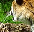
 An otter with facial whiskers.
 -
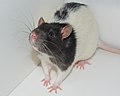 Macrovibrissae of a Lister hooded laboratory rat.
 -
 A cat's prominent macrovibrissae.
 -
 Micrograph cross section of an equine vibrissa.
 -
 Macrovibrissae of a tiger.
 -
 Laboratory mouse (C57BL/6) showing macrovibrissae.
 -
 Prominent immotile vibrissae on a horse's muzzle.
 -
 Closeup picture of the vibrissae of a black and white house cat.
 -
 Supraorbital vibrissae and mystacial macrovibrissae of a house cat.
 -
 The vibrissae of an eastern gray squirrel are normally black.
 -
 Whiskers of the brown thrasher near the head.
.jpg)


References
[edit]- ^ Feldhamer, George A.; Drickamer, Lee C.; Vessey, Stephen H.; Merritt, Joseph H.; Krajewski, Carey (2007). Mammalogy: Adaptation, Diversity, Ecology (3 ed.). Baltimore: Johns Hopkins University Press. p. 99. ISBN 978-0-8018-8695-9. OCLC 124031907.
- ^ Van Horn, R.N. (1970). "Vibrissae Structure in the Rhesus Monkey". Folia Primatol. 13 (4): 241–285. doi:10.1159/000155325. PMID 5499675.
- ^ Old, J.M.; Sheehan, K.H.; Le Breton, N.; Dege, A.M.; Yaghi, L. (2025). "Whisker morphology and distribution in wombats: a comparative anatomical study". Australian Mammalogy. 47. doi:10.1071/AM25025.
- ^ Attard, M.R.G.; Lewis, A.; Wroe, S.; Channing, H.; Rogers, T.L. (2021). "Whisker growth in Tasmanian devils (Sarcophilus harrisii) and applications for stable isotope studies". Ecosphere. 25. doi:10.1002/ecs2.3846.
- ^ Andrew M. Baker (2023). Strahan's Mammals of Australia. Bloomsbury Publishing. p. 31. ISBN 978-1-39941-420-3. Retrieved 10 December 2024.
- ^ a b c Grant, Robyn A.; Breakell, Vicki; Prescott, Tony J. (13 June 2018). "Whisker touch sensing guides locomotion in small, quadrupedal mammals". Proceedings of the Royal Society B: Biological Sciences. 285 (1880) 20180592. doi:10.1098/rspb.2018.0592. PMC 6015872. PMID 29899069.
- ^ "Vibrissae". The Free Dictionary's Medical dictionary. Farlex, Inc. April 14, 2009. Retrieved April 29, 2009.
- ^ Grant, Robyn A.; Haidarliu, Sebastian; Kennerley, Natalie J.; Prescott, Tony J. (1 January 2013). "The evolution of active vibrissal sensing in mammals: evidence from vibrissal musculature and function in the marsupial opossum Monodelphis domestica" (PDF). Journal of Experimental Biology. 216 (Pt 18): 3483–3494. doi:10.1242/jeb.087452. PMID 23737559. S2CID 207170888.
- ^ a b c d e f Grant, Robyn; Mitchinson, Ben; Prescott, Tony (2011). "Vibrissal behaviour and function". Scholarpedia. 6 (10): 6642. Bibcode:2011SchpJ...6.6642P. doi:10.4249/scholarpedia.6642.
- ^ Vincent, S. B. (1913). "The tactile hair of the white rat". The Journal of Comparative Neurology. 23 (1): 1–34. doi:10.1002/cne.900230101. S2CID 86132752.
- ^ a b Wineski, Lawrence E. (1983). "Movements of the cranial vibrissae in the Golden hamster (Mesocricetus auratus)". Journal of Zoology. 200 (2): 261–280. doi:10.1111/j.1469-7998.1983.tb05788.x.
- ^ Thé, L.; Wallace, M. L.; Chen, C. H.; Chorev, E.; Brecht, M. (2013). "Structure, function, and cortical representation of the rat submandibular whisker trident" (PDF). The Journal of Neuroscience. 33 (11): 4815–4824. doi:10.1523/jneurosci.4770-12.2013. PMC 6619006. PMID 23486952.
- ^ a b c Brecht, Michael; Preilowski, Bruno; Merzenich, Michael M. (1997). "Functional architecture of the mystacial vibrissae". Behavioural Brain Research. 84 (1–2): 81–97. doi:10.1016/S0166-4328(97)83328-1. PMID 9079775. S2CID 3993159.
- ^ Beddard, Frank E. (1902). "Observations upon the carpal vibrissae in mammals". Journal of Zoology. 72 (1): 127–136. doi:10.1111/j.1469-7998.1902.tb08213.x.
- ^ Kulikov, V. F. (2011). "A new vibrissa group in insectivores (Mammalia, Insectivora) and its role in orientation". Doklady Biological Sciences. 438 (1): 154–157. doi:10.1134/s0012496611030021. PMID 21728125. S2CID 27361386.
- ^ a b "Whiskers! A Feel For The Dark".
- ^ Reep, R. L.; Marshall, C. D.; Stoll, M. L. (2002). "Tactile Hairs on the Postcranial Body in Florida Manatees: A Mammalian Lateral Line?". Brain, Behavior and Evolution. 59 (3): 141–154. doi:10.1159/000064161. PMID 12119533. S2CID 17392274.
- ^ a b Weldon Owen Pty Ltd. (1993). Encyclopedia of animals – Mammals, Birds, Reptiles, Amphibians. Reader's Digest Association. p. 18. ISBN 1-875137-49-1.
- ^ a b Rice, Frank L.; Mance, Ajuan; Munger, Bryce L. (8 October 1986). "A comparative light microscopic analysis of the sensory innervation of the mystacial pad. I. Innervation of vibrissal follicle-sinus complexes". The Journal of Comparative Neurology. 252 (2): 154–174. doi:10.1002/cne.902520203. PMID 3782505. S2CID 8228091.
- ^ a b c Ebara, Satomi; Kumamoto, Kenzo; Matsuura, Tadao; Mazurkiewicz, Joseph E.; Rice, Frank L. (22 July 2002). "Similarities and differences in the innervation of mystacial vibrissal follicle–sinus complexes in the rat and cat: A confocal microscopic study". The Journal of Comparative Neurology. 449 (2): 103–119. doi:10.1002/cne.10277. PMID 12115682. S2CID 6428629.
- ^ Halata, Z.; Baumann, K. I.; Grim, M. (2008-01-01). "6.02 – Merkel Cells". In Masland, Richard H.; Albright, Thomas D.; Albright, Thomas D.; Masland, Richard H. (eds.). The Senses: A Comprehensive Reference. New York: Academic Press. pp. 33–38. doi:10.1016/b978-012370880-9.00341-8. ISBN 978-0-12-370880-9. Retrieved 2020-12-09.
- ^ Stuttgen, M. C.; Rüter, J.; Schwarz, C. (July 26, 2006). "Two Psychophysical Channels of Whisker Deflection in Rats Align with Two Neuronal Classes of Primary Afferents". The Journal of Neuroscience. 26 (30): 7933–7941. doi:10.1523/JNEUROSCI.1864-06.2006. PMC 6674210. PMID 16870738.
- ^ Starostin, E. L.; et al. (15 January 2020). "The Euler spiral of rat whiskers". Science Advances. 6 (3) eaax5145. Bibcode:2020SciA....6.5145S. doi:10.1126/sciadv.aax5145. PMC 6962041. PMID 31998835.
- ^ Marshall, CD; Amin, H.; Kovacs, K. M.; Lydersen, C. (January 2006). "Microstructure and innervation of the mystacial vibrissal follicle sinus complex in bearded seals, Erignathus barbatus (Pinnipedia: Phocidae)". The Anatomical Record Part A: Discoveries in Molecular, Cellular, and Evolutionary Biology. 288 (1): 13–25. doi:10.1002/ar.a.20273. PMID 16342212.
- ^ Spotorno, Angel E.; Zuleta, Carlos A.; Valladares, J. Pablo; Deane, Amy L.; Jiménez, Jaime E. (15 December 2004). "Chinchilla Laniger". Mammalian Species. 758: 1–9. doi:10.1644/758.
- ^ a b Dörfl, J (1982). "The musculature of the mystacial vibrissae of the white mouse". Journal of Anatomy. 135 (Pt 1): 147–154. PMC 1168137. PMID 7130049.
- ^ Hill, D. N.; Bermejo, R.; Zeigler, H. P.; Kleinfeld, D. (2008). "Biomechanics of the Vibrissa Motor Plant in Rat: Rhythmic Whisking Consists of Triphasic Neuromuscular Activity". The Journal of Neuroscience. 28 (13): 3438–3455. doi:10.1523/JNEUROSCI.5008-07.2008. PMC 6670594. PMID 18367610.
- ^ RoyalSociety (Oct 2, 2011). "CIA-Rat". Youtube. Archived from the original on Jul 3, 2013. Retrieved 2013-06-24.
- ^ Jin, T.-E.; Witzemann, V.; Brecht, M. (March 31, 2004). "Fiber Types of the Intrinsic Whisker Muscle and Whisking Behavior". The Journal of Neuroscience. 24 (13): 3386–3393. doi:10.1523/JNEUROSCI.5151-03.2004. PMC 6730039. PMID 15056718.
- ^ Mugnaini, Matias; Mehrotra, Dhruv; Davoine, Federico; Sharma, Varun; Mendes, Ana Rita; Gerhardt, Ben; Concha-Miranda, Miguel; Brecht, Michael; Clemens, Ann M. (2023). "Supra-orbital whiskers act as wind-sensing antennae in rats". PLOS Biology. 21 (7) e3002168. doi:10.1371/journal.pbio.3002168. ISSN 1545-7885. PMC 10325054. PMID 37410722.
- ^ a b McSporran, Keith. "Just the cat's whiskers". Archived from the original on 2012-02-12.
- ^ Wolfe, Jason; Mende, Carolin; Brecht, Michael (2011). "Social facial touch in rats". Behavioral Neuroscience. 125 (6): 900–910. doi:10.1037/a0026165. ISSN 1939-0084. PMID 22122151.
- ^ a b Dehnhardt, G. (2001). "Hydrodynamic trail-following in harbor seals (Phoca vitulina)". Science. 293 (5527): 102–104. doi:10.1126/science.1060514. PMID 11441183. S2CID 9156299.
- ^ Ahissar, E.; Knutsen, P. M. (2011). "Vibrissal location decoding". Scholarpedia. 6 (10): 6639. Bibcode:2011SchpJ...6.6639A. doi:10.4249/scholarpedia.6639.
- ^ Diamond, M.; von Heimendahl, P; Knutsen, P.; Kleinfeld, D.; Ahissar, A. (2008). "'Where' and 'what' in the whisker sensorimotor system". Nature Reviews Neuroscience. 9 (8): 601–612. doi:10.1038/nrn2411. PMID 18641667. S2CID 6450408.
- ^ Brecht, Michael (September 2004). "What Makes Whiskers Shake?". Journal of Neurophysiology. 92 (3): 1265–1266. doi:10.1152/jn.00404.2004. PMID 15331639.
- ^ Souza, Dorothy (2007). Look What Whiskers Can Do. Lerner Publishing Group. p. 17. ISBN 978-0-761-39459-4.
- ^ "Focus Magazine Q&A". Archived from the original on 2018-07-18. Retrieved 2012-04-13.
- ^ Krupa, David J.; Matell, Matthew S.; Brisben, Amy J.; Oliveira, Laura M.; Nicolelis, Miguel A. L. (2001). "Behavioral Properties of the trigeminal somatosensory system in rats performing whisker-dependent tactile discriminations". The Journal of Neuroscience. 21 (15): 5752–5763. doi:10.1523/JNEUROSCI.21-15-05752.2001. PMC 6762640. PMID 11466447.
- ^ For example: "Cops save kitten with head stuck in can". Toronto Sun. 2011-01-25. Retrieved 2013-06-24.
- ^ R. J., Schusterman; D., Kastak; D. H., Levenson; C. J., Reichmuth; B. L., Southall (2000). "Why pinnipeds don't echolocate". The Journal of the Acoustical Society of America. 107 (4): 2256–64. Bibcode:2000ASAJ..107.2256S. doi:10.1121/1.428506. PMID 10790051. S2CID 17153968.
- ^ a b Miersch, L.; Hanke, W.; Wieskotten, S.; Hanke, F. D.; Oeffner, J.; Leder, A.; Brede, M.; Witte, M.; Dehnhardt, G. (2011). "Flow sensing by pinniped whiskers". Philosophical Transactions of the Royal Society B: Biological Sciences. 366 (1581): 3077–84. doi:10.1098/rstb.2011.0155. PMC 3172597. PMID 21969689.
- ^ Schulte-Pelkum, N.; Wieskotten, S.; Hanke, W.; Dehnhardt, G. & Mauck, B. (2007). "Tracking of biogenic hydrodynamic trails in harbour seals (Phoca vitulina)". Journal of Experimental Biology. 210 (5): 781–787. doi:10.1242/jeb.02708. PMID 17297138.
- ^ Grant R, Wieskotten S, Wengst N, Prescott T, Dehnhardt G (2013). "Vibrissal touch sensing in the harbor seal (Phoca vitulina): how do seals judge size?". Journal of Comparative Physiology A. 199 (6): 521–531. doi:10.1007/s00359-013-0797-7. PMID 23397461. S2CID 14018274.
- ^ Hyvärinen H (1989). "Diving in darkness: whiskers as sense organs of the ringed seal (Phoca hispida saimensis)". Journal of Zoology. 218 (4): 663–678. doi:10.1111/j.1469-7998.1989.tb05008.x.
- ^ a b Murphy, T. C.; Eberhardt, W. C.; Calhoun, B. H.; Mann, K. A.; Mann, D. A. (2013). "Effect of Angle on Flow-Induced Vibrations of Pinniped Vibrissae". PLOS ONE. 8 (7) e69872. Bibcode:2013PLoSO...869872M. doi:10.1371/journal.pone.0069872. PMC 3724740. PMID 23922834.
- ^ Ginter CC, Fish FE (2010). "Morphological analysis of the bumpy profile of phocid vibrissae". Marine Mammal Science. 26: 733–743. doi:10.1111/j.1748-7692.2009.00365.x.
- ^ Mynett, Natasha; Mossman, Hannah L.; Huettner, Tim; Grant, Robyn A. (2022). "Diversity of vibrissal follicle anatomy in cetaceans" (PDF). The Anatomical Record. 305 (3): 609–621. doi:10.1002/ar.24714. PMID 34288543. S2CID 236158643.
- ^ Deschenes, Martin; Urbain, Nadia (2009). "Vibrissal afferents from trigeminus to cortices". Scholarpedia. 4 (5): 7454. Bibcode:2009SchpJ...4.7454D. doi:10.4249/scholarpedia.7454.
- ^ Kleinfeld, Rune w. Berg (1999). "Anatomical loops and their electrical dynamics in relation to whisking by rat". Somatosensory & Motor Research. 16 (2): 69–88. CiteSeerX 10.1.1.469.3914. doi:10.1080/08990229970528. PMID 10449057.
- ^ a b Mitchinson, B.; Grant, R. A.; Arkley, K.; Rankov, V.; Perkon, I.; Prescott, T. J. (12 November 2011). "Active vibrissal sensing in rodents and marsupials". Phil. Trans. R. Soc. B. 366 (1581): 3037–3048. doi:10.1098/rstb.2011.0156. PMC 3172598. PMID 21969685.
- ^ Tamatsu, Yuichi; Tsukahara, Kazue; Hotta, Mitsuyuki; Shimada, Kazuyuki (August 2007). "Vestiges of vibrissal capsular muscles exist in the human upper lip". Clin Anat. 20 (6): 628–31. doi:10.1002/ca.20497. PMID 17458869. S2CID 21055062.
- ^ "Invention: Artificial whiskers".
- ^ Costandi, Mo (2006-10-05). "Sculpted Face". Neurophilosophy.wordpress.com. Retrieved 2013-06-24.
- ^ Fend, Miriam; Bovet, Simon; Hafner, Verena Vanessa (2004). The Artificial Mouse - A Robot with Whiskers and Vision. 35th International Symposium on Robotics. CiteSeerX 10.1.1.58.6535.
- ^ Archived at Ghostarchive and the Wayback Machine: "Bristol Robotics Lab - Scratchbot". YouTube. 2009-07-01. Retrieved 2013-06-24.
- ^ Archived at Ghostarchive and the Wayback Machine: "SCRATCHbot - A Rat like Robot". YouTube. 2011-09-15. Retrieved 2013-06-24.
- ^ Archived at Ghostarchive and the Wayback Machine: "Whiskerbot". YouTube. 2011-09-03. Retrieved 2013-06-24.
- ^ Archived at Ghostarchive and the Wayback Machine: "A robot inspired by the Etruscan shrew called Shrewbot". YouTube. 2012-01-19. Retrieved 2013-06-24.
- ^ Brown, S. (2008). "Bird uses 'whiskers' like a cat". Nature. doi:10.1038/news.2008.674. Retrieved September 28, 2013.
- ^ Bennett et al 2007b
- ^ Wilton, Mark P. (2013). Pterosaurs: Natural History, Evolution, Anatomy. Princeton University Press. ISBN 978-0-691-15061-1.
- ^ Benton, Michael J.; Xu, Xing; Orr, Patrick J.; Kaye, Thomas G.; Pittman, Michael; Kearns, Stuart L.; McNamara, Maria E.; Jiang, Baoyu; Yang, Zixiao (2019). "Pterosaur integumentary structures with complex feather-like branching" (PDF). Nature Ecology & Evolution. 3 (1): 24–30. doi:10.1038/s41559-018-0728-7. hdl:1983/1f7893a1-924d-4cb3-a4bf-c4b1592356e9. PMID 30568282. S2CID 56480710.
External links
[edit]- A night in the life of a rat, Annals of the New York Academy of Sciences, 1225:110–118, April 2011.
- The Mysterious Whiskers of Cats, Blog-post about the functions of cat whiskers, April 7, 2012.
Whiskers
View on GrokipediaOverview
Definition and Terminology
Whiskers, scientifically known as vibrissae, are specialized, elongated sensory hairs found primarily in mammals, serving roles in tactile sensing that extend beyond the functions of typical fur.[2] These vibrissae are prominent sinus hairs that act as mechanosensory organs, enabling animals to detect environmental stimuli through touch, and are present in most therian mammals, though absent in humans and monotremes.[5] Unlike ordinary pelage hairs, vibrissae are characterized by their length, thickness, and association with large follicles containing blood-filled sinus tissues, which facilitate heightened sensitivity.[2] Vibrissae differ fundamentally from guard hairs or underfur in structure and purpose; while guard hairs form the coarser outer layer of the coat for physical protection and underfur provides insulation for thermoregulation, vibrissae are rooted in deep follicles richly innervated by sensory nerve fibers, prioritizing sensory input over protective or thermal roles.[2] This rich innervation, including mechanoreceptors within the follicle-sinus complex, allows vibrissae to transmit detailed tactile information to the brain, distinguishing them as active sensory tools rather than passive coverings.[5] In scientific literature, the term "vibrissae" (plural; singular "vibrissa") is standard for these sensory hairs, derived from the Latin "vibrissa," referring to nostril hairs that vibrate, with the zoological usage emphasizing their vibratory movement in certain species.[2] Common names include "whiskers" for the facial vibrissae in animals like cats, while specific groupings are termed "mystacial vibrissae" for those on the snout's mystacial pad.[5] The term entered zoological contexts in the 19th century, as anatomists distinguished these specialized hairs from general pelage.[6]Etymology
The term "whiskers" originates from Middle English "whisker," an agent noun derived from the verb "whisk," denoting something that moves quickly or sweeps, akin to a fan or fly-swatter.[7] This usage traces back to the early 15th century, with the earliest recorded instance around 1425, initially referring to objects that whisk or stir, and linked etymologically to Old Norse "visk," meaning to wipe or stir.[8] By the 17th century, the word had evolved to describe human facial hair, particularly the long hairs on the cheeks and chin, reflecting its connotation of swift, sweeping motion.[7] In scientific nomenclature, "vibrissa" (plural: vibrissae) entered English in the late 17th century from Latin "vibrissae," originally denoting the stiff hairs inside the nostrils, derived from "vibrare," meaning to vibrate, shake, or wave.[9] This anatomical term was adopted in the 1700s for human nasal hairs and later extended in zoology to describe specialized sensory hairs in mammals in the 19th century, amid advances in comparative anatomy.[6] "Vibrissa" specifically signified elongated, tactile whiskers in animals like rodents and cats, emphasizing their vibrational sensitivity rather than mere cosmetic resemblance.[9] Related terms include "mystacial," referring to the whisker pad on the muzzle, derived from Ancient Greek "mústax," meaning upper lip or moustache, combined with the suffix "-ial" to denote location.[10] Historically, "whiskers" appeared in 18th-century English literature to describe feline facial features, as in natural history accounts portraying cats' "long whiskers" for vivid depiction, often in a ornamental rather than functional context.[11]Anatomy
Structure of Vibrissae
Vibrissae, or whiskers, consist of keratin-based shafts that form a tapered, cylindrical structure optimized for mechanical signal transmission. The primary structural protein, alpha-keratin, provides the necessary rigidity and elasticity, with the shaft narrowing progressively from base to tip in a conical or Euler spiral profile. This tapering design allows for differential bending responses along the length, where proximal sections resist deformation more effectively than distal ones.[12][13][14] In mammals, vibrissal lengths typically range from 1 to 10 cm, varying by species, position on the face, and individual factors such as age or environment; for instance, rat mystacial vibrissae measure approximately 4-5 cm on average. A key mechanical feature is the stiffness gradient, with the base exhibiting greater flexural rigidity—up to five orders of magnitude higher than the tip—due to the increasing cross-sectional area and material density toward the follicle. This gradient ensures that subtle deflections at the tip amplify into detectable signals at the base without structural failure.[15][14][16] The cross-section of a vibrissa reveals a circular profile with a central hollow or medullated core, which enhances flexibility by reducing weight and allowing compressive deformation. The outer cuticle comprises overlapping imbricated scales that form a protective sheath, while some species, particularly aquatic mammals, display longitudinal ridges on the surface to minimize hydrodynamic drag and facilitate airflow or water current interactions. These microstructural elements collectively support the vibrissa's role in propagating tactile and aerodynamic cues.[17][18] Vibrissae undergo cyclic growth independent of the pelage hair cycle, progressing through anagen (growth), catagen (regression), and telogen (rest) phases, with hormonal regulation—such as thyroid and sex steroids—influencing initiation and duration. In rodents like rats and mice, this cycle results in an average vibrissal lifespan of 1-3 months, enabling continuous renewal without synchronized shedding.[19][20][21] Innervation at the base integrates up to 200 nerve fibers per vibrissa, predominantly myelinated axons from the trigeminal ganglion that terminate in mechanoreceptors such as Merkel cells, lanceolate endings, and reticular networks within the follicle-sinus complex. This dense afferent supply, estimated at 100-200 fibers in rodents, encodes mechanical stimuli through specialized endings without extending along the shaft itself.[17][22][23]Vibrissal Follicles and Groups
Vibrissal follicles represent highly specialized, elongated structures that anchor and mechanosensorily equip the vibrissae in mammals. These follicles are deeply embedded in the dermis, typically extending 2–5 mm in depth in rodents, and are richly vascularized to support their sensory functions. The follicle wall features a thick collagenous capsule enclosing the hair shaft, which is surrounded by fluid-filled sinuses that amplify mechanical signals. Specifically, the upper portion contains a ring sinus, a narrow cavity filled with blood and lymph, while the lower portion includes a broader cavernous sinus, both contributing to the transduction of vibrissal movements into neural signals.[24][25][26] Embedded within the follicle's outer root sheath are Merkel cell-neurite complexes, densely clustered sensory endings that detect sustained deflections of the vibrissa shaft with high sensitivity. These complexes, along with other mechanoreceptors like lanceolate and Ruffini endings, are uniquely concentrated in vibrissal follicles compared to ordinary hair follicles, enabling precise tactile discrimination. The overall architecture forms a follicle-sinus complex that acts as a mechanotransducer, with the sinuses' hydrostatic properties facilitating signal propagation.[26][24] Vibrissae emerge from organized groups or arrays distributed across the mammalian body, with the most prominent being the mystacial pad on the upper lip, comprising macrovibrissae (longer, primary whiskers for exploration) and microvibrissae (shorter, for fine texture sensing). In rodents like rats, the mystacial pad typically hosts 20–40 vibrissae, arranged in 4–5 rostrocaudal rows with bilateral symmetry; for instance, rats exhibit exactly 31 vibrissae per side in this pad. Other facial arrays include supraorbital vibrissae above the eyes, genal vibrissae on the cheeks, mandibular vibrissae under the snout, and interramal tufts between the jaw rami. Beyond the face, body arrays such as carpal vibrissae at the wrists aid in locomotion-related sensing.[27][28][17] Species variations in vibrissal groups reflect ecological adaptations, with rodents showing consistent bilateral symmetry and row-based arrangements, though total numbers differ; mice, for example, have fewer mystacial vibrissae (around 30 total) than rats. Embryonic development begins with follicle primordia forming as placodes or ridges on the snout at approximately embryonic day 11.5 in mice, patterning the mystacial array through epithelial-mesenchymal interactions that establish the symmetric grid before birth. These primordia deepen and vascularize rapidly, with innervation following shortly after.[29][30] Associated with these follicles is a specialized musculature unique to vibrissal systems, enabling controlled movements. Intrinsic muscles, such as the follicular sling muscles, encircle individual follicles to protract or retract single vibrissae, while extrinsic muscles attach to the mystacial pad to coordinate array-wide whisking. In rats, these include protractor colli and retrahens auris muscles, forming a motor plant that integrates with the follicle-sinus complex for active sensing. This musculature is absent in non-vibrissal hairs, underscoring the follicles' role in dynamic tactile exploration.[31][32][33]Physiology and Function
Sensory Mechanisms and Movement
Whiskers in mammals, especially rodents, engage in both active and passive movements to detect environmental stimuli. Active whisking consists of rhythmic protraction-retraction cycles, occurring at frequencies of 5-20 Hz in rodents, driven by intrinsic mystacial muscles to explore surroundings.[34] This motion is typically bilateral and symmetric for coordinated scanning, but unilateral or asymmetric patterns emerge during tasks like head turning or unilateral object contact, allowing targeted sensing.[5] In contrast, passive deflection arises from external forces such as direct touch or air currents impinging on stationary whiskers, providing sensory feedback without motor involvement.[35] Sensory transduction within whisker follicles relies on mechanoreceptors that convert mechanical deflections into neural signals. These include rapidly adapting (RA) receptors, which fire phasically to detect vibrations and transient movements, and slowly adapting (SA) receptors, which maintain firing during sustained contact to signal pressure or texture onset.[36] The follicle's structure amplifies deflections through a piezoelectric-like mechanical response, where bending stresses activate mechanosensitive ion channels, such as Piezo2, to generate ionic currents.[37] Deflection of whiskers produces signals proportional to the stimulus intensity; angles up to 90° elicit graded increases in afferent firing rates, with velocity and direction further modulating response magnitude.[38] Airflow from nearby objects induces resonant oscillations in the whisker, whose amplitude scales with proximity, enabling distance estimation through vibratory cues.[39] Neural signals originate from primary afferents in the follicle, which convey information via the trigeminal nerve to cell bodies in the ipsilateral trigeminal ganglion, marking the first central relay without detailed cortical projection here.[40]Detection and Navigation Roles
Whiskers enable rodents to perform precise tactile sensing, allowing discrimination of object shape, texture, and size through active whisker sweeps. In rats, this involves palpating surfaces to detect micro-textures, where neural responses in the barrel cortex encode roughness via variations in whisker vibrations during contact. For instance, rats can distinguish fine textures through differential spike rates in primary sensory neurons. Shape and size discrimination occur via the geometric deflection patterns of whiskers against contours, enabling identification of objects as small as 1 mm in diameter by integrating contact points across multiple whiskers during exploratory movements.[41][42][43] In navigation, whiskers facilitate head-centered spatial mapping, particularly in dark or cluttered environments where vision is limited. Rodents construct egocentric representations of nearby space by tracking whisker contacts relative to head orientation, allowing obstacle avoidance during locomotion. For example, mice adjust gait and limb placement in real-time to step over barriers using whisker feedback, achieving sub-millimeter precision in trajectory corrections at speeds up to 30 cm/s. Burrowing species like moles and voles integrate whisker-derived path information with self-motion cues for efficient tunnel navigation, preventing collisions and enabling spatial updating over distances of several body lengths. This tactile guidance is especially critical in subterranean habitats, where whiskers detect wall textures and openings to maintain orientation.[44][45][46] Whiskers also contribute to social and feeding behaviors through direct contact. In rodents, whisker-to-whisker or whisker-to-body interactions during social encounters convey identity and intent, with protraction patterns differing between affiliative and aggressive contexts to facilitate recognition of familiar conspecifics, including potential mates. For feeding, terrestrial mammals like rodents use whiskers to probe for food items in low-light conditions, discriminating edible textures from non-edible ones. In seals, whiskers detect hydrodynamic wakes from swimming prey, allowing tracking over distances of several meters even in turbid waters, though this sensory role extends briefly to terrestrial phases for environmental scanning during haul-outs.[47][45][48] Quantitatively, whisker resolution reaches approximately 0.5 mm in mice for object localization along a single whisker axis, limited by follicle mechanics and neural tuning. This precision integrates with other senses for enhanced multisensory cues; for example, whisker signals in the barrel cortex are modulated by concurrent olfactory inputs during active exploration, improving object discrimination in complex scenes. Vision complements whisker data in lit environments, with superior colliculus neurons combining tactile and visual inputs for faster localization. Such integration ensures robust behavioral outcomes, as rodents without whiskers show deficits in both solitary navigation and social discrimination tasks.[49][50][51][52]Adaptations in Specific Environments
In marine mammals, particularly pinnipeds like harbor seals, vibrissae exhibit specialized streamlined morphology with flattened, undulating shafts that minimize vortex-induced vibrations during swimming, thereby enhancing the detection of hydrodynamic flows from distant prey trails.[53] This adaptation suppresses self-generated noise by up to sixfold compared to smooth cylinders, allowing seals to sense low-velocity disturbances as faint as 245 µm/s and track wakes over distances exceeding 180 meters in low-visibility conditions.[54] The vibrissae vibrate across a broad frequency spectrum (10–1,000 Hz, peaking at 20–250 Hz) when encountering prey-induced vortices, providing encoded cues on object size, shape, and direction that inform foraging behaviors.[53] Sea otters demonstrate aquatic adaptations through their macrovibrissae, which are richly innervated with an average of 1,339 axons per follicle-sinus complex, supporting high tactile sensitivity comparable to that of other pinnipeds.[55] These vibrissae enable texture discrimination thresholds of approximately 0.50 mm underwater, facilitating the detection and manipulation of cryptic benthic prey like invertebrates during foraging dives in turbid environments.[56] Unlike paws, which offer finer resolution, vibrissae provide broader scanning for initial prey location, with performance remaining consistent across air and water media due to the follicle's tripartite blood sinus system.[56][55] Walruses, as semi-aquatic pinnipeds specialized for benthic feeding, possess up to 350 mystacial vibrissae per side, each with large follicle-sinus complexes (up to 2 cm) innervated by 1,000–1,600 nerve fibers, optimizing tactile discrimination of buried prey in soft sediments.[57] During bottom foraging, they root through the seafloor with their snouts, using vibrissae to detect bivalve siphons and other slow-moving organisms, a behavior observed in 43% of underwater sequences and comprising 29% of bottom contact time.[58] These smooth, slightly flattened vibrissae differ from the undulating forms in phocids by prioritizing direct-contact sensitivity over hydrodynamic trail tracking, reflecting adaptations to shallow, sediment-rich habitats.[57] Aquatic transitions in mammals involve reduced whisking compared to terrestrial counterparts, where cyclic protraction-retraction movements are common for active scanning; instead, aquatic species like pinnipeds employ static positioning or limited protraction to sense flows without the high energetic costs of whisking in denser water.[1] This modification aligns with longer, thicker vibrissae arrays that facilitate passive hydrodynamic reception during dives, contrasting the shorter, mobile whiskers of terrestrial mammals optimized for rapid environmental probing.[1] Whisker integrity is critical for survival in pinnipeds, as their role in prey detection directly influences foraging success; experimental evidence from hydrodynamic tracking studies indicates that vibrissae enable seals to follow trails with 80–90% accuracy, implying that trimming or ablation would severely impair this capability and elevate predation vulnerability in predatory-prey interactions.[48][54]Evolutionary History
Origins in Mammals
The evolutionary origins of whiskers, or vibrissae, in mammals trace back to the early Mesozoic era among advanced synapsids (cynodont therapsids), the ancestral group to mammals. Fossil evidence indicates that vibrissae-like structures may have first appeared in advanced therapsids (cynodonts) around 241 million years ago during the Middle Triassic, with more definitive indications in advanced cynodont therapsids by the Middle Triassic, approximately 241 million years ago. These early structures are inferred from enlarged infraorbital foramina in skulls, which housed nerves for sensory innervation similar to those in modern vibrissae, suggesting that whisker-like tactile organs aided in navigation and foraging in dimly lit or burrowing environments. By the Cretaceous period, around 100-66 million years ago, vibrissae were fully developed in early mammals such as multituberculates, as evidenced by their large infraorbital canals and sensitive snouts, which supported dense whisker arrays for precise object manipulation in nocturnal habitats. Although vibrissae are a therian synapomorphy, the associated active whisking behavior has evolved convergently at least seven times in therian lineages, particularly in small nocturnal species.[59][60][1][1] Developmental genetics underlying vibrissae formation show deep conservation across mammalian lineages, involving key signaling pathways that specify follicle types. Hox genes, particularly Hoxd cluster members, regulate the collinear patterning of skin appendages, including vibrissae, by controlling ectodermal gene expression during embryogenesis; this mechanism is preserved in both placentals and marsupials, as demonstrated by comparative analyses of murine and avian models that highlight mammalian-specific enhancers. Wnt signaling plays a central role in follicle induction and differentiation, where β-catenin stabilization activates downstream targets like Lef/Tcf transcription factors to promote vibrissal follicle specification distinct from pelage hair; disruptions in Wnt/β-catenin pathways lead to agenesis of vibrissae in experimental models, underscoring its essentiality. This genetic framework, shared between placentals (e.g., rodents) and marsupials (e.g., opossums), indicates that vibrissae development evolved early in the therian mammal lineage and remained stable despite divergences in reproductive strategies.[61][62] Following the Cretaceous-Paleogene extinction event 66 million years ago, which eliminated non-avian dinosaurs, mammals underwent adaptive radiation into diverse niches, with vibrissae expanding in importance for crepuscular and nocturnal species. Early mammals, constrained to nocturnal lifestyles during the Mesozoic to avoid diurnal predators, relied on enhanced somatosensation via vibrissae for obstacle detection and prey localization in low-light conditions; post-extinction, this trait facilitated rapid diversification into forested and underground habitats, as seen in the proliferation of rodent-like forms with elaborate whisker systems. However, vibrissae have been lost or reduced in certain lineages adapting to specialized environments, such as fully aquatic cetaceans, where hair including vibrissae was secondarily lost around 50 million years ago to streamline bodies for swimming, though vestigial follicles persist embryonically. Independent losses also occur in some subterranean species, like certain moles with minimized external pelage, reflecting trade-offs against constant soil contact.[63] Phylogenetically, vibrissae are distributed across therian mammals (marsupials and placentals), but absent in monotremes, attesting to their origin near the base of Theria. This near-universal presence in therians underscores vibrissae as a synapomorphy of therian mammals, with losses confined to derived aquatic (cetaceans) and select fossorial taxa, where alternative sensory modalities like echolocation or amplified audition compensate. Comparative genomic and fossil data confirm no reversals, indicating vibrissae as a conserved adaptation that underpinned mammalian success in sensory-challenging environments.[64][60][65]Comparative Development Across Species
Whisker development exhibits significant diversity across mammalian taxa, reflecting adaptations to varied ecological niches and sensory demands. In many species, vibrissae emerge early in embryonic stages, with follicles forming before birth, and continue to elongate and specialize postnatally to support functions like tactile exploration and environmental sensing. This comparative variation in morphology, such as array organization and follicle complexity, and distribution, including the number and positioning of vibrissae, underscores evolutionary divergences while maintaining core mechanosensory roles. Ontogenetically, whiskers often reach functional maturity by weaning, though their growth and innervation patterns differ by taxon, influencing behaviors from hunting to navigation.[64] In carnivores, mystacial vibrissae form elaborate arrays that enhance predatory efficiency. Felids and canids typically possess four rows of mystacial whiskers on each side of the snout, with lengths varying to detect subtle air currents and prey movements during hunting; these vibrissae protract and retract actively, allowing precise localization of objects in low-light conditions. For instance, in domestic cats, whiskers serve as radar-like sensors, alerting to approaching threats or aiding in pouncing accuracy by sensing vibrations from up to 12 inches away. Pinnipeds, such as seals and sea lions, feature specialized vibrissal pads on the snout with undulated, flattened whiskers that minimize hydrodynamic drag while detecting water flow trails from fish, facilitating underwater foraging; these vibrissae are among the largest in mammals, often exceeding 30 cm in length, and are richly innervated for fine-scale tactile discrimination.[66][67][57] Rodents display highly organized whisker grids that support intricate palpation behaviors, making them ideal for laboratory studies of somatosensation. In mice, vibrissae are arranged in five curvilinear rows (A to E) bilaterally on the mystacial pad, with up to 31 principal whiskers forming a precise array for sweeping and contacting surfaces during exploration; this grid-like structure maps directly to barrel cortex organization, enabling whisker-driven object identification and texture discrimination. These behaviors, such as rhythmic whisking at 5-20 Hz, emerge postnatally and are crucial for sensorimotor development, with deafferentation studies in lab models revealing deficits in navigation and social interaction. Whiskers in rodents grow continuously from birth, reaching adult length by postnatal day 21, supporting their role as primary tactile sensors in nocturnal, burrow-dwelling lifestyles.[68][69][70] Among primates, whisker development shows reduction in higher taxa but retention in prosimians, correlating with sensory ecology. In humans, vibrissae are vestigial, manifesting as sinus hairs in the upper lip and nostrils without active movement or extensive innervation, remnants of ancestral capsular muscles that once controlled protraction; these sparse hairs provide minimal tactile feedback compared to other mammals. In contrast, prosimians like lemurs retain prominent, mobile vibrissae with well-developed intrinsic musculature, aiding arboreal navigation through dense foliage by detecting branch textures and gaps during nocturnal leaps; histological comparisons reveal thicker follicle walls and richer innervation in these species, enabling active whisking for spatial orientation. Ontogenetically, primate vibrissae form early in gestation but elaborate less postnatally than in rodents, aligning with visual dominance in diurnal lineages.[71][72] Anomalies in whisker development highlight genetic influences on morphology across breeds and species. In hairless cat breeds like the Sphynx, hypotrichosis results in absent or short, curled vibrissae due to mutations in keratin genes, impairing tactile sensing and requiring environmental adjustments for navigation; these cats retain follicle remnants but lack full elongation, a condition inherited recessively. Hypertrichosis, excessive hair growth including vibrissae, occurs rarely in mammals but can lead to denser whisker arrays in affected individuals, as seen in some congenital syndromes altering follicle cycling. Ontogenetically, whisker anomalies often manifest from birth, with normal mammals showing rapid postnatal growth—e.g., in rodents, follicles initiate prenatally and whiskers functional by day 7—while delays or absences in carnivores like cats correlate with broader hair coat defects, emphasizing whiskers' sensitivity to developmental pathways.[73][74][75]Whiskers in Non-Mammals
In Fish and Aquatic Animals
In fish, barbels serve as elongated, fleshy appendages primarily functioning as chemosensory and mechanosensory organs, analogous to whiskers in mammals but not composed of true hair structures. These appendages, often extending from the mouth, chin, or nostrils, are equipped with taste buds for gustation and neuromasts for detecting mechanical stimuli such as water flow and vibrations. For instance, in catfish species like the red-tail catfish (Hemibagrus wyckioides), the maxillary barbels can reach lengths representing about two-thirds of the fish's body length, allowing them to probe substrates effectively.[76] These structures are innervated by branches of the facial and trigeminal nerves, enabling the integration of chemical and tactile information to locate food in low-visibility environments.[77] The primary functions of barbels in fish center on prey detection and environmental navigation, particularly in murky or turbid waters where vision is limited. Taste buds distributed along the barbel epithelium detect amino acids and other chemical cues from potential prey, while neuromasts—clusters of hair cells—sense subtle water movements and pressure changes, facilitating the localization of hidden food items on the bottom. In species such as channel catfish (Ictalurus punctatus), barbels are crucial for bottom-foraging behaviors, allowing the fish to "taste" the substrate and respond to nearby stimuli. Additionally, in some elasmobranchs like nurse sharks (Ginglymostoma cirratum), barbel-like nasal extensions integrate with electroreceptive ampullae of Lorenzini, enhancing the detection of bioelectric fields from buried prey in sandy habitats.[77][78][79] Evolutionarily, barbels in teleost fish arose independently around 200 million years ago during the diversification of this group in the Mesozoic era, likely through co-option of developmental pathways involved in fin formation, such as those regulated by Fgf and Hox genes. This adaptation provided a selective advantage in aquatic niches with poor visibility, leading to diverse forms across taxa; for example, goatfish (Mullidae) possess highly mobile pectoral-fin barbels for active foraging, while some loaches exhibit reduced nasal barbels for stream-dwelling navigation. Although analogous structures exist in aquatic invertebrates, such as the tactile antennae of shrimp used for detecting water flow, vertebrate barbels represent a specialized convergent evolution focused on integrated sensory processing.[80][76]In Birds and Reptiles
In birds, bristle-like structures known as rictal bristles, which are modified hair-like feathers surrounding the beak, serve tactile sensory roles analogous to whiskers, particularly in low-light foraging and navigation. These bristles contain mechanoreceptors such as Herbst corpuscles that detect vibrations and contact, aiding in prey capture and obstacle avoidance; for instance, in nightjars (Caprimulgiformes), they facilitate insect detection during nocturnal or crepuscular flight in cluttered environments.[81] In species like kiwis, sensory pits at the bill tip house similar mechanoreceptors (Herbst and Grandry corpuscles), enabling remote tactile probing of soil for invertebrates without reliance on vision, a function enhanced by the bill's enlarged trigeminal sensory nucleus.[82] Rictal bristles are ancestrally present in birds, likely evolving around 108 million years ago, with their prevalence and length correlating to nocturnality across approximately 10% of avian species.[83] Feather-associated structures further contribute to whisker-like sensing in birds, particularly for aerodynamic feedback during flight. Filoplumes, slender feathers attached to the base of contour and flight feathers, act as vibration-sensitive mechanoreceptors that detect airflow velocity and direction over the wing surface, allowing birds to monitor airspeed, stall risk, and separation points for precise control.[84] In owls, semiplumes—downy feathers beneath contour layers—provide positional feedback on underlying feather alignment, potentially contributing to silent flight by sensing subtle airflow disruptions and maintaining low-noise wing configurations during hunting.[85] In reptiles, whisker-like tactile functions arise from specialized scale-associated structures rather than follicles, with mechanoreceptors embedded in the integument for environmental sensing. Squamate reptiles (lizards and snakes) possess scale sensilla, small tactile organs on head scales that detect mechanical stimuli like touch and vibration, aiding in prey localization and navigation; these are particularly numerous in ventral tail scales of geckos, where spinulated bristles enhance sensitivity.[86] In crocodilians, integumentary sensory organs (ISOs)—dome-shaped pits distributed across jaws (up to 9,000 in Nile crocodiles) and body scales—function as highly sensitive mechanoreceptors, detecting water movements at 20–35 Hz, direct contact, and pressure waves with thresholds finer than human fingertips (0.078 mN), supporting prey capture and social interactions.[87] These structures in birds and reptiles represent convergent adaptations derived from reptilian scales and proto-feather filaments, distinct from mammalian vibrissae. Fossil evidence from Jurassic forms like Archaeopteryx indicates early filamentous feathers on wings and tails served sensory roles before aerodynamic specialization, evolving from scale hinge regions in reptilian ancestors around 150–200 million years ago.[88]Research and Applications
Neuroscience Investigations
Neuroscience investigations into whisker sensory systems have primarily focused on rodents as model organisms, leveraging the precise somatotopic organization of the barrel cortex in the primary somatosensory cortex (S1). In layer IV of the rodent barrel cortex, each mystacial whisker is represented by a discrete cytoarchitectonic unit known as a "barrel," forming a one-to-one topographic map that mirrors the arrangement of whiskers on the snout.[89] These barrels, with diameters typically ranging from 100 to 300 μm depending on whisker position and species, receive segregated thalamocortical inputs from the ventral posteromedial nucleus of the thalamus, enabling high-fidelity representation of individual whisker deflections.[90] Developmental plasticity in this mapping is evident during early postnatal periods, where sensory experience refines barrel boundaries and connectivity; for instance, neonatal whisker trimming disrupts barrel formation, leading to fused or malformed structures that persist into adulthood.[91] Neural coding in the barrel cortex emphasizes spike timing over mere rate for discriminating tactile features like texture. Neurons respond to whisker vibrations with precise temporal patterns, where rough surfaces elicit higher-frequency spike bursts (often >100 Hz) compared to smooth ones, allowing texture identity to be encoded in the relative timing of action potentials across neuronal populations.[92] This temporal code is particularly prominent in layer IV granular cells and is preserved through thalamocortical relays. Post-2010 optogenetic studies have illuminated the trigeminal pathway's role in this coding; for example, channelrhodopsin-2 expression in trigeminal ganglion neurons enables patterned activation of whisker afferents, revealing how synchronized inputs drive barrel cortex responses with millisecond precision and how pathway-specific inhibition alters sensory discrimination.[93] Such manipulations confirm that the lemniscal trigeminal pathway conveys rapidly adapting mechanosensory signals, while non-lemniscal routes modulate gain during active whisking.[94] Experimental models, particularly whisker deprivation in rats, have demonstrated profound cortical reorganization. Chronic trimming of all but one whisker row (e.g., row C sparing) during critical developmental windows leads to expanded receptive fields in spared barrel columns, with neurons increasingly responsive to the intact whiskers and suppressed for deprived ones, reflecting unmasking of lateral inhibitory connections.[90] In adults, prolonged deprivation induces similar plasticity, albeit slower, involving dendritic spine remodeling and shifts in cytochrome oxidase activity. Functional magnetic resonance imaging (fMRI) in awake, head-fixed rats has visualized these changes, showing BOLD signal expansion in spared barrels during whisker stimulation, with enhanced activation in secondary somatosensory areas.[95] Recent advances up to 2025 have employed two-photon calcium imaging to capture whisker-evoked activity at cellular resolution in behaving rodents. These techniques reveal sparse, columnar activation patterns in layer 2/3 of the barrel cortex during naturalistic whisking, with population synchrony correlating to object localization accuracy.[50] Emerging findings link whisker sensory processing to pain and itch via C-fiber innervation of the follicle; a small proportion of unmyelinated C-fibers in whisker pad afferents respond to noxious mechanical or thermal stimuli, projecting to the spinal trigeminal nucleus and influencing barrel cortex via brainstem feedback.[96] Moreover, 2020s studies highlight multisensory integration in the barrel cortex, where olfactory cues modulate whisker responses—odors enhance or suppress calcium transients in up to 20% of neurons—facilitating context-dependent tactile perception during exploration.[50]Bioinspired Technologies
Bioinspired technologies draw from the sensory capabilities of natural whiskers, particularly their roles in tactile and flow detection, to develop artificial vibrissae for robotics. Since the 2000s, micro-electro-mechanical systems (MEMS) have enabled the creation of compact artificial whisker sensors, often using polymer materials like polydimethylsiloxane (PDMS) or SU-8 integrated with strain gauges for precise tactile feedback.[97] These sensors mimic the bending mechanics of biological vibrissae, detecting forces and vibrations at the base to provide robots with enhanced environmental interaction, such as in obstacle avoidance and surface exploration.[98] In underwater applications, artificial whiskers inspired by the undulated structure of seal vibrissae have been developed to sense hydrodynamic flows, reducing drag and enabling trail tracking for autonomous underwater vehicles (AUVs).[99] U.S. Navy projects in the 2010s explored these designs for improving navigation in submerged environments, where the whisker geometry minimizes self-induced vibrations while detecting minute water movements from nearby objects.[100] For planetary exploration, multifunctional whisker arrays mounted on rovers facilitate terrain mapping and obstacle detection by measuring deflection patterns during contact, allowing distance estimation up to 20 cm and feature extraction like ridges or slopes without relying solely on vision.[101] Advancements in the 2020s have integrated artificial intelligence with whisker arrays, particularly in soft robotics, where machine learning processes sensor data for real-time object recognition and navigation. Tapered whisker systems employing reservoir computing—a form of machine learning—achieve up to 87% accuracy in classifying eight terrain types by analyzing vibration signatures, enabling adaptive path planning on uneven surfaces.[102] Biomimetic materials, such as electroactive polymers, further enhance these sensors by providing both actuation for active whisking and integrated sensing, allowing flexible, low-power operation in dynamic environments.[103] Key challenges in scaling these technologies include replicating the stiffness gradients of natural tapered whiskers, which span orders of magnitude in flexibility to enable slip-off during contact and prevent sensor stalling.[104] Achieving energy efficiency in active whisking remains difficult, as motorized deflection consumes power without the biological advantages of muscle-driven motion. Recent work from 2023 to 2025 on bioinspired gradient whiskers using 3D-printed functionally graded materials addresses these issues for haptic feedback in prosthetics, improving tactile discrimination by modulating stiffness for finer touch resolution.[105]References
- https://en.wiktionary.org/wiki/mystacial