Hubbry Logo
Broken toeBroken toeMain
Open search
Broken toe
Community hub
Broken toe
logo
8 pages, 0 posts
0 subscribers
Be the first to start a discussion here.
Broken toe
Broken toe
from Wikipedia
Broken toe
Other namesBedroom fracture, nightstand fracture, nightwalker fracture[1][2]
X-ray shows a small portion broken off the corner of the distal bone, and a [more longitudinal fracture in the bone in the middle of the toe?},
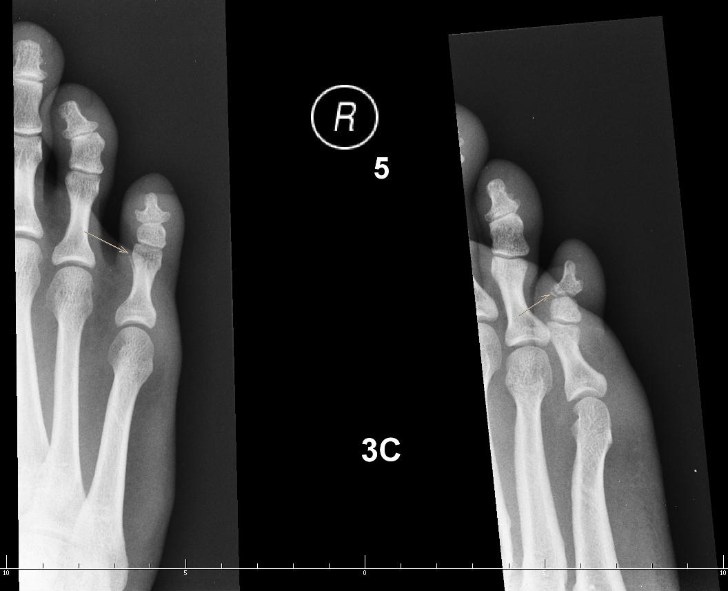
X-rays of fractures of the proximal (left) and distal (right) phalanges in the little toe.
SpecialtyEmergency medicine
SymptomsPain, tenderness, bruising, swelling, displacement of the bones.[3]
ComplicationsCompromised blood circulation; malunion, long-term pain, degenerative joint disease, infection[3]
Usual onsetSudden[3]
CausesStubbing or crushing[3] over-extending a toe joint, stress fracture[3][4]
Diagnostic methodVisualisation, X-rays[3]
TreatmentFor pain and swelling,[3] rest, icing, elevation and pain medication; wearing wide, flat, comfortable, stiff-soled shoes; for smaller toes, buddy wrapping (taping the toe to the nearest toe, with some absorbent padding in-between);[4][5] rarely, a cast or surgery[4]
MedicationOver-the-counter painkillers[4]
Prognosis4 to 8 weeks for full healing; pain lessens within days[4]
FrequencyCommon,[4] 8–9% of all fractures[6]

A broken toe is a type of bone fracture.[6] Symptoms include pain when the toe is touched near the break point, or compressed along its length (as if gently stubbing the toe).[3] There may be bruising, swelling, stiffness, or displacement of the broken bone ends from their normal position.[4]

Toes usually break because they have been stubbed or crushed.[3][4] Crushing breaks are often caused by dropping something on the toe.[3][4] More rarely, over-extending a toe joint can break off a portion of the bone, and stress fractures are possible,[3] especially just after a sudden increase in activity.[7] Diagnosis can be based on symptoms and X-rays.[4][8]

Fractures of the smaller toes are usually treated with rest, buddy taping (taping the toe to the nearest toe, with some absorbent padding in-between), and wearing comfortable, wide-toed, flat, stiff-soled shoes.[5][4] For pain and swelling of all toes,[3] rest, icing, elevation and pain medication are used. Pain usually decreases significantly within a week, but the toe may take 4–6 weeks to heal fully.[4] As activity is slowly increased to normal levels, the toe may be a bit sore and stiff. If the bone heals crooked, it may be relocated with or without surgery.[4] Broken toes can usually be cared for at home, unless the break is in the big toe, there is an open wound, or the broken ends of the bone are displaced.[4] In high-force crushing and shearing injuries, especially those with open wounds, blood circulation (tested by capillary refill) can be impaired, which needs urgent professional treatment.[3] More serious broken toes may need to be re-aligned or put in a cast; surgery is rarely needed. These cases may take longer (six to eight weeks) to heal fully.[4]

Broken toes are one of the most common types of fracture seen in doctor's offices, and make up just under 10% of fractures in some offices.[3]

Definition and classification

[edit]

Toe bones or phalanges of the foot. Note the big toe has no middle phalanx.
People vary; sometimes the smallest toe also has none (not shown).[3]

  Distal phalanges of the foot
  Middle phalanges of the foot
  Proximal phalanges of the foot

Big toe fractures are treated differently from fractures of the lesser toes.[1][9] Toe fractures may be articular (affecting the joint surfaces at the ends of the bone) or diaphyseal (between the ends).[10] They can be displaced, non-displaced, closed or open.[1]

The AO Foundation/Orthopaedic Trauma Association (AO/OTA) classification generates numeric codes for describing broken toes.[10] They run 88[meaning a fracture of the phalanges].[number-code of toe, with the big toe=1 and the little toe=5].[number-code of phalanx, counting 1-3 outwards from the foot].[number-code of location on the bone, with 1 being the inner end, 3 the outer, and 2 in-between].[10] So, for instance, 88.1.2.1 means a fracture to the big toe's innermost bone, at the proximal end.[10][11] A letter can be added to describe the fracture pattern.[11]

Signs and symptoms

[edit]
Broken toe with bruising and swelling

Symptoms include pain when the site of the fracture is gently pressed,[8] or when the toe is gently compressed along its length[3] or moved.[7] There may be bruising or swelling;[8] sometimes there is a crackling sound.[8] There may be displacement of the bones; the alignment of the nail bed is compared to the same toe on the uninjured foot to check if the toe has rotated (see spiral fracture).[8]

Injuries to the nail bed and neurovascular bundles may be present.[8]

Complications

[edit]

Diabetes can make foot injuries more serious, and those with diabetes are advised to have broken toes seen by a doctor.[5] Peripheral arterial disease can also be a complication.[7]

Malunion, healing with the bones out-of-place, can cause long-term pain and significant disability. Malunion of joint surfaces may cause degenerative joint disease.[3] Malunions may be corrected with or without surgery.[4]

When a toe is broken by crushing, there is often also a subungual hematoma (bleeding/bruising of the nail bed, under the toenail).[4] If there is enough blood to cause pain, it can be drained to relieve the pain and avoid (temporarily) losing the nail.[4] Draining is usually done if the injury is less than 24 hours old. Preserving the nail helps splint the broken toe.[3] Contaminated wounds are more serious; the wound should be kept clean.[10]

Broken toes with open wounds, especially if there is necrosis, can lead to osteomyelitis.[3] Joint problems are more likely in cases of involvement/possible displacement of the joint surface and, in children, involvement of the growth plate.[3] Degenerative arthritis of the distal (outer) big toe joint can occur as a complication of fractures, especially fractures to the proximal (inner) end and diaphysis (midsection) of the proximal bone.[10] If the proximal phalanx of the big toe is broken, hallux valgus (bunion) is a frequent complication.[10]

In high-force crushing and shearing injuries, especially those with open wounds, blood circulation can be impaired.[3]

Causes

[edit]

Toes usually break because they have been stubbed or crushed.[3][4] Crushing breaks are often caused by dropping something on the toe.[3][4] More rarely, over-extending a toe joint can break off a portion of the bone, and stress fractures are possible,[3] especially just after a sudden increase in activity.[7]

Risk factors

[edit]

Kicking the ground during sports may result in "turf toe" with an associated broken toe.[12]

Getting up suddenly at night, particularly when barefoot, and having a forceful impact with furniture may lead to a broken toe, also called a "bedroom fracture" "nightstand fracture" or "nightwalker fracture".[1][2] Although generally associated with the fifth toe and big toe, it can occur in any toe.[1][13] In such a fracture, the hard blow to the tip of the distal phalanx typically results in a transverse or oblique fracture in the proximal phalanx (base of toe), but can occur in any phalanx.[1][13]

An open wound toe fracture may result from an injury from a lawn mower.[6]

Although broken toes in horse riders are uncommon, riders are most likely to get broken toes when standing next to their horse.[14]

Mechanism

[edit]

Because the big toe is more important for weight-bearing, balance, walking, and running, breaks to the big toe are more likely to be problematic.[8][10] If the big toe is stubbed and breaks, it usually breaks the distal (outermost) bone. A crushing injury can break both big-toe bones.[10]

If the joint was bent too far (i.e. either hyperextended or hyperflexed) then spiral fractures and avulsion fractures are common. Spiral fractures with displacement make the toe rotate and shorten. With transverse fractures (i.e. across the toe), the toe may bend abnormally.[8]

Diagnosis

[edit]

Broken toes are diagnosed by physically examining the toe, gently pressing on it, asking how it feels, and comparing it to the toe on the opposite foot[8] (see signs and symptoms, above). Blood circulation may be tested by capillary refill.[3]

It may be unclear whether the toe has a bone fracture or just a soft-tissue injury[5] (such as bruising,[5] sprains and tendon injuries[15]). In these cases, it doesn't matter, because the treatment is usually the same for both types of injury. There is no need to figure out whether the toe is actually broken.[5] Follow-up X-rays also generally have no effect on treatment, and are unnecessary.[3]

X-rays are usually only taken if there are complications, or the toe is not healing as expected.[5] If X-rays are taken, the neighbouring toes and joints are also imaged.[8] If there is diffuse pain and tenderness across the foot, it may be necessary to X-ray the whole foot.[8] For displaced fractures, follow-up X-rays may be taken 3-6 weeks after injury.[3]

If a dislocated toe (a joint dislocation) is suspected, an X-ray may needed.[7]

In people with multiple traumas, foot trauma is often neglected.[10]

Treatment

[edit]

It may not be clear whether the toe is broken or just bruised.[5] In such cases the treatment is usually the same in either case.[5]

Removing rings

[edit]

Any rings on the toes are removed immediately, before the toe starts to swell.[16][17] Pulling rings off forcefully may worsen the swelling. Relaxation, elevation, icing, lubrication (e.g. soapy water or oil), and rotating the ring as if unscrewing it may help. If these methods don't work, it may be possible to remove the ring by temporarily wrapping the toe with a slick thread (something like dental floss), passing the inner end of the thread under the ring and then unwrapping it, pushing the ring ahead of the unwrapping thread. Failing that, the ring may need to be cut off by a doctor.[18]

Reduction

[edit]

If the fracture displaces the bones from their proper position (which it often doesn't), it needs to be reduced. Reduction puts the toe back into alignment, with all parts of the bones in the correct anatomical position. This is usually done by pulling gently along the length of the toe, then gently pulling the toe back into place. If there are multiple displaced bone fragments, shuffling them back into the correct positions may be more complicated. The fracture will usually stay in the correct position once re-aligned; if not, it needs more specialized treatment to hold the parts in place so that it heals straight.[3]

Stabilization and protection

[edit]
Buddy-taping toes, using the most suitable adjacent toe to splint the broken toe.
Wider shoes allow the toes to lie straight. If the toes overhang when standing on the removed insole (right), then they will be cramped inside the shoe (left).
A walking boot

Fractures of the smaller toes are commonly treated by buddy taping (see image). Padding is used between the toes to keep the space dry[4] and the toes aligned comfortably. If the toes are less comfortable when buddy-taped, the buddy tape should be removed.[7] Taping is not recommended for those with diabetes or peripheral arterial disease.[7]

It is also helpful for shoes to be stiff-soled (to protect the toe from bending),[4] low at the heel[5] (even a <2cm heel can increase pressure on the forefoot by over 20%[19]), and wide.[20][5] Most shoes, especially women's shoes, have a toe box which is more than a centimeter too tight; the width of the foot should be measured standing, with weight on it.[19] Comfortable shoes are recommended; tight, pointy shoes are undesirable.[5] If the top of the shoe is making the broken toe more painful, it is should be changed for something that won't, like open-toe sandals or old sneakers with the toe cut away.[16]

Somewhat more serious fractures which affect a joint, but with less than 2mm displacement and less than 25% of the area of the joint surface on the broken part, are generally also be treated with buddy taping and suitable shoes; the evidence on this treatment is not extensive.[8]

Fractures with displacement at the break, including rotation, can often be reduced (re-aligned) by a family doctor. Some broken toes may need to be put in casts, especially if the fracture is unstable (it won't stay reduced on its own).[3][4] If more than 25% of the area of the joint surface was on the broken-loose part, or the break had to be reduced, follow-up X-rays are done 7–10 days afterwards.[8]

Fractures of the big toe are treated with a short-leg orthopedic walking boot, or a short-leg walking cast with a sole that protrudes beyond the big toe. These are worn for 2–3 weeks. Buddy taping and a rigid sole are then used for 3–4 weeks, if symptoms allow. At four weeks, range-of-motion exercises can start. If the joint was involved or the break had to be reduced, follow-up X-rays are done a week afterwards.[8]

Pain and swelling

[edit]

Activities which cause pain should be avoided, and resumed slowly as the toe heals.[4]

To reduce pain and swelling,[3] rest, ice, elevation and over-the-counter pain medication are used. The toe is chilled with ice 20 minutes of every hour for the first waking day, and 2-3 times a day afterwards. Ice is not put directly on the skin.[4]

Surgical

[edit]

Surgery is not needed for most broken toes,[4] but may involve fastening bits of toe bone together with wires, screws, or screwed plates.[10] Such procedures are within the scope of orthopaedic surgery.

Prognosis

[edit]

Complete healing may take four to six weeks, and complex cases may take up to eight weeks.[4] Some athletes may need longer.[8] Long-term disability is rare.[3] (see complications section).

Epidemiology

[edit]

Approximately 8 to 9% of all broken bones are of a toe.[6] Studies have varied as to whether broken big toes are more or less common than broken lesser toes.[1] In a UK study involving nearly 6000 fractures seen in hospital, 3.6% were broken toes.[10] Fractures of big toes make up about a fifth[3] or third[8] of all toe fractures, and 5.5% of all foot and ankle fractures in major US trauma hospitals.[10] Toe fractures are the most common foot fractures.[8] About 20% of broken toes involve open wounds.[10]

Other animals

[edit]

Buddy strapping can be used for toe fractures in big birds.[21] Sometimes a ball bandage can be used, where the bird curls its toes over it.[21] Due to pneumatic bones in birds, washing an open toe fracture may be harmful.[21] Broken toes in grebes can be splinted but if dislocated, often require amputation.[22] A toe fracture in an elephant may go unnoticed.[23][24] Knocked-up toes in racing greyhounds may be mistaken for a toe fracture.[25]

See also

[edit]

References

[edit]
Revisions and contributorsEdit on WikipediaRead on Wikipedia
from Grokipedia
A broken toe, also known as a , is a common injury involving the breakage of one or more of the phalanges—the small bones that make up the s of the foot—typically resulting from acute trauma such as stubbing the against a hard surface or dropping a heavy object on the foot. Symptoms often include immediate throbbing pain, swelling, bruising or discoloration of the skin, , and difficulty walking or wearing shoes, with the pain intensifying when weight is placed on the foot. Most broken toes heal without within 6 to 8 weeks, though recovery can extend to 3 to 6 months for full comfort and up to a year for complete . Toe fractures are classified as nondisplaced (where the bone remains aligned but cracked) or displaced (where bone fragments separate and may misalign), and as closed (skin intact) or open (skin broken, increasing infection risk); the proximal phalanx, the bone closest to the foot, is the most frequently affected. Causes include direct impacts from falls or heavy objects, twisting injuries during sports, or repetitive stress leading to stress fractures, particularly in high-impact activities like running. Risk factors include participation in contact sports, osteoporosis, or occupations involving heavy lifting. Complications such as infection, chronic pain, or post-traumatic arthritis may develop if the injury is not properly managed. Diagnosis typically begins with a to assess tenderness, , blood flow, and nerve function, followed by X-rays to confirm the , though stress fractures may require MRI if initial imaging is inconclusive. Treatment focuses on immobilization through buddy taping (securing the injured toe to an adjacent one with padding), use of a stiff-soled or walking boot, rest, application for 15-20 minutes every few hours, , and over-the-counter relievers like ibuprofen or acetaminophen; severe cases involving the big toe or misalignment may necessitate , bone reduction, or rarely with pins or screws. Patients should seek immediate medical attention for open wounds, numbness, fever, or worsening symptoms to prevent long-term issues.

Definition and Classification

Overview

A broken toe, also known as a , is a disruption in the continuity of one or more of the phalanges, the small long bones that form the toes of the foot. The human foot contains 14 phalanges in total: the hallux (big toe) consists of two phalanges—a proximal and a distal—while each of the four lesser toes has three phalanges—proximal, middle, and distal. These fractures typically result from direct trauma and can range from simple cracks to complete breaks, affecting the structural integrity of the toe. Unlike other foot injuries, a broken toe specifically involves damage, distinguished from sprains (which stretch or tear ligaments connecting bones at s), dislocations (where bones are forced out of their normal positions), or injuries such as contusions or lacerations that do not affect bony structure. This differentiation is crucial for accurate diagnosis, as it relies on to confirm bone discontinuity rather than or assessment alone. Toe fractures represent a common minor injury, accounting for approximately 9% of fractures managed in settings, with an estimated annual incidence of 14 to 39.6 cases per 10,000 individuals. While often resolving with conservative care, untreated cases can lead to complications such as or joint degeneration, potentially impairing mobility and daily function. Specific case reports of phalangeal fractures appeared by the late 1800s. Various types of these fractures, including displaced and nondisplaced patterns, are detailed in subsequent classifications.

Types of Fractures

Toe fractures are classified based on the location within the phalanges (the bones of the toes), the pattern of the break, and whether they involve growth plates in children or in adults. In pediatric cases, the Salter-Harris classification system is commonly used to categorize fractures involving the phalangeal growth plates, dividing them into five types based on the injury's relationship to the growth plate: Type I (separation through the growth plate), Type II (fracture through the and growth plate), Type III (intra-articular fracture through the growth plate), Type IV (fracture crossing the , growth plate, and ), and Type V (crush injury to the growth plate). For adults, the AO/OTA classification is applied to phalangeal fractures, grouping them into Type A (extra-articular fractures, such as simple transverse or spiral breaks), Type B (partial articular fractures, involving one portion of the surface), and Type C (complete articular fractures, disrupting the entire surface), which helps guide surgical decisions regarding stability and alignment. Specific fracture types in toes are often named by their anatomical location and mechanism. Tuft fractures occur at the distal tip of the distal phalanx and are typically caused by crush injuries, such as dropping a heavy object on the toe; these are usually stable but can lead to nail bed involvement. Shaft fractures affect the middle portion of the phalanx and may present as transverse (straight across the bone, often unstable), oblique (angled break, potentially displacing with rotation), or spiral (twisting pattern from rotational forces). Base fractures involve the proximal end of the phalanx, particularly common in the hallux (big toe), where they can disrupt the joint and affect weight-bearing stability. Stress fractures, resulting from repetitive microtrauma, commonly occur in the metatarsals or proximal phalanges of runners or athletes, appearing as linear cracks without acute displacement. Special considerations apply to certain toe fractures due to unique . Sesamoid fractures involve the small sesamoid bones embedded in the flexor hallucis brevis tendon under the big 's metatarsophalangeal joint, typically from direct trauma or chronic stress, leading to during flexion. Anatomical variations influence fracture implications between the hallux and lesser toes (digits 2-5). Fractures in the hallux are more likely to impact and balance due to its role in propulsion, often requiring immobilization or for displacement, whereas lesser toe fractures tend to be less symptomatic and heal with conservative management, though multiple phalanges in these toes increase the risk of complex patterns like comminuted breaks.

Clinical Presentation

Signs and Symptoms

A broken toe typically presents with acute pain at the injury site, which is often throbbing and intensifies with movement, pressure, or weight-bearing activities. Swelling and bruising usually develop within hours of the injury, with discoloration appearing under the skin or toenail due to . Patients often report difficulty walking or bearing weight on the affected foot, along with stiffness that limits normal toe function. Physical examination reveals tenderness upon of the , particularly at the site, and may show such as angulation, shortening, or an unnatural bend in the . Limited is common, with pain or inability to bend the fully. Associated features include redness around the injury site and, in cases of open fractures, a visible laceration or protruding bone. Symptoms generally peak in severity within the first few days, with initial pain and swelling subsiding over about a week, though discomfort may persist for 1 to 2 weeks without treatment. Worsening symptoms, such as increased redness or swelling, may signal potential infection and require prompt medical attention.

Complications

A broken toe can lead to various acute complications if not managed promptly, particularly in cases involving open fractures where the skin is breached. Infection, such as , may occur when bacteria enter the wound, potentially causing bone inflammation and requiring antibiotics or surgical intervention. Compartment syndrome, though rare in isolated toe fractures, can develop from excessive swelling that increases pressure within the foot's fascial compartments, compromising blood flow and necessitating urgent to prevent tissue damage. Early immobilization and are crucial to mitigate these risks by reducing swelling and preventing bacterial ingress. Chronic complications often arise from improper healing, including , where the bone fails to mend, and , resulting in or misalignment that alters foot mechanics. These issues can cause persistent and limit mobility, sometimes necessitating corrective . , characterized by joint degeneration, is particularly common in fractures involving the big toe due to its role, leading to stiffness and during movement. and underscore the importance of timely alignment and follow-up care to promote proper union. Other risks include , especially in sesamoid fractures of the big toe, where disrupted blood supply leads to death and chronic forefoot . Delayed is more prevalent in individuals with or who smoke, as these factors impair circulation and regeneration. Prompt medical evaluation, including risk factor management like , can significantly reduce the likelihood of these long-term sequelae.

Etiology

Causes

Toe fractures most commonly result from traumatic events involving direct force to the foot. These include direct impacts, such as stubbing the against hard furniture or dropping heavy objects like a the foot, which deliver sudden axial or bending forces to the phalanges. Crush injuries, often from slamming a on the toe or a falling object compressing the forefoot, are another frequent traumatic cause that can lead to multiple phalangeal breaks. In sports, toe fractures arise from specific mechanisms like hyperextension of the great toe, as seen in soccer where the toe is forcibly bent upward during tackling or pushing off (commonly termed turf toe, which may involve associated fractures). Repetitive stress from activities like running can cause stress fractures in the metatarsals or phalanges due to cumulative microtrauma. Axial loading occurs in high-impact sports such as , where jumping and landing transmit compressive forces through the toes to the ground. Accidental events also contribute significantly to toe fractures. Falls, particularly tripping or landing awkwardly on the foot, can produce bending or twisting forces sufficient to fracture toes. accidents may cause crush injuries when a runs over the foot or during pedal impacts. Occupational hazards, such as in where workers risk heavy materials like bricks falling on their feet, heighten exposure to these traumatic causes. Non-traumatic causes include pathological fractures, which occur when underlying bone weakness leads to breaks from minimal or no external force, such as in where reduced predisposes the phalanges to spontaneous fracturing. These events trigger the injury mechanisms detailed in the section.

Risk Factors

Certain demographic factors increase the susceptibility to toe fractures. Older adults, particularly those over 65, face heightened risk due to age-related bone density loss and conditions like , which weaken bones and make them more prone to fractures from minor trauma. Children and adolescents, especially aged 10-14, experience higher incidence rates from active play and , with toe fractures peaking in this group at an incidence rate of 56.7 per 100,000 for females and 57.7 for males. Gender differences show females comprising about 59% of cases overall, though males may have elevated risk in contact due to participation patterns. Lifestyle elements also contribute significantly. Participation in high-impact sports such as football, running, , and soccer increases the likelihood of both acute and stress fractures through repetitive forefoot stress or direct impacts. Occupations involving heavy machinery, construction, or work on uneven surfaces, like or , elevate risk by exposing workers to falling objects or unstable footing. Medical conditions further predispose individuals. reduces bone strength, making toe fractures more likely even with low-force injuries, particularly in postmenopausal women. heightens vulnerability through impaired bone quality and healing, with associated with an increased fracture risk, including a 2- to 3-fold increase for hip fractures, in older adults. , often linked to diabetes, diminishes foot sensation, leading to unnoticed repetitive microtrauma that can result in fractures. Environmental factors play a key role as well. Ill-fitting shoes, including those with narrow toe boxes or inadequate support, apply uneven pressure and contribute to stress fractures by altering foot . walking on rough terrain heightens injury risk by lacking protective cushioning against impacts. adds mechanical stress to the feet, increasing the of foot and ankle fractures by up to threefold in some cases due to excess body weight. These factors can exacerbate complications, such as elevating risk post-fracture.

Pathophysiology

Mechanism of Injury

A broken toe, or phalangeal fracture, often results from direct trauma involving compressive or shearing forces applied to the toe bones. These forces typically occur when an object impacts the toe, such as stubbing it against a hard surface or dropping a heavy item, leading to axial loading on the tip of the toe that transmits force along the . This can cause transverse or oblique fractures in the distal or middle phalanges due to the bone's inability to absorb the sudden energy. Indirect forces contribute to fractures through abnormal motion at the metatarsophalangeal (MTP) joint, such as hyperextension or hyperflexion, which stretches or tears supporting structures and secondarily fractures the proximal . A classic example is the turf toe mechanism, where forceful dorsiflexion of the great toe beyond approximately 78 degrees applies tensile stress to the plantar plate and sesamoids, potentially avulsing bone fragments from the phalanx base. These injuries are common in sports involving pushing off from the forefoot, like football or soccer, where the toe is fixed while the body moves forward. Repetitive stress leads to microfractures in the phalanges or adjacent metatarsals through cyclic loading that exceeds the bone's remodeling capacity, eventually resulting in insufficiency fractures. This process begins with subchondral bone weakening under repeated compressive forces during activities like running or dancing, progressing to complete breaks if unaddressed. Such fractures are more prevalent in or third toes, where biomechanical alignment distributes higher loads. The biomechanical vulnerability of toe bones stems from their small size, thin cortical structure, and minimal surrounding protection, making them susceptible to comminuted or transverse fractures under moderate impact forces. Cadaveric studies show that proximal phalanges can fail at loads of 100-200 N when compromised, highlighting how even everyday mishaps in or occupational settings can overwhelm these delicate structures.

Diagnostic Approach

Clinical Assessment

The clinical assessment of a broken toe begins with a detailed history-taking to understand the injury's context and potential contributing factors. Patients typically report an acute onset of pain following a traumatic event, such as a direct blow or stubbing the , though stress fractures may present with a more gradual onset related to repetitive loading in activities like running. The mechanism of injury is crucial, often involving axial loading, crushing forces from heavy objects, or hyperextension leading to spiral or avulsion patterns. Inquiry should also cover associated injuries, such as lacerations or concurrent ankle trauma, and relevant , including conditions like that may impair healing due to neuropathy or poor circulation. Physical examination follows, starting with inspection of the affected toe and foot for visible signs of injury. Swelling, bruising, ecchymosis, and —such as angulation or shortening—are common, particularly in displaced fractures, while open wounds or subungual hematomas may indicate more severe involvement. is performed gently to localize tenderness over the fracture site, assess for (a sensation from bone fragments), and evaluate , avoiding excessive manipulation to prevent further damage. A neurovascular assessment is essential, checking distal pulses, , and sensation to rule out vascular compromise or ; additionally, a test assesses the patient's ability to ambulate without severe pain or instability. Red flags during assessment warrant immediate attention, including open wounds suggesting an , persistent numbness indicating possible nerve damage, or complete inability to move the , which may signal severe displacement or other complications requiring urgent intervention. involves distinguishing a from other causes of acute toe pain, such as (evidenced by abnormal alignment), (with intact bone but soft tissue tenderness), or inflammatory conditions like (particularly in the first metatarsophalangeal with rapid swelling). If clinical findings strongly suggest a , confirmation via may be pursued subsequently.

Imaging and Tests

The primary imaging modality for diagnosing toe fractures is plain radiography, typically involving anteroposterior (AP), lateral, and oblique views of the foot to visualize the line, displacement, alignment, and any associated swelling. views may be obtained to assess stability, particularly in cases of suspected stress injuries or subtle displacements. These X-rays allow identification of key features such as cortical disruption in acute fractures and periosteal reaction in healing or stress fractures. For more complex cases, computed tomography (CT) scans are indicated when evaluating intra-articular or comminuted fractures, providing detailed three-dimensional images of fragments and involvement that are not fully appreciated on plain radiographs. (MRI) is particularly useful for assessing involvement, such as ligament damage in turf toe injuries, and for detecting fractures or not visible on X-rays. serves as a non-invasive option to evaluate swelling and can aid in detection, demonstrating high sensitivity (96.7%) and negative predictive value (98.3%) for metatarsal fractures compared to . In suspected stress fractures, a bone scan is employed when initial X-rays are negative, revealing increased uptake in areas of bone repair due to injected radioactive tracers. (DEXA) may be recommended to evaluate underlying in patients with insufficiency-type stress fractures of the toes, as low contributes to such injuries. Interpretation of imaging focuses on fracture characteristics to guide management; for instance, displacements greater than 2 mm often necessitate reduction, while non-displaced fractures may be managed conservatively. These findings directly inform treatment decisions, distinguishing stable, non-displaced fractures amenable to immobilization from those requiring surgical intervention.

Management

Initial Care

Upon suspecting a broken toe, immediate first-aid measures focus on reducing , swelling, and further injury while preparing for professional evaluation. The protocol is the standard initial approach recommended by medical authorities. involves avoiding weight-bearing on the affected foot to prevent additional stress on the ; this can be achieved by using crutches or keeping weight off the as much as possible. should be applied using a cloth-wrapped pack for 15 to 20 minutes every hour during the first 24 to 48 hours to minimize swelling and numb , ensuring the skin is protected to avoid . Compression is initiated through buddy taping, where the injured is gently taped to an adjacent uninjured with padding such as between them to provide stability and limit movement. requires positioning the foot above heart level whenever sitting or lying down to facilitate drainage and reduce . Any constricting items, such as rings or tight jewelry on the affected toe or nearby fingers, should be removed immediately to prevent complications from impending swelling that could impede circulation. If the ring cannot be easily removed, seek urgent assistance to cut it off without delay. Medical attention is essential in cases of severe pain that does not subside with initial measures, an open wound suggesting a compound fracture, or numbness and tingling indicating possible nerve involvement; for an open fracture, proceed directly to the emergency room to mitigate infection risk. Initial self-care with the RICE protocol should be maintained for 48 to 72 hours, after which reassessment by a healthcare provider is advised to confirm the diagnosis and plan further treatment, potentially transitioning to more structured buddy taping.

Non-Surgical Treatment

Non-surgical treatment, also known as conservative management, is the standard approach for most broken toe fractures, particularly stable, non-displaced fractures of the lesser toes. This method focuses on immobilization to promote healing, control, and supportive care to reduce swelling, with full recovery typically occurring in 4 to 8 weeks. Fractures of the big toe may require additional rigid support, such as a splint or short walking cast, due to its role in weight-bearing and balance. Immobilization is essential to stabilize the and prevent further displacement. For lesser toes, buddy taping—securing the injured to an adjacent healthy with medical tape and placing or between them to avoid —is commonly used and maintained for 4 to 6 weeks. The should be changed daily to prevent moisture buildup and soreness. Patients are advised to wear a stiff-soled or postoperative with a rigid sole and open to limit flexion while accommodating swelling; a walking boot may be recommended for added protection in more unstable cases. Pain management involves over-the-counter medications to alleviate discomfort and . Nonsteroidal anti-inflammatory drugs (NSAIDs) such as ibuprofen or naproxen sodium, or acetaminophen, are typically recommended at standard dosages as directed on the packaging. Supportive measures include continuing ice application—wrapped in a cloth for 15 to 20 minutes every 1 to 2 hours initially, then as needed—and elevating the foot above heart level whenever possible to minimize swelling. Rehabilitation emphasizes gradual return to activity to restore function without reinjury. Weight-bearing is limited initially, with patients using crutches if necessary, but progressive loading is encouraged as pain and swelling subside, often within the first 1 to 2 weeks. Once immobilization is discontinued, transitioning to a supportive, stable shoe allows for normal walking, though full comfort in regular footwear may take 6 to 8 weeks. Residual stiffness or soreness can persist for several months but generally improves with daily use. If conservative treatment fails to achieve alignment or healing after 4 to 6 weeks, surgical evaluation may be considered.

Surgical Interventions

Surgical interventions for broken toes are rarely indicated, primarily when conservative management is inadequate or the fracture presents specific risks for poor healing. These include fractures with displacement greater than 2 mm, intra-articular involvement, open fractures, or those resulting from failed non-surgical approaches. Additional criteria encompass fracture-dislocations, significant angulation (e.g., >20° dorsoplantar or >10° mediolateral in lesser toes), or rotational deformities exceeding 20°, particularly in the great toe where stability is critical. Common procedures involve closed reduction followed by percutaneous pinning using Kirschner (K)-wires to stabilize displaced fractures and maintain alignment without extensive dissection. For more complex cases, especially in the great toe, open reduction and internal fixation (ORIF) employs screws or small plates to secure fragments, particularly when intra-articular extension or substantial displacement (>25% joint surface involvement) threatens joint function. In instances of fragmented sesamoid fractures in the great toe, sesamoidectomy—excision of the affected sesamoid bone—may be performed to alleviate pain and prevent chronic issues, with surrounding tendons reattached to preserve toe mechanics. Postoperative management typically includes immobilization in a stiff-soled shoe, walking , or short leg for 4 to 6 weeks to protect the repair site and promote union, with non- or partial weight-bearing as tolerated. Prophylactic antibiotics are administered for open fractures to mitigate risk, and patients receive instructions for wound care and elevation to control swelling. Complications such as pin-site occur in up to 9% of cases involving K-wires, potentially leading to loosening or if untreated, though most resolve with local care or antibiotics. In rare scenarios, such as severe crush injuries with vascular compromise or uncontrollable , partial or complete toe may be necessary to preserve overall foot viability and prevent systemic spread. This intervention is reserved for cases where or fails, emphasizing the importance of early vascular assessment in high-energy trauma.

Prognosis and Epidemiology

Recovery and Outcomes

The healing process for a broken toe typically involves bony union within 4 to 6 weeks, as confirmed by follow-up imaging, after which patients can gradually resume activities. For lesser toes, full return to normal activities often occurs in 6 to 8 weeks, while fractures of the big toe may require 8 to 12 weeks due to its greater role and potential for more complex involvement. During this period, immobilization with buddy taping or a stiff-soled supports alignment and reduces stress on the site. Functional recovery is generally favorable, with most patients regaining pre-injury toe function and returning to daily activities without significant limitations. However, —where the bone heals in a misaligned position—can result in persistent pain, stiffness, or that limits future mobility, particularly in active individuals. Complications such as delayed union may extend recovery timelines beyond the typical range. Monitoring involves serial clinical assessments and X-rays, typically at 2 to 4 weeks to evaluate alignment and formation, and again at 6 weeks to confirm union. Favorable prognostic factors include younger age, which supports robust , and non-smoking status, as impairs vascular supply and osteogenesis essential for healing. As of 2025, advancements in bioabsorbable pins and screws, such as magnesium-based implants, offer promising alternatives for in fractures, providing stable support during healing while eliminating the need for secondary removal surgeries and reducing associated complications.

Incidence and

fractures account for approximately 5% to 9% of all fractures presenting to departments and settings globally, comprising about 9% of fractures evaluated in . Recent epidemiological studies estimate the annual incidence of fractures at 14 to 39.6 cases per 10,000 individuals, or roughly 140 to 396 per 100,000 person-years, though these rates vary by region and reporting standards. In a 2021 global analysis of 2019 data, fractures overall numbered 178 million new cases, underscoring the relative scale of fractures within this burden, particularly as the most common type of podiatric . Demographically, toe fractures show distinct patterns by , age, and activity. Overall incidence is higher among females (32.8 per 100,000 person-years) compared to males (23.0 per 100,000), but males predominate in sports-related cases due to higher participation in contact activities. Incidence peaks in children aged 10 to from play and recreational injuries, with rates reaching 57 per 100,000 for both sexes in this group; it then rises again in the elderly due to falls, where fractures account for a notable portion of low-impact injuries in osteoporotic bones. , approximately 92,000 visits for fractures occur annually, based on data from 2013 to 2022, with females comprising 59% of cases and younger patients more affected by trauma. Trends indicate a potential rise in sports-related toe fractures, including turf toe, attributed to the increased use of artificial turf surfaces, which are more rigid and elevate injury risk compared to natural grass. From 2013 to 2022, sports and recreation accounted for 19% of U.S. emergency visits for toe fractures, with no significant overall trend but a marked decline in 2020 likely due to pandemic-related activity reductions. Data suggest underreporting in developing regions, where global burden analyses show lower documented incidence rates (e.g., age-standardized rates decreasing in low-income areas from 1990 to 2019) compared to high-income countries, possibly due to limited healthcare access. Risk factors include athletic participation, with 19% of toe fractures occurring during , and , which elevates complication rates in fractures by impairing healing and increasing neuropathy-related oversight.

Comparative Aspects

Injuries in Other Animals

Phalangeal fractures occur frequently in domestic animals, particularly in dogs and cats, where they are often caused by external trauma such as vehicular accidents, falls, or altercations with other animals. In , these injuries are commonly associated with high-impact stresses from , , or kicks during social interactions or accidents. Such mechanisms can lead to comminuted or avulsion fractures in animal digits. Key differences exist between and toe fractures due to anatomical and variations. Animals generally lack protective , heightening their vulnerability to , rough , or machinery in outdoor environments. Furthermore, the analog to the big toe—such as the in dogs—is homologous as the first digit but bears less weight and contributes minimally to propulsion in quadrupedal compared to the hallux's role in bipedal balance. Veterinary treatment for phalangeal fractures in small animals like dogs and cats emphasizes conservative approaches for nondisplaced cases, including bandaging or splinting to promote immobilization and healing. External skeletal fixators are a common surgical option for unstable or open fractures, allowing for precise stabilization while accommodating the animal's mobility needs. In horses, management may involve with lag screws or plates for articular fractures, though conservative rest is viable for simple distal injuries. Healing durations mirror those in humans, generally spanning 4 to 8 weeks with appropriate care. These fractures represent a common subset of orthopedic presentations in , particularly elevated in working or athletic animals such as farm dogs or racing Greyhounds, where trauma exposure is greater. In one survey of racing Greyhounds, phalangeal injuries comprised a notable portion of digit-related cases, underscoring their relevance in high-activity populations.

References

Add your contribution
Related Hubs
User Avatar
No comments yet.