Hubbry Logo
March fractureMarch fractureMain
Open search
March fracture
Community hub
March fracture
logo
8 pages, 0 posts
0 subscribers
Be the first to start a discussion here.
March fracture
March fracture
from Wikipedia

March fracture
Other namesFatigue fracture or Stress fracture of metatarsal bone
Stress fracture of the second metatarsal bone
SpecialtyOrthopedic

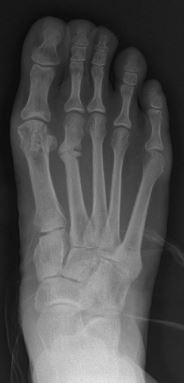
March fracture is the fracture of the distal third of one of the metatarsal bones occurring because of recurrent stress. It is more common in soldiers, but also occurs in hikers, organists, and other people whose duties entail much standing (such as hospital doctors). March fractures most commonly occur in the second and third metatarsal bones of the foot.[1][2][3] It is a common cause of foot pain, especially when people suddenly increase their activities.[4]

Signs and symptoms

[edit]

The onset is not dramatic. When the boot or shoes are taken off, there is a cramp-like pain in the affected forefoot, and moderate local edema appears on the dorsal aspect. On moving each toe in turn, that of the involved metatarsal causes pain, and when the bone is palpated from the dorsal surface, a point of tenderness is found directly over the lesion. Radiography at this stage is negative, but the condition is diagnosed correctly by military surgeons without the aid of x-rays. In civil life, it is seldom diagnosed correctly for a week or two, when, because of lack of immobilization, there is an excessive deposit of callus (which may be palpable) around the fracture.[1]

Diagnosis

[edit]

X-ray is seldom helpful, but a CT scan and an MRI study may help in diagnosis.[5] Bone scans are positive early on. Dual energy X-ray absorptiometry is also helpful to rule out comorbid osteoporosis.[6]

Differential diagnosis

[edit]

Treatment

[edit]

The first line treatment should be reduction of movements for 6 to 12 weeks. Wooden-soled shoes or a cast should be given for this purpose. In rare cases in which stress fracture occurs with a cavus foot, plantar fascia release may be appropriate.[6]

Occurrence

[edit]

Stress fractures can occur at many sites in the body; "march fracture" simply refers to a stress fracture specifically of the metatarsals, so named because the injury is sometimes sustained by soldiers during sustained periods of marching.[2] Although march fractures can occur to the 5th metatarsal, fractures of this bone are more likely to be trauma-related fractures to the diaphysis, termed Jones fractures. In runners, march fracture occurs most often in the metatarsal neck, while in dancers it occurs in the proximal shaft. In ballet dancers, fracture mostly occurs at the base of the second metatarsal and at Lisfranc joints. This fracture always occurs following a prolonged stress or weight bearing, and the history of direct trauma is very rare. Consideration should always be given to osteoporosis and osteomalacia. Cavus feet are a risk factor for march fracture.[6] [8]

References

[edit]
[edit]
Revisions and contributorsEdit on WikipediaRead on Wikipedia
from Grokipedia
A March fracture, also known as a , is a small crack or fatigue break in one of the long of the foot that connect the ankle to the toes, most commonly affecting the second or third due to repetitive mechanical rather than a single traumatic event. This injury derives its name from its historical association with prolonged marching in recruits, where enforced repetitive foot loading leads to bone overload. March fractures typically arise from overuse, such as a sudden increase in intensity, duration, or frequency—common in runners, dancers, athletes, and soldiers—or from underlying factors like reduced (e.g., ), nutritional deficiencies, hormonal imbalances (e.g., in women), or biomechanical issues like or improper footwear. Women are at higher risk due to factors including lower bone mass and menstrual irregularities, and the condition accounts for a significant portion of foot injuries in high-impact sports and military training. Symptoms usually begin insidiously with localized pain in the midfoot that intensifies during weight-bearing activities and eases with rest, progressing to constant aching, point tenderness, mild swelling, and possible bruising if untreated. Diagnosis often involves clinical evaluation, including a history of recent activity changes and physical exam for tenderness, supplemented by imaging: initial X-rays may be negative (as fractures can take 2-6 weeks to appear), so MRI or bone scans are used for confirmation. Treatment is primarily conservative, emphasizing and to allow , typically involving 4-8 weeks of non-weight-bearing with crutches, a walking boot or cast, , elevation, and anti-inflammatory medications like ibuprofen for relief. Full recovery requires gradual return to activity, for strengthening and balance, and addressing risk factors through nutrition (e.g., calcium and ) and proper training progression; surgical intervention is rare and reserved for non-healing or displaced cases. Prevention focuses on avoiding abrupt activity escalations, using supportive footwear, and incorporating to reduce repetitive stress.

Overview

Definition

A March fracture is a that specifically affects the distal third of one or more in the foot. These fractures are characterized by small cracks or breaks in the bone resulting from repetitive mechanical applied below the threshold required for an acute, traumatic . The metatarsals are the five long bones that form the forefoot, connecting the midfoot (tarsal bones) to the phalanges of the toes; March fractures most commonly involve the second or third metatarsal due to their anatomical positioning and load-bearing role during activities. Unlike traumatic fractures, which arise from a single high-energy impact on otherwise normal bone, March fractures represent a fatigue type of occurring in structurally normal bone subjected to cyclical, submaximal loading over time. In contrast, insufficiency stress fractures develop in bones weakened by underlying pathology, such as , where even normal physiological stresses exceed the bone's compromised capacity. This condition is classically linked to prolonged marching, especially in contexts, though it can occur in various overuse scenarios.

Etymology and History

The term "march fracture" originated in the , derived from its frequent occurrence among soldiers engaged in prolonged marching, which imposed repetitive stress on the of the foot. This nomenclature reflected the condition's association with military training and campaigns, where recruits unaccustomed to such demands developed painful foot injuries. The first detailed description of march fracture is attributed to Carl August von Breithaupt, a Prussian military surgeon, in 1855. In his publication, Breithaupt reported observing focal forefoot swelling and pain in military recruits following strenuous marches, coining the term "Fussgeschwulst" (swollen foot) to describe the phenomenon, though the English equivalent "march fracture" emerged later in . Subsequent reports appeared in the and among military surgeons in British and American armies during conflicts such as the and the , highlighting similar patterns of overuse injuries in infantry units subjected to extended foot marches with heavy loads. By the late 19th and early 20th centuries, radiographic confirmation advanced recognition, with the term "march fracture" formalized in 1897 through imaging studies of affected soldiers. The understanding evolved from isolated anecdotal cases in military settings to a more comprehensive view of stress fractures by the mid-20th century, as clinicians identified analogous injuries in civilians, particularly runners and athletes undertaking repetitive loading activities. This shift paralleled broader advancements in orthopedics and , emphasizing the condition's beyond wartime contexts.

Etiology and Pathophysiology

Causes

March fracture, also known as a metatarsal stress fracture, primarily arises from repetitive submaximal loading on the , particularly the second and third metatarsals, during prolonged activities such as marching or running. This cumulative mechanical stress leads to microtrauma in the , where normal physiological is overwhelmed by the frequency and intensity of the loading. In recruits, for instance, the term "March fracture" originated from the high incidence observed during extended marches, highlighting how unaccustomed repetitive impact exacerbates this process. The underlying pathophysiology involves an imbalance in , where osteoclastic resorption outpaces osteoblastic formation, resulting in localized weakening and eventual . Repetitive loading disrupts signaling, which normally coordinates repair, leading to accumulation of microdamage without sufficient time for recovery. This imbalance is particularly pronounced in the metatarsals due to their role in absorbing ground reaction forces during propulsion. Stress fracture progression in March fracture typically occurs in stages: initial periosteal reaction as the bone attempts to repair microdamage, followed by a cortical break if loading continues, and potential propagation to a complete if unaddressed. Early stages may show no radiographic changes, with a radiolucent resorption gap appearing only after 10-14 days, reflecting the delayed visibility of the remodeling process. Biomechanical factors contribute significantly, including increased ground reaction forces from high-impact activities, altered patterns that unevenly distribute load, and sudden increases in activity intensity without gradual , all of which amplify stress on the metatarsals. during prolonged exertion can further exacerbate these forces by reducing shock absorption.

Risk Factors

March fracture, a type of metatarsal stress fracture, is influenced by a combination of intrinsic and extrinsic risk factors that increase susceptibility to bone stress injuries. Intrinsic factors include female sex, which is associated with higher incidence due to lower bone mineral density and hormonal influences such as amenorrhea in athletes or menstrual dysfunction in military recruits. Low (BMI) and smaller body frames also predispose individuals by reducing the mechanical buffering capacity of bones during repetitive loading. Nutritional deficiencies, particularly in and calcium, further compromise bone health and remodeling, elevating risk in populations like recruits or endurance athletes. Additionally, or heightens vulnerability, as seen in studies of stress fracture cohorts where low correlates with fracture occurrence. Extrinsic factors primarily involve training-related variables, such as sudden increases in , duration, or volume, which overwhelm in unconditioned individuals. Inappropriate lacking adequate cushioning or support exacerbates metatarsal loading during activities. on hard surfaces amplifies impact forces, while recruit programs, with their rapid escalation of marching and running, represent a classic high-risk scenario due to repetitive submaximal stress. Biomechanical risks encompass structural variations like (high-arched feet), which alter load distribution across the metatarsals, increasing localized stress. Leg length discrepancies and muscle imbalances, such as weakness or inflexibility in the lower leg muscles, can further disrupt force absorption and contribute to uneven metatarsal strain. Evidence from cohort studies indicates a higher incidence among those with prior stress injuries, as previous microdamage impairs subsequent bone recovery and resilience. These factors often interact; for instance, in military settings, combinations of low fitness, nutritional deficits, and intensive training regimens amplify overall risk.

Clinical Features

Signs and Symptoms

March fracture typically presents with an insidious onset of dull, aching pain in the forefoot, particularly over the second or third metatarsal, that worsens with weight-bearing activities such as walking or running and improves with rest. Patients often report the pain beginning after recent increases in , such as prolonged marching or running on hard surfaces, without a history of acute trauma. Associated symptoms include localized swelling and tenderness in the affected area. Bruising is uncommon, particularly in hairline stress fractures. Swelling on the top of the foot without visible bruising can occur in march fractures but is not necessarily indicative of a fracture, as similar presentations can result from other conditions such as sprains, tendonitis, gout, arthritis, or infection. These symptoms are nonspecific, and definitive diagnosis requires medical evaluation, typically including imaging such as X-ray (though early stress fractures may not be visible) or MRI. In early stages, the pain is often diffuse over the forefoot and occurs primarily during or immediately after activity, but it may progress to a constant ache even at rest if the stress continues. Night pain may occur, particularly as the condition progresses. Functionally, individuals experience limping, difficulty bearing full weight on the affected foot, and challenges wearing regular shoes due to pressure on the painful area. These symptoms can significantly impair daily activities and mobility, often leading patients to seek medical attention after 2-6 weeks of worsening discomfort.

Physical Examination

The physical examination for suspected March fracture focuses on identifying localized signs of stress injury in the , typically the second or third, through systematic assessment of the foot. Inspection reveals mild swelling or over the dorsum of the foot, particularly in the midfoot region, without gross deformity in the early stages of the injury. Bruising or ecchymosis may occasionally be present but is often subtle. elicits point tenderness along the affected metatarsal shaft, most commonly in the distal third, serving as a hallmark finding. Compression of the forefoot, by squeezing the metatarsal heads together, reproduces sharp pain localized to the fracture site, helping differentiate from injuries. is rare but may occur if there is periosteal reaction. Special tests include the hop test, where pain on single-leg hopping on the affected foot indicates loading intolerance at the stress site. The forefoot loading test, involving axial compression of the metatarsals while the patient is seated, further provokes focal if a fracture is present. typically shows an pattern, characterized by shortened stance phase and reduced push-off on the affected side to minimize . Clinicians should also assess for underlying biomechanical abnormalities, such as excessive supination or , which may contribute to uneven forefoot loading. These objective findings correlate with patient-reported but emphasize clinician-elicited signs.

Diagnosis

Diagnostic Approach

The diagnostic approach to March fracture begins with a thorough history taking to identify potential triggers and risk factors associated with this metatarsal stress injury. Clinicians should inquire about recent changes in , such as sudden increases in training intensity, duration, or , which are common precipitants in individuals engaging in repetitive exercises. Specific attention is given to training history, including the type of used—poorly cushioned or ill-fitting shoes can exacerbate mechanical stress on the metatarsals—and screening for modifiable risk factors like nutritional deficiencies (e.g., low or calcium intake), hormonal imbalances (e.g., amenorrhea in female athletes), or prior lower extremity injuries. Integrating the patient's history with findings is crucial for raising clinical suspicion. An insidious onset of forefoot pain, often worsening with activity and improving with rest, combined with localized tenderness over the metatarsal shafts during or the hop test (where pain is elicited on single-leg hopping), strongly suggests a when no acute trauma is reported. This holistic assessment helps differentiate March fracture from overuse syndromes by focusing on focal rather than diffuse symptoms. Suspicion for March fracture should be heightened in high-risk contexts, such as new military recruits undergoing intensive marching or runners presenting with unexplained forefoot pain after escalating mileage beyond 25 miles per week. If initial clinical findings indicate a likely , multidisciplinary input is recommended, with prompt referral to an orthopedist for further management, particularly in cases involving persistent pain or high-risk anatomical locations like the second metatarsal base. Advanced imaging may be pursued under specialist guidance to confirm the .

Imaging

Plain radiography serves as the initial imaging modality for suspected March fracture due to its accessibility and low cost. However, it has low sensitivity in the early stages, often appearing normal within the first 2-3 weeks of symptom onset, as radiographic changes lag behind clinical presentation. Later findings may include periosteal reaction, cortical thickening, a sclerotic line, or a faint fracture lucency across the metatarsal neck, with sensitivity improving to 30-70% after three weeks. The "gray cortex" sign, representing early , can occasionally be detected on serial radiographs. Magnetic resonance imaging (MRI) is considered the gold standard for early detection of March fracture, offering high sensitivity (68-100%) and specificity (up to 100%) without ionizing radiation. It excels at visualizing bone marrow edema as a low-signal intensity band on T1-weighted images and high-signal intensity on T2-weighted or STIR sequences, often before radiographic changes appear. MRI also delineates associated soft tissue injuries, periosteal reaction, and fracture lines, making it ideal for confirming occult metatarsal stress fractures. Bone scintigraphy, using technetium-99m, provides high sensitivity (74-100%) for detecting early stress reactions through focal increased uptake at the site, useful when MRI is unavailable. However, it lacks specificity, potentially showing uptake in infections, tumors, or , and involves radiation exposure, limiting its use for follow-up. Computed (CT) is employed in equivocal cases to assess cortical disruption, sclerosis, or subtle lines, with specificity around 88-98% but lower sensitivity (32-38%) compared to MRI. It is particularly helpful for evaluating healing progress or when metallic implants preclude MRI. Ultrasound offers a non-invasive, radiation-free option for assessing metatarsal stress fractures, demonstrating sensitivity of approximately 83% and specificity of 75%, with findings such as cortical irregularities or periosteal thickening. It is most useful for evaluation and dynamic assessment but is operator-dependent and less effective for deep changes. Severity grading of March fractures on imaging aids in prognosis and management decisions. The Fredericson classification, originally for tibial stress injuries but applicable to metatarsals, uses MRI to grade from 1 (periosteal only) to 4 (severe marrow with fracture line). The Arendt system, tailored for foot stress fractures, similarly progresses from grade 1 (marrow signal change on STIR) to grade 4 (visible fracture line on multiple sequences). These scales correlate imaging findings with clinical severity and return-to-activity timelines.

Differential Diagnosis

The differential diagnosis for March fracture, a stress fracture typically affecting the second or third metatarsal shaft, includes several conditions presenting with forefoot pain that must be distinguished based on clinical history, , and findings. Common mimics encompass overuse-related disorders and bony pathologies that overlap in location but differ in onset and pathophysiology. Metatarsalgia involves chronic pain at the metatarsal heads due to mechanical overload, often from ill-fitting footwear or high-impact activities, contrasting with the diaphyseal location and insidious progression of March fracture; it lacks focal bony tenderness and shows no fracture line on imaging, instead revealing soft tissue thickening or fat pad atrophy on MRI. Plantar fasciitis presents with heel and arch pain exacerbated by initial weight-bearing, differing from the midfoot pain of March fracture by its plantar fascia origin and absence of metatarsal edema on MRI, where fasciitis demonstrates enthesophyte formation or fascial thickening without cortical disruption. Freiberg's disease, an osteochondrosis of the metatarsal head (usually second), affects adolescents with subacute onset and swelling, distinguished by epiphyseal avascular necrosis on radiographs showing flattening and sclerosis, unlike the linear periosteal reaction of stress fractures. Sesamoiditis causes localized pain under the first metatarsal head from repetitive hyperextension, differentiated by tenderness over the sesamoids rather than the shaft and normal metatarsal alignment on imaging, often with sesamoid fragmentation visible on oblique views. Traumatic fractures must be excluded due to their acute presentation versus the repetitive microtrauma history of March fracture. , at the fifth metatarsal base, arises from inversion injury or stress, featuring immediate swelling and a transverse line on radiographs with potential risk, unlike the subtle, longitudinal stress reaction in central metatarsals that may initially appear normal on plain films. involves midfoot instability from axial loading or twisting, with acute severe pain and ecchymosis across the tarsometatarsal joints, identified by diastasis (>2 mm) on weight-bearing radiographs or CT, contrasting the isolated metatarsal and no joint disruption in stress fractures on MRI. Systemic conditions can mimic insufficiency-type stress fractures in March fracture presentations. Osteoporosis-related insufficiency fractures occur in low bone density states, often postmenopausal, with minimal trauma history and multifocal involvement; confirms reduced density, and MRI shows diffuse marrow without the focal cortical stress response. Inflammatory arthropathies, such as , present with symmetric polyarticular pain, morning stiffness, and systemic features, differentiated by metatarsophalangeal joint erosions and on MRI or , absent in isolated stress fractures which respond to rest without anti-inflammatory needs. Dorsal foot swelling without ecchymosis — Swelling on the top of the foot without visible bruising is not diagnostic of a march fracture. Metatarsal stress fractures frequently present with localized dorsal swelling and pain without bruising, particularly in early or hairline stages, but several other conditions can produce similar findings. These include midfoot sprains, extensor tendonitis, gout, arthritis (such as rheumatoid arthritis or osteoarthritis), and infections (e.g., cellulitis or osteomyelitis). Distinction requires careful history (e.g., repetitive activity in tendonitis, acute onset in gout or infection), physical examination (e.g., tenderness along tendon sheaths or joints rather than focal bony sites), and imaging. March fracture remains a key consideration in patients with repetitive forefoot loading, but swelling alone does not confirm it; diagnosis typically requires X-ray (which may be normal initially) or MRI to demonstrate bone marrow edema or fracture line. Key differentiators across these conditions include the insidious onset and activity-related progression in March fracture versus acute trauma in fractures like Jones or Lisfranc; imaging patterns reveal and periosteal reaction on MRI for stress injuries compared to displacement or widening in traumatic cases; and clinical response, where March fracture improves with 2-6 weeks of rest, unlike persistent symptoms in arthropathies or neuromas.

Management

Treatment

The primary treatment for March fracture, a stress fracture typically affecting the second or third metatarsal , involves conservative management to promote healing and prevent progression. This approach centers on the protocol—rest, ice application for 15-20 minutes several times daily, compression with a wrap to reduce swelling, and elevation of the foot above heart level when possible—to alleviate pain and inflammation in the acute phase. Non-weight-bearing status is enforced using crutches or a walking boot for approximately 4-6 weeks, allowing the to rest while maintaining mobility; a stiff-soled or short-leg cast may also be used for immobilization if symptoms persist. Adjunctive therapies support symptom relief and recovery. Nonsteroidal anti-inflammatory drugs (NSAIDs), such as ibuprofen, are commonly prescribed to manage pain and reduce swelling, typically for the first 1-2 weeks or as needed. Following the initial immobilization period, is initiated to focus on retraining, strengthening exercises, and , facilitating a gradual return to weight-bearing activities over 6-12 weeks. Surgical intervention is rare for March fractures, as most heal without operative management, but it is indicated in cases of non-union after 3-6 months of conservative treatment or for high-risk fractures prone to displacement, such as those at the base of the fifth metatarsal. Procedures may include intramedullary screw fixation to stabilize the bone and promote union, particularly in athletes or individuals with delayed . Follow-up care emphasizes monitoring healing progress through serial , such as radiographs every 4-6 weeks, to confirm formation and resolution of the fracture line. Return to full activity is guided by pain-free milestones, typically allowing progression from partial to full once symptoms subside, with full recovery expected in 6-12 weeks for low-risk cases.

Prevention

Preventing March fracture, a type of metatarsal often linked to repetitive loading in activities like marching, involves targeted strategies to mitigate overuse and biomechanical risks in susceptible individuals, particularly military recruits and athletes. These approaches focus on modifiable factors to reduce incidence rates, which can reach 5-20% in high-risk training environments. Activity modification is a of prevention, emphasizing gradual progression of training volume and intensity to allow without overload. The "10% rule"—limiting weekly increases in running or marching distance to no more than 10%—has been shown to lower risk by promoting physiological acclimation. , such as incorporating low-impact activities like or , helps distribute mechanical stress across different muscle groups and reduces repetitive metatarsal loading, thereby decreasing rates in programs. Appropriate and play a critical role in shock absorption and biomechanical alignment. Cushioned running or marching shoes with adequate arch support can attenuate ground reaction forces on the metatarsals. Custom or insoles correct foot deformities like high arches or pronation, improving load distribution and lessening fatigue-related stress on the forefoot bones. Nutritional and lifestyle interventions address bone health vulnerabilities, especially in populations with suboptimal intake. Ensuring adequate calcium (1,000-2,000 mg/day) and (800-1,000 IU/day) through diet or supplementation strengthens and has been demonstrated to decrease rates by 20-21% in recruits undergoing intense . Screening for deficiencies, particularly in or those with low baseline fitness, allows for early correction via fortified foods or supplements, while maintaining overall caloric balance prevents energy deficits that exacerbate vulnerability. In military contexts, structured protocols during boot camp acclimation are essential for high-risk groups. Programs that phase in marching loads over 12-16 weeks, combined with regular monitoring for early pain symptoms, have reduced March fracture occurrences by integrating periods and fitness assessments. Pre-enlistment conditioning, including aerobic and strength exercises, further prepares recruits by enhancing musculoskeletal resilience before exposure to rigorous demands.

Epidemiology

Incidence and Prevalence

March fractures, also known as metatarsal stress fractures, account for approximately 20-25% of all stress fractures encountered in . In military recruits, these fractures constitute up to 5% of cases during initial training periods, reflecting their prominence among overuse injuries in high-impact environments. Incidence rates vary by population, with lifetime prevalence estimates of 5-13% among recreational runners, rising to 15-20% of musculoskeletal injuries in competitive runners. In basic training programs, rates escalate significantly, reaching 10-30% in some cohorts due to intense repetitive loading. U.S. data indicate approximately 20 stress fractures per 1,000 recruits during basic training, underscoring the condition's impact in structured physical regimens. The overall incidence of March fractures has remained stable over time in military settings as of 2023 reviews, though increased participation in endurance sports has led to greater recognition among civilian athletes, with potential rises in civilian cases following post-2020 fitness trends. Demographic patterns reveal a peak in young adults aged 18-25 years, aligning with the typical age of recruits and novice athletes. In contexts, occurrences exhibit seasonal patterns tied to cycles, with higher rates during early phases of intensive programs.

Affected Populations

March fractures, a type of metatarsal , predominantly affect individuals engaged in activities involving repetitive high-impact loading on the feet. , especially recruits undergoing basic , represent the highest-risk group due to the sudden increase in marching and load-bearing exercises. In this population, march fractures constitute approximately 9% of all stress fractures, with incidence rates during training reaching up to 5-10% overall for stress injuries, disproportionately impacting females at 2-10 times the rate of males. Athletes participating in sports with repetitive impact, such as runners, dancers, and gymnasts, are also highly susceptible, as these activities impose cyclic stress on the metatarsals. Runners commonly experience fractures at the second metatarsal , while dancers frequently sustain them in the same due to pointe work and jumps. athletes face an elevated risk, approximately 2-4 times higher than males, often linked to the female athlete triad involving energy deficiency, menstrual irregularities, and low . Beyond these primary groups, march fractures occur in elderly individuals with , where reduced bone density exacerbates vulnerability to even moderate repetitive stress, and in obese persons, as excess body weight amplifies mechanical loading on the feet. Additionally, people in occupations requiring prolonged standing or walking on hard surfaces, such as teachers, nurses, and factory workers, experience increased risk from cumulative daily strain. Geographic variations show higher reporting of march fractures in countries with mandatory , such as and , where leads to abrupt training intensities among young adults unaccustomed to such demands.

References

Add your contribution
Related Hubs
User Avatar
No comments yet.