Hubbry Logo
Catheterization laboratoryCatheterization laboratoryMain
Open search
Catheterization laboratory
Community hub
Catheterization laboratory
logo
8 pages, 0 posts
0 subscribers
Be the first to start a discussion here.
Catheterization laboratory
from Wikipedia

Catheterization laboratory
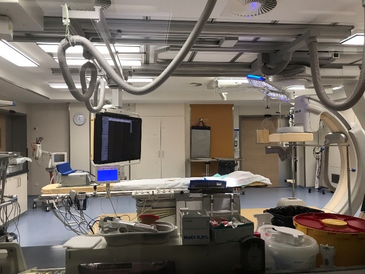
German cardiac catheterization laboratory
Purposevisualize the arteries and chambers of the heart

A catheterization laboratory, commonly referred to as a cath lab, is an examination room in a hospital or clinic with diagnostic imaging equipment used to visualize the arteries of the heart and the chambers of the heart and treat any stenosis or abnormality found.

Equipment

[edit]

Most catheterization laboratories are "single plane" facilities, those that have a single X-ray generator source and an X-ray image intensifier for fluoroscopic imaging.[1] Older cath labs used cine film to record the information obtained, but since 2000, most new facilities are digital. The latest digital cath labs are biplane (have two X-ray sources) and use flat panel detectors.[2]

Staff

[edit]

Cardiac catheterization laboratories are usually staffed by a multidisciplinary team. This may include a medical practitioner (normally either a consultant cardiologist or radiologist), cardiac physiologist, radiographer and nurse.[3][4]

Medical practitioner (interventional cardiologist/electrophysiologist)

[edit]

The consultant cardiologist is responsible for gaining arterial access, inserting a sheath into either the radial or femoral artery, passing a wire and catheter into the coronary artery and selectively injecting contrast media into the coronary arteries. They then interpret the images taken to ascertain where the narrowed or blocked artery has the problem. They use a variety of techniques and imaging tools to measure the size of things such as balloons and stents.

Cardiac physiologist

[edit]

Cardiac physiologists usually set up what is known as a transducer to monitor pressure in the arteries. They also have a live view of the patients ECG so they can tell whether or not there is a problem caused by the insertion of the catheter into the heart to the electrical pathways.[5][6] The physiologist will also set up a temporary pacemaker if the procedure is an angioplasty or a percutaneous coronary intervention (PCI). Finally, they also set up defibrillators on to the patient for emergency use if needed. In some locations, some of these responsibilities may be carried out by other personnel, such as trained nurses or technologists.[7]

Procedures

[edit]

Cardiac catheterization is a general term for a group of procedures that are performed in the cath lab, such as coronary angiography. Once a catheter is in place, it can be used to perform a number of procedures including angioplasty, PCI (percutaneous coronary intervention) angiography, transcatheter aortic valve replacement, balloon septostomy, and an electrophysiology study or catheter ablation. Devices such as pacemakers may be fitted or rotablation to remove plaque can be performed.[8][9]

See also

[edit]

References

[edit]
[edit]
Revisions and contributorsEdit on WikipediaRead on Wikipedia
from Grokipedia
A catheterization laboratory, commonly known as a cath lab, is a specialized medical facility within a hospital or clinic designed for performing diagnostic and interventional procedures on the heart and vascular system using thin, flexible tubes called catheters inserted into blood vessels. These procedures allow physicians to visualize and treat conditions such as coronary artery disease, heart valve disorders, and congenital heart defects with minimal invasiveness, often under local anesthesia and fluoroscopic guidance. Cath labs are equipped with advanced imaging systems, including fluoroscopy units for real-time visualization, hemodynamic monitoring devices to measure heart pressures and oxygen levels, and sterile environments to minimize infection risks during catheter insertion, typically via the femoral or . Procedures performed in these labs range from diagnostic , which assesses blockages in heart arteries, to therapeutic interventions like () involving balloon and placement to restore blood flow. Additional treatments may include transcatheter repairs or replacements, endomyocardial biopsies for tissue analysis, and electrophysiological studies for management. Operated by multidisciplinary teams led by interventional cardiologists, cath labs facilitate over one million procedures annually in the United States alone, contributing significantly to cardiovascular care by enabling rapid diagnosis and treatment that reduces the need for open-heart surgery. Many facilities, such as those at major medical centers, have pioneered innovations like radial access techniques and advanced catheter-based therapies, enhancing and recovery times. Despite their benefits, procedures carry low risks of complications like or vessel damage, which are managed through rigorous protocols.

Overview

Definition and Purpose

A catheterization laboratory, commonly referred to as a cath lab, is a specialized, sterile room within a or medical facility designed for performing minimally invasive cardiac procedures using thin, flexible catheters inserted into blood vessels to access the heart and vascular system. These laboratories resemble small operating rooms equipped with advanced imaging technology to enable precise navigation and real-time visualization during interventions. The primary purposes of a cath lab include diagnosing cardiovascular conditions by visualizing coronary arteries, heart chambers, and blood flow patterns, as well as identifying issues such as arterial blockages, valve malfunctions, or congenital defects through techniques like and pressure measurements. Additionally, cath labs facilitate therapeutic interventions, such as to widen narrowed arteries, to maintain vessel openness, or valvuloplasty to repair faulty heart valves, often allowing treatment during the same session as diagnosis. Key benefits of procedures in a cath lab include significantly reduced recovery times compared to traditional open-heart , typically enabling patients to resume normal activities within days rather than weeks. Real-time fluoroscopic provides high precision, minimizing tissue damage, while the integration with hybrid operating rooms supports complex cases combining catheterization with surgical elements for enhanced safety and efficiency. Over the past half-century, cath labs have evolved from basic rooms focused on diagnostic into multifunctional suites capable of supporting a broad range of procedures, including studies for arrhythmias and structural heart interventions like transcatheter valve replacements. This progression reflects advancements in and technologies, transforming them from specialized research environments into essential clinical hubs for comprehensive cardiac care.

Historical Development

The origins of the catheterization laboratory trace back to pioneering experiments in cardiac catheterization during the early 20th century. In 1929, German surgeon Werner Forssmann performed the first documented right heart catheterization on himself, inserting a ureteral catheter through his arm vein to reach the right atrium and ventricle, an act of bold self-experimentation that demonstrated the feasibility of accessing the heart via peripheral vessels despite initial controversy and professional repercussions. This breakthrough laid the groundwork for safer diagnostic applications, though it was not immediately adopted. In the 1940s, André Cournand and Dickinson W. Richards at Columbia University advanced the technique by establishing systematic right heart catheterization for diagnosing congenital and acquired heart diseases, opening the first dedicated laboratory at Bellevue Hospital in 1945 to refine pressure measurements and oxygen saturation assessments. Their collaborative work, building on Forssmann's innovation, earned the trio the Nobel Prize in Physiology or Medicine in 1956 for transforming cardiac catheterization into a clinical tool essential for understanding hemodynamics. The post-World War II era saw rapid expansion in the 1950s and 1960s, driven by innovations that necessitated specialized facilities for safer, more precise procedures. In 1958, F. Mason Sones Jr. at the Cleveland Clinic serendipitously developed selective coronary angiography during a routine cardiac catheterization of a young adult patient, injecting contrast directly into the coronary arteries to visualize blockages, which revolutionized the diagnosis of coronary artery disease and prompted the establishment of dedicated catheterization laboratories in hospitals to handle the increased complexity and radiation exposure risks. By the 1970s, these labs had become standard in major medical centers, incorporating advanced fluoroscopy and cineangiography to support growing volumes of diagnostic studies, with Mayo Clinic's early lab from the 1940s evolving into a model for integrated hemodynamic monitoring. This period marked a shift from ad-hoc procedures in operating rooms to purpose-built environments optimized for cardiac interventions. The and brought interventional capabilities to the forefront, further evolving lab design and technology. In 1977, Andreas Grüntzig performed the first percutaneous transluminal coronary angioplasty (PTCA) in Zurich, using a to dilate a stenotic coronary , ushering in the era of (PCI) and expanding cath labs to include therapeutic roles beyond diagnostics. The witnessed a transition from analog film-based to digital systems, enabling real-time processing, reduced radiation doses, and easier storage, with becoming widespread by the decade's end. Entering the 2000s, systems gained adoption for enhanced visualization in complex cases like congenital heart disease, while flat-panel detectors replaced traditional image intensifiers around 2000, improving image quality and workflow efficiency in modern labs. In the 2010s and up to 2025, catheterization laboratories have integrated multidisciplinary approaches, exemplified by the rise of hybrid operating rooms that combine catheterization capabilities with surgical suites for seamless transitions in procedures like transcatheter valve replacements. The from 2020 onward prompted stringent infection control protocols in cath labs, including pre-procedure testing, PPE enhancements, and prioritization of emergent over elective cases, resulting in temporary volume reductions of up to 50% in many facilities while underscoring the need for resilient operational standards. As of 2025, further advancements include novel radiation shielding devices and AI-powered diagnostic tools to enhance safety and efficiency. These adaptations have solidified the cath lab's role as a versatile hub for minimally invasive .

Facility Design

Layout and Environment

A catheterization laboratory, or cath lab, typically features a sterile procedure room measuring 400 to 600 square feet, designed to accommodate the patient table, imaging equipment such as a or single-plane arch, and space for up to six to eight staff members while maintaining clear pathways for movement. This room is adjacent to a separate of 150 to 200 square feet, shielded by lead-lined glass windows for real-time monitoring and communication via , with direct access corridors to preparation and recovery areas to streamline flow. The layout emphasizes sterility and efficiency, with piped medical gases routed through ceiling or floor booms to minimize clutter and trip hazards, and integrated storage for essential supplies near the procedure area. Environmental controls in the cath lab are critical for maintaining a sterile field and comfort, featuring HEPA-filtered air systems with positive to prevent airborne contamination, alongside at least 15 . Temperature is regulated between 70°F and 75°F (21°C to 24°C), and relative is kept at 30% to 60% to inhibit microbial growth while supporting staff performance during lengthy procedures. Emergency provisions include unobstructed access to defibrillators, crash carts, and power backups, often located within or immediately adjacent to the room for rapid response. Hybrid cath labs differ from traditional setups by integrating operating room functionalities into a larger space, often 800 square feet or more, to support complex interventions like (TAVR) without patient transfer. These rooms incorporate distinct zoning for clean and dirty pathways—separating sterile supply routes from waste disposal areas—to reduce infection risks during hybrid surgical-interventional procedures, while retaining core cath lab elements like imaging arches. Accessibility features enhance operational and compliance, including wide doors (at least 48 inches or 1.2 meters) for gurney transport, automatic operation where feasible, and seamless, non-slip vinyl flooring with coved bases to facilitate cleaning and prevent falls. The design also integrates with hospital-wide imaging networks via dedicated cable ducts, ensuring seamless data transfer without compromising the controlled environment. shielding, such as lead-lined walls and , is incorporated into the structure to protect adjacent areas, as detailed in specialized guidelines.

Radiation Safety Measures

Radiation safety in the catheterization laboratory is paramount due to the extensive use of and cineangiography, which expose patients and staff to . The primary goal is to minimize deterministic and stochastic effects through a multifaceted approach encompassing structural shielding, , dose monitoring, and optimized procedural techniques. These measures adhere to the ALARA (As Low As Reasonably Achievable) principle, which emphasizes justification, optimization, and dose limitation to reduce exposure risks. Structural shielding in cath labs includes lead-lined walls and ceilings to contain primary and scattered , preventing leakage to adjacent areas. Protective for staff typically consists of lead aprons with 0.25-0.5 mm lead equivalence, which can attenuate 96-98% of scattered , along with ceiling-suspended transparent shields and mobile barriers such as table skirts to block scatter from the patient. Additional items like lead-equivalent and gloves protect sensitive areas, with reducing lens dose by up to 80% and gloves providing hand protection during close-proximity tasks. For patients, lead drapes are applied to non-imaged areas, such as the gonadal region, to limit unnecessary exposure. Dose monitoring involves real-time personal dosimeters worn by staff at collar and waist levels to track exposure and ensure compliance with limits. The (ICRP) recommends an effective dose limit of 20 mSv per year, averaged over five consecutive years, without exceeding 50 mSv in any single year for occupationally exposed workers. Facilities maintain records of cumulative doses and conduct regular audits to enforce ALARA, often targeting staff exposures below 5 mSv annually in high-volume labs. Procedural safeguards focus on minimizing beam output and exposure time. Collimation restricts the field to the , reducing scatter by up to 50-70%. Pulsed modes, operating at 7.5-15 frames per second, lower patient and staff doses by 50-75% compared to continuous modes without significant loss in image quality. Patient positioning away from the beam's primary path, combined with last-image-hold techniques to avoid unnecessary acquisitions, further optimizes . Regulatory compliance is governed by standards from the U.S. (FDA) and the (IAEA), which mandate equipment performance criteria for fluoroscopic systems, including dose rate limits and . Post-2020, there has been increased adoption of AI-assisted tools for real-time dose optimization in cath labs, enabling significant dose reductions through automated collimation adjustments and frame rate modulation while preserving diagnostic accuracy.

Equipment

Imaging Systems

The primary imaging modality in catheterization laboratories is fluoroscopy, which provides real-time visualization of vascular structures and device navigation during procedures. This system typically employs a C-arm gantry configuration, supporting either image intensifiers or modern flat-panel detectors to convert into visible images. Image intensifiers, historically dominant, amplify low-intensity X-ray signals but can introduce geometric distortions and higher doses; flat-panel detectors, increasingly standard since the early 2000s, offer superior spatial uniformity, wider , and dose reduction of up to 50% compared to intensifiers while maintaining high contrast sensitivity. Cath lab fluoroscopy systems operate in single-plane or biplane configurations, distinguished by the number of sources and detectors. Single-plane systems use one and detector assembly, providing two-dimensional (2D) imaging from a single projection, which suffices for routine diagnostic but limits multi-angle views. Biplane systems incorporate dual orthogonal sources and detectors mounted on independent gantries, enabling simultaneous 2D imaging from two perpendicular planes and facilitating rudimentary 3D visualization without patient repositioning; this configuration may slightly increase procedure time but provides a modest reduction in contrast use, though it increases by approximately 36% compared to single-plane setups. Advancements in have transformed cath lab capabilities, shifting from analog -based to fully digital systems by the early 2000s, with (DSA) as a cornerstone technique since its commercial introduction in the 1980s. DSA enhances vessel conspicuity by subtracting pre-contrast "mask" images from post-contrast frames, minimizing bone and soft-tissue overlay; this transition eliminated processing, enabling immediate image review and archival. Modern DSA in cath labs achieves resolutions of 1024×1024 matrices with 8-10 bit depth, supporting frame rates of 15-30 frames per second (fps) for cine acquisition to capture dynamic vascular flow while balancing radiation dose. Integration of advanced features like rotational angiography further enhances imaging precision, allowing from a single contrast-enhanced rotation of the C-arm gantry over 5-8 seconds. This technique acquires 100-200 projections at 15-30 fps, enabling software-based volumetric rendering of cardiac with sub-millimeter accuracy, which aids in complex structural interventions by reducing the need for multiple static angiograms. Iodine-based contrast agents, typically non-ionic monomers like or iopamidol, are essential for opacification, injected at rates of 3-5 mL/s via automated injectors to achieve peak arterial enhancement without ; total volumes per run range from 20-50 mL, optimized to minimize risk. Maintenance and of imaging systems are critical to ensure diagnostic quality and , with annual (QA) programs recommended by professional societies. These include testing for image uniformity, low-contrast detectability, and using phantoms, alongside of automatic brightness control to maintain signal-to-noise ratios above 5:1 and minimize artifacts. Adherence to guidelines from the American Association of Physicists in Medicine (AAPM) and Society of Cardiovascular Angiography and Interventions (SCAI) involves physicist-led surveys verifying dose-area product accuracy within 10% and fluoroscopic output below 5 R/min at 80 kVp, preventing degradation in image clarity or excessive noise.

Catheterization Tools

Catheterization tools encompass a range of specialized instruments designed for vascular access and manipulation during procedures in the catheterization laboratory. Guidewires serve as foundational tools, typically ranging from 0.014 to 0.035 inches in diameter, enabling navigation through the vascular system. These wires often feature hydrophilic coatings, such as (PVP) or similar polymers, which reduce friction and enhance lubricity when activated with saline, facilitating smoother advancement in tortuous vessels. Diagnostic catheters, used for selective coronary artery engagement, include pre-shaped designs like the Judkins left and right configurations for standard aortic root anatomies, and Amplatz shapes for more challenging accesses, such as in dilated aortas or anomalous origins. These catheters allow contrast injection for while minimizing trauma to vessel walls. Interventional devices extend these basic tools for therapeutic applications. Balloon catheters, essential for percutaneous transluminal coronary angioplasty (PTCA), are available in diameters from 1.5 to 4.0 mm, with inflation pressures commonly reaching 12 to 20 atmospheres to dilate stenotic lesions. Stents, deployed over these balloons or via self-expanding mechanisms, include bare-metal stents (BMS) made primarily from or cobalt-chromium alloys, and drug-eluting stents (DES) coated with antiproliferative agents like or to inhibit neointimal and reduce restenosis rates compared to BMS. Self-expanding stents, often constructed from nitinol, rely on the material's superelastic properties to radially expand upon deployment from the delivery sheath, conforming to vessel contours without requiring high-pressure assistance. Accessories support safe and efficient tool delivery. Vascular access sheaths, sized from 4 to 8 French (Fr), provide a stable conduit for introducing catheters and guidewires while sealing the arteriotomy site. Integrated hemostatic valves in these sheaths, featuring designs like rotating or cross-cut seals, minimize blood loss by maintaining around passing devices. For heavily calcified lesions resistant to standard dilatation, rotational atherectomy devices such as the Rotablator employ diamond-coated burrs, typically 1.25 to 2.0 mm in diameter, rotating at speeds of 140,000 to 180,000 (rpm) to ablate plaque and create a smoother pathway for subsequent interventions. Material properties are critical for tool performance and . Biocompatible polymers, including or , form the shafts and coatings of catheters and balloons, offering flexibility and radiopacity while resisting . Nitinol, a nickel-titanium , imparts shape memory and superelasticity to guidewires and self-expanding stents, allowing deformation up to 8-10% strain without permanent kinking. These devices undergo sterilization, predominantly via gas, which penetrates complex geometries to achieve high-level disinfection without damaging heat-sensitive components, followed by to remove residuals below safe thresholds.

Personnel

Medical Professionals

The medical professionals in a catheterization laboratory primarily consist of specialized physicians who lead and execute invasive cardiac and vascular procedures. Interventional cardiologists, board-certified by the American Board of Internal Medicine after completing a three-year cardiovascular disease fellowship followed by a one-year interventional cardiology fellowship, perform percutaneous coronary interventions (PCI) and diagnostic coronary angiography to treat and assess coronary artery disease. Clinical cardiac electrophysiologists, who undertake an additional two-year fellowship in clinical cardiac electrophysiology after cardiovascular training per ACC/AHA/HRS guidelines, specialize in catheter-based arrhythmia ablations to manage heart rhythm disorders within the electrophysiology segment of the cath lab. Interventional radiologists, trained through a one-year preliminary internship, four-year diagnostic radiology residency, and subsequent one- to two-year interventional radiology fellowship as outlined by the Society of Interventional Radiology and ACGME, conduct peripheral vascular procedures such as angioplasty and stenting for arterial occlusions outside the coronary system. These professionals bear key responsibilities during procedures, including establishing arterial or venous access through femoral or radial approaches, frequently employing guidance to enhance precision and reduce risks like formation. They conduct real-time decision-making based on fluoroscopic and angiographic imaging to navigate catheters, select appropriate devices, and optimize therapeutic outcomes. Complication management forms a critical aspect of their role, encompassing prompt recognition and intervention for issues such as vessel perforation, occlusion, or bleeding, often utilizing vascular closure devices or anticoagulation adjustments to achieve . Training for these specialists follows rigorous ACC/AHA/SCAI guidelines, which mandate minimum procedural volumes—a minimum of 250 interventional procedures, including at least 200 coronary interventions, during the fellowship to achieve competency in complex cases. Simulation-based training has gained prominence post-2020, enabling fellows to rehearse access techniques, device deployment, and emergency scenarios on high-fidelity models, as evidenced by SCAI surveys indicating widespread adoption for skill acquisition despite barriers like access limitations. Ongoing (CME) and maintenance of certification (MOC) are required to stay abreast of new devices and technologies, with organizations like SCAI offering targeted modules on innovations in PCI and peripheral interventions. In their capacity as team leaders, these physicians direct pre-procedure planning, incorporating risk stratification tools like the SCAI/ACC PCI Risk Calculator to inform shared decision-making and obtain tailored to patient-specific factors. They also manage post-procedure follow-up, evaluating recovery, monitoring for access-site complications, and coordinating multidisciplinary care. These leaders collaborate briefly with support staff, including physiologists for hemodynamic monitoring (detailed in ### Support Staff), to ensure seamless execution.

Support Staff

Support staff in the catheterization laboratory, including registered nurses, cardiovascular technicians, and radiographers, play essential roles in facilitating safe and efficient procedures by handling patient monitoring, equipment preparation, and sterile environment maintenance. Registered nurses, often specialized in critical care, are responsible for administering , monitoring such as , , and , and ensuring the sterile field remains intact during interventions. Cardiovascular technicians, frequently certified as Registered Cardiovascular Invasive Specialists (RCIS), manage ECG and hemodynamic monitoring, including the setup and calibration of pressure transducers to (0 mmHg) for accurate readings, and assist with device programming for pacemakers and implantable cardioverter-defibrillators (ICDs). Radiographers optimize imaging systems, operating equipment to provide clear visualization while minimizing to the team and patient. These professionals undertake a range of responsibilities that support procedural flow, such as preparing and administering contrast media, documenting intra-procedural events like vital sign changes and doses, and performing post-procedure tasks including sheath removal and initial recovery monitoring. In high-acuity scenarios, such as primary (PPCI), they assess puncture sites, anticipate complications, and coordinate emergency responses to maintain patient stability. is typically flexible, with a minimum of one circulating technician and one per procedure, adjusted based on case complexity to ensure comprehensive support under cardiologist oversight. Training for support staff emphasizes certification and ongoing education to meet safety standards, including (BLS) and Advanced Cardiovascular Life Support (ACLS) for , alongside specialized credentials like RCIS for technicians, which covers invasive cardiovascular procedures and patient care competencies. The Society for Cardiovascular Angiography and Interventions (SCAI) promotes multidisciplinary teams through educational guidelines that include at least one RCIS-trained individual per lab, fostering expertise in aseptic techniques, radiation safety, and hemodynamic assessment. In integration, support staff perform scrub and circulating duties to uphold sterility—scrub roles involve direct assistance with instruments and catheters, while circulating roles handle supplies, positioning, and —enabling seamless procedure execution and rapid to intra-procedural needs. This coordinated approach, aligned with SCAI best practices, enhances team efficiency and outcomes in the dynamic cath lab environment.

Procedures

Diagnostic Procedures

Diagnostic procedures in the catheterization laboratory utilize specialized catheters to evaluate cardiac and vascular function without therapeutic intervention, providing critical on flow, pressures, and electrical activity to guide clinical management of conditions such as and arrhythmias. These techniques leverage fluoroscopic imaging and contrast agents to assess and in real-time, enabling precise of ischemia, valvular issues, and conduction abnormalities. Performed under , they typically involve femoral or radial access, with catheters advanced to the heart for targeted measurements. Coronary is a cornerstone diagnostic method where a is advanced to the coronary ostia, and is injected to opacify the lumen, allowing visualization of , occlusions, and collateral circulation via cineangiography. This procedure identifies the and severity of atherosclerotic lesions, aiding in the assessment of . is often graded using the Thrombolysis in (TIMI) flow scale, which categorizes antegrade flow from grade 0 (no beyond the occlusion) to grade 3 (complete with normal speed and clearance). Right and left heart catheterization measures intracardiac and pulmonary pressures to evaluate hemodynamic status, such as in suspected or valvular disease. In right heart catheterization, a is floated through the venous system to record right atrial, right ventricular, , and wedge pressures; normal pulmonary artery systolic pressure ranges from 15 to 25 mmHg. Left heart catheterization accesses the left ventricle via arterial approach to assess aortic and left ventricular pressures. Ventriculography, involving contrast injection into the ventricle, delineates chamber volumes for using : EF=EDVESVEDV×100EF = \frac{EDV - ESV}{EDV} \times 100, where EDV is and ESV is end-systolic volume, providing insight into systolic function. Electrophysiology studies employ multipolar electrode catheters positioned in the high right atrium, His bundle, , and right ventricular apex to map intracardiac electrograms, identifying origins and conduction pathways through bipolar or unipolar recordings. These catheters detect local sequences and far-field signals to differentiate mechanisms like reentry or . Programmed electrical stimulation, such as incremental pacing or extra stimuli (up to three after a drive train of 400-600 ms), induces under controlled conditions to replicate clinical , facilitating based on cycle length, ventriculoatrial intervals, and patterns. Fractional flow reserve (FFR) assesses the functional significance of intermediate coronary stenoses by measuring the translesional during maximal hyperemia, induced via intravenous or intracoronary to simulate stress conditions and reveal ischemia potential. A pressure-sensing wire is advanced distal to the , yielding FFR as the of distal (Pd) to proximal (Pa) (FFR = Pd / Pa); values below 0.80 signify hemodynamically significant lesions warranting further evaluation, as they indicate reduced maximal blood flow to 80% or less of normal capacity.

Interventional Procedures

Interventional procedures in the catheterization laboratory represent a of minimally invasive , enabling therapeutic interventions for , valvular heart conditions, arrhythmias, and peripheral vascular pathologies. These techniques typically involve catheter-based access through arterial or venous routes, guided by real-time and intravascular imaging to deliver devices that restore blood flow, repair structural defects, or ablate aberrant electrical pathways. Unlike diagnostic evaluations, interventional approaches aim to achieve immediate hemodynamic improvements and long-term symptom relief, with procedural success often exceeding 90% in appropriately selected patients. Percutaneous coronary intervention (PCI) is a primary therapeutic modality for obstructive , involving to dilate stenotic lesions followed by deployment of a self-expanding or -expandable to maintain vessel patency. The procedure begins with guidewire advancement across the occlusion, inflation to compress plaque, and subsequent placement, which reduces the risk of and restenosis compared to alone. Acute procedural success rates for PCI surpass 99% in uncomplicated cases, as demonstrated in primary PCI for acute , where timely reperfusion minimizes myocardial damage. According to (ACC) data, overall PCI success exceeds 90%, with deployment facilitating durable revascularization in over 93% of interventions. Transcatheter aortic valve replacement (TAVR) addresses severe in patients across surgical categories, deploying a collapsible prosthetic via , most commonly through transfemoral access accounting for over 95% of procedures to minimize invasiveness. The procedure entails crossing the stenotic native with a guidewire, valvuloplasty for predilation, and precise positioning of the transcatheter using rapid ventricular pacing to ensure deployment without . selection relies on stratification tools such as the Society of Thoracic Surgeons (STS) Predicted of Mortality (PROM) score, which integrates comorbidities to guide selection across a range of levels. Femoral access predominates and has been associated with reduced vascular complications when combined with advanced imaging for iliofemoral assessment. Long-term outcomes show sustained function, with 30-day mortality rates approximately 2-3% in contemporary cohorts as of 2025, aligning with or below STS predictions. Electrophysiology ablations target (AFib) by isolating arrhythmogenic foci, particularly through pulmonary vein isolation using or delivered via intracardiac catheters. employs heat to create circumferential lesions around ostia, disrupting ectopic triggers, while uses extreme cold for precise, single-shot applications that minimize esophageal injury. Emerging techniques include pulsed field ablation (PFA), a non-thermal method using electrical pulses that offers high efficacy and safety profiles. These techniques achieve initial success in restoring in 70-80% of paroxysmal AFib cases, but recurrence rates range from 20-40% within the first year due to reconnection or extra-pulmonary triggers. Repeat procedures address reconnections, improving freedom from AFib to over 80% in selected patients, with and PFA showing comparable efficacy to in randomized trials. Peripheral interventions manage limb-threatening ischemia from atherosclerotic disease, utilizing to excise plaque or to occlude aneurysmal segments, often with adjunctive balloon or stenting. Directional or rotational devices debulk calcified lesions in femoropopliteal arteries, achieving procedural success rates of 97.6% and reducing the need for stenting in over 90% of cases by minimizing risk. For acute limb ischemia, embolic protection devices such as distal filters capture debris during , preventing distal occlusion and lowering periprocedural complications to under 5%. These approaches restore inline flow in critical limb ischemia, with 12-month patency rates exceeding 80% when embolic safeguards are employed.

Advancements and Safety

Technological Innovations

Recent advancements in (AI) and have transformed catheterization laboratories by enabling automated lesion detection in and for procedure outcomes. Quantitative Flow Ratio (QFR), a non-hyperemic computational method, uses AI to derive functional assessments from standard angiographic images, improving diagnostic accuracy without the need for invasive wires or pharmacological agents. Clinical studies post-2020 have validated QFR's in identifying hemodynamically significant stenoses, with diagnostic comparable to (FFR) in over 90% of cases. algorithms further enhance by analyzing imaging data to forecast procedural risks, such as malapposition or restenosis, achieving up to 85% accuracy in outcome models derived from large registries. Robotic systems have gained prominence for their precision in catheter navigation, minimizing operator exposure to and fatigue during complex interventions. The CorPath GRX platform, developed by Corindus Vascular Robotics (now ), allows remote control of catheters with sub-millimeter accuracy, facilitating percutaneous coronary interventions (PCI) and peripheral procedures. Initially granted FDA clearance in 2012 for coronary use and expanded to peripheral applications in 2018, the system has seen increasing adoption. Studies report exposure reductions of 80-95% for primary operators compared to manual techniques, attributed to the system's tabletop design that positions the physician outside the direct beam. Advanced imaging modalities continue to evolve, providing detailed plaque characterization to guide therapeutic decisions. (IVUS) offers cross-sectional views of vessel walls with resolutions around 150-200 μm, enabling assessment of plaque burden and composition, while (OCT) achieves superior axial resolution below 20 μm (typically 10-15 μm), allowing visualization of fine structures like fibrous caps and microcalcifications. Post-2020 innovations, such as high-definition IVUS, have improved tissue differentiation, correlating strongly with histopathological findings in plaque vulnerability assessment. Integration of 3D and 4D imaging fusion with pre-procedural computed tomography (CT) or (MRI) enhances spatial orientation during catheterization, overlaying volumetric data onto live for real-time guidance in complex anatomies like congenital heart defects. Emerging future trends in catheterization laboratory technology emphasize minimally invasive and immersive solutions. Nanotechnology enables targeted drug delivery via catheter-deployed nanoparticles, such as lipid-based carriers for site-specific release of anti-restenotic agents, reducing systemic toxicity and improving endothelial repair in post-PCI settings. Wireless sensors, including batteryless implantable devices integrated into stents, provide continuous real-time hemodynamic monitoring of and flow without additional invasiveness, transmitting data via for remote oversight. Additionally, (VR) integration supports procedural training through immersive simulations of catheterization environments, enhancing skills in radiation safety and device manipulation while reducing reliance on animal models or live cases.

Quality Assurance and Risks

Quality assurance in catheterization laboratories is governed by accreditation standards from organizations such as The Joint Commission (formerly JCAHO) and the American College of Cardiology (ACC), which require facilities to demonstrate compliance with evidence-based protocols for procedural safety, staff credentials, and equipment maintenance to achieve certification. These guidelines emphasize continuous quality improvement, including regular audits of procedural outcomes and adherence to minimum volume requirements for operators to maintain proficiency. Mandatory reporting to the National Cardiovascular Data Registry (NCDR), managed by the ACC, tracks key performance indicators such as door-to-balloon times for ST-elevation myocardial infarction (STEMI) patients, with a target of less than 90 minutes to optimize reperfusion and reduce mortality. Infection control protocols in catheterization laboratories prioritize strict aseptic techniques and antibiotic prophylaxis to minimize catheter-related bloodstream infections (CLABSIs). is commonly administered as perioperative prophylaxis, typically as a single intravenous dose of 1-2 grams prior to procedures involving vascular access, due to its efficacy against and low risk of resistance. Laboratories aim for a zero-tolerance approach to CLABSIs, with benchmarks targeting rates below 1 per 1,000 central line device-days through bundle interventions like skin preparation, maximal barrier precautions, and prompt catheter removal. Common procedural risks in catheterization laboratories include contrast-induced nephropathy (CIN), vascular complications, and arrhythmia induction, each managed through targeted preventive and responsive strategies. CIN, characterized by a rise in serum of at least 0.5 mg/dL or 25% within 48-72 hours post-procedure, occurs in approximately 5-10% of patients, particularly those with pre-existing renal impairment, and is primarily prevented by intravenous hydration with isotonic saline to maintain renal . Vascular complications, such as hematomas at the access site, affect 2-6% of cases, more commonly with femoral access, and are mitigated by ultrasound-guided puncture, proper techniques, and close post-procedure monitoring. s induced by manipulation, often ventricular in origin, are typically self-limiting and benign but may require immediate in hemodynamically unstable cases to restore . Quality metrics in catheterization laboratories incorporate regular processes for device tracking to ensure , , and recall management, integrating these into broader continuous improvement frameworks to identify procedural variations and enhance safety. Following , there has been increased emphasis on equity in access to catheterization services, addressing disparities in underserved populations through targeted , alongside the integration of telemedicine for post-procedure follow-up to improve adherence and reduce readmissions without compromising care .

References

Add your contribution
Related Hubs
User Avatar
No comments yet.