Hubbry Logo
First-degree atrioventricular blockFirst-degree atrioventricular blockMain
Open search
First-degree atrioventricular block
Community hub
First-degree atrioventricular block
logo
8 pages, 0 posts
0 subscribers
Be the first to start a discussion here.
First-degree atrioventricular block
from Wikipedia
First-degree AV block
Other namesFirst degree heart block, PR prolongation
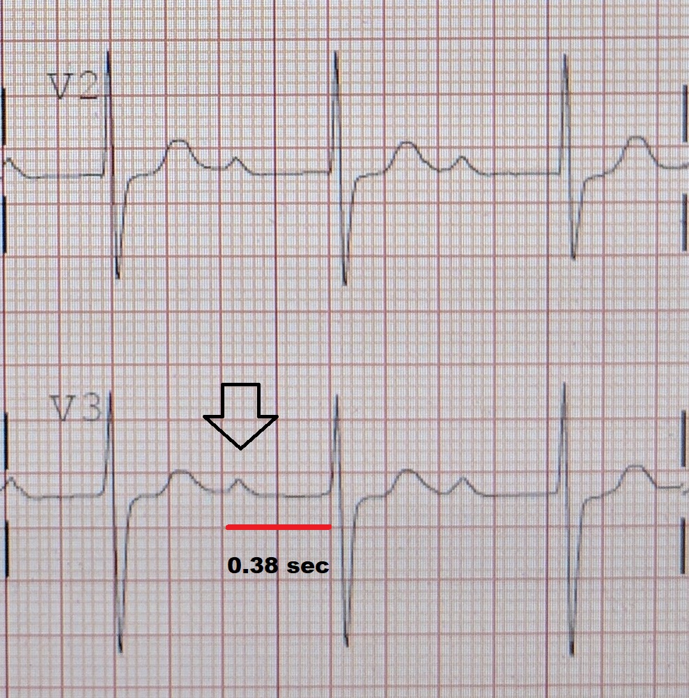
An ECG showing a first degree AV block of greater than 300 ms
SpecialtyCardiology
SymptomsAsymptomatic
ComplicationsProgression to second or third degree AV block
CausesFibrosis in AV node, medication, vagal tone, electrolyte disturbances
Diagnostic methodElectrocardiogram
TreatmentAvoidance of AV-nodal-blocking medication

First-degree atrioventricular block (AV block) is a disease of the electrical conduction system of the heart in which electrical impulses conduct from the cardiac atria to the ventricles through the atrioventricular node (AV node) more slowly than normal. First degree AV block does not generally cause any symptoms, but may progress to more severe forms of heart block such as second- and third-degree atrioventricular block. It is diagnosed using an electrocardiogram, and is defined as a PR interval greater than 200 milliseconds.[1] First degree AV block affects 0.65-1.1% of the population with 0.13 new cases per 1000 persons each year.

Causes

[edit]

The most common causes of first-degree heart block are AV nodal disease, enhanced vagal tone (for example in athletes), myocarditis, acute myocardial infarction (especially acute inferior MI), electrolyte disturbances and medication. The medications that most commonly cause first-degree heart block are those that increase the refractory time of the AV node, thereby slowing AV conduction. These include calcium channel blockers, beta-blockers, cardiac glycosides, and anything that increases cholinergic activity such as cholinesterase inhibitors.[2]

Diagnosis

[edit]

In normal individuals, the AV node slows the conduction of electrical impulses through the heart. This is manifest on a surface electrocardiogram (ECG) as the PR interval. The normal PR interval is from 120 ms to 200 ms in length. This is measured from the initial deflection of the P wave to the beginning of the QRS complex.[3]

In first-degree heart block, the AV node conducts the electrical activity more slowly. This is seen as a PR interval greater than 200 ms in length on the surface ECG. It is usually an incidental finding on a routine ECG.[4]

First-degree heart block does not require any particular investigations except for electrolyte and drug screens, especially if an overdose is suspected.[5]

In comparison to second-degree atrioventricular block, in first-degree block there is an absence of non-conduction or "dropped beats."

In an electrophysiology study, this corresponds to a prolonged A-H interval that shows the time between atrial depolarization and His bundle depolarization near the AV node.

Treatment

[edit]

The management includes identifying and correcting electrolyte imbalances and withholding any offending medications. This condition does not require admission unless there is an associated myocardial infarction. Even though it usually does not progress to higher forms of heart block, it may require outpatient follow-up and monitoring of the ECG, especially if there is a comorbid bundle branch block. If there is a need for treatment of an unrelated condition, care should be taken not to introduce any medication that may slow AV conduction. If this is not feasible, clinicians should be very cautious when introducing any drug that may slow conduction; and regular monitoring of the ECG is indicated.[6]

Prognosis

[edit]

Isolated first-degree heart block has no direct clinical consequences. There are no symptoms or signs associated with it. It was originally thought of as having a benign prognosis. In the Framingham Heart Study, however, the presence of a prolonged PR interval or first degree AV block doubled the risk of developing atrial fibrillation, tripled the risk of requiring an artificial pacemaker, and was associated with a small increase in mortality. This risk was proportional to the degree of PR prolongation.[7]

A subset of individuals with the triad of first-degree heart block, right bundle branch block, and either left anterior fascicular block or left posterior fascicular block (known as trifascicular block) may be at an increased risk of progression to complete heart block.[8]

See also

[edit]

References

[edit]
[edit]
Revisions and contributorsEdit on WikipediaRead on Wikipedia
from Grokipedia
First-degree atrioventricular block is a conduction delay in the or His-Purkinje system, defined electrocardiographically by a prolonged greater than 200 milliseconds with every followed by a , indicating intact but slowed atrioventricular conduction. This abnormality reflects slowed impulse transmission from the atria to the ventricles without interruption, often resulting from increased , medications such as beta-blockers or , myocardial ischemia, , or inflammatory conditions like . Prevalence increases with age, affecting approximately 1-4% of the general population, with higher rates in older adults and those with comorbidities like or structural heart disease. Typically and benign, it requires no specific treatment beyond addressing reversible causes, though "marked" first-degree block with PR intervals exceeding 300 milliseconds may cause symptoms such as fatigue or due to ineffective atrial contribution to ventricular filling. Emerging longitudinal data indicate associations with increased risks of , , pacemaker requirement, and mortality, challenging the traditional view of it as inconsequential and warranting monitoring in select patients.

Pathophysiology

Definition and electrocardiographic features

First-degree atrioventricular block is characterized by a delay in conduction from the atria to the ventricles, manifested on the electrocardiogram (ECG) as a prolonged exceeding 0.20 seconds while maintaining 1:1 atrioventricular conduction, with every followed by a . The normal ranges from 0.12 to 0.20 seconds, measured from the onset of the to the beginning of the ; prolongation beyond this threshold indicates first-degree block without interruption in impulse transmission. Electrocardiographic features include a consistently extended across consecutive beats, with no progressive lengthening or dropped QRS complexes as seen in higher-degree blocks. P waves remain normal in morphology and precede each QRS, which typically exhibits normal duration unless confounded by unrelated conduction abnormalities such as . Atrial and ventricular rates are usually preserved, reflecting intact overall without evidence of atrioventricular dissociation. When the surpasses 0.30 seconds, it is termed marked first-degree block, potentially leading to P waves overlapping preceding T waves, though conduction remains complete. relies on standard 12-lead ECG, with confirmation in multiple leads to exclude artifacts or measurement errors.

Mechanisms of conduction delay

The conduction delay in first-degree atrioventricular (AV) block manifests as prolongation of the beyond 200 milliseconds on , reflecting slowed impulse propagation from atria to ventricles while maintaining 1:1 conduction. This delay most commonly localizes to the AV node, where nodal cells exhibit inherently slow conduction velocities due to reliance on calcium-dependent action potentials and sparse intercellular connections, but it can also arise intra-atrially, within the His bundle, or in the Purkinje system. Infra-nodal delays, indicated by widened QRS complexes, are less frequent in isolated first-degree block and carry higher risk of progression to advanced blocks. Physiologically, enhanced parasympathetic (vagal) tone contributes to delay by acetylcholine-mediated activation of muscarinic receptors on AV nodal cells, leading to membrane hyperpolarization, reduced phase 0 upstroke velocity, and prolonged refractoriness. Sympathetic withdrawal or dominance inversely shortens the PR interval by increasing cyclic AMP and enhancing calcium influx. Pharmacological agents exacerbate delay via similar ion channel effects: beta-blockers and non-dihydropyridine calcium channel blockers suppress nodal excitability, while digoxin increases vagal tone indirectly through enhanced parasympathetic activity. Pathologically, structural alterations such as or sclerosis of the AV nodal region—prevalent in aging—increase conduction resistance by disrupting cell-to-cell coupling and replacing excitable tissue with non-conductive . Ischemia, particularly from inferior affecting the AV nodal artery, impairs ATP-dependent ion handling, while inflammatory or infiltrative processes (e.g., , ) cause or cellular infiltration that slows propagation. Electrolyte derangements like further depress nodal excitability by altering resting . In rare cases, congenital anomalies or congenital heart defects contribute to intra-atrial delays.

Epidemiology

Prevalence and incidence

First-degree atrioventricular block exhibits a that varies by age, sex, and population studied, typically ranging from 1% to 7% in adults, with higher rates observed in older individuals due to age-related degenerative changes in the conduction . In general population cohorts, prevalence approximates 3-4%, as documented in electrocardiographic screenings of adults. Males demonstrate consistently higher prevalence than females, with ratios around 2:1 in large-scale studies (e.g., 5.1% in men versus 2.2% in women among over 10,000 screened individuals). Age is the strongest correlate, with rates below 1% in those under 60 years rising to 6% or more in those over 60, reflecting cumulative fibrotic infiltration of the . In younger populations, such as children and adolescent athletes, remains low at approximately 1%, substantially less than in older adults or non-athletic cohorts. Ethnic variations show minor differences, with some evidence of slightly higher rates in African-American versus Caucasian groups across age strata, though data are limited and confounded by comorbidities. Incidence data are scarcer, as the condition is often and detected incidentally, but estimates indicate about 0.13 new cases per 1,000 person-years in general populations, underscoring its gradual onset rather than acute emergence. Longitudinal studies confirm low annual progression rates from normal conduction, primarily driven by aging or underlying cardiac pathology rather than isolated incidents.

Associated demographic and risk factors

First-degree atrioventricular block demonstrates a marked increase in with advancing age, remaining relatively uncommon at 1.0-1.5% in individuals under 60 years but rising to approximately 6.0% in those over 60 years, reflecting cumulative degenerative changes in the . Males exhibit a higher prevalence than females, with one population-based study reporting rates of 5.1% in men versus 2.2% in women, and multivariate analyses consistently identifying male sex as an independent risk factor alongside older age. Additional demographic correlates include greater body height, observed as an independent association in cohort studies, potentially linked to variations in cardiac anatomy or vagal tone. Limited evidence suggests elevated risk in white populations compared to others, though data on racial and ethnic disparities remain sparse and require further validation across diverse cohorts. Beyond demographics, familial aggregation contributes to risk, with first-degree relatives of affected individuals facing a significantly elevated for , modulated by the proband's age at and suggesting heritable components in conduction pathway integrity. Comorbid conditions such as , , , and overweight status independently predict development, with population-attributable fractions indicating that modifiable metabolic factors like elevated and glucose may underlie over half of cases in some analyses.

Etiology

Intrinsic causes

Idiopathic fibrosis and sclerosis of the conduction system constitutes the predominant intrinsic cause of first-degree atrioventricular (AV) block, responsible for approximately 40% of cases. This degenerative process, prevalent in older adults, entails progressive fibrotic replacement of specialized conducting tissues, including the AV node and His-Purkinje system. Specific variants include Lev's disease, characterized by annular fibrocalcific changes encroaching on the AV junction, and Lenegre's disease, involving idiopathic sclerosis of the infranodal conduction pathways. These conditions arise from age-related wear, leading to conduction delay without external provocation, and prevalence of first-degree AV block rises to about 6% in those over 60 years. Ischemic heart disease accounts for roughly 20% of intrinsic etiologies, where coronary artery occlusion or infarction impairs blood supply to the AV node or bundle branches, resulting in localized and . Inferior particularly affects the AV node due to its dominance, prolonging the in up to 10-20% of such events. Infiltrative disorders, such as cardiac or , deposit abnormal proteins or granulomas within the conduction tissues, disrupting impulse propagation. , for instance, underlies up to one-third of new-onset AV blocks in adults aged 18-60 without evident structural heart disease upon targeted evaluation, including cardiac MRI or . Similarly, amyloid infiltration in or light-chain stiffens and replaces nodal tissue, manifesting as conduction abnormalities in advanced stages. Congenital and hereditary forms represent rarer intrinsic causes, often linked to genetic mutations affecting connexins or sodium channels essential for conduction. Progressive familial heart block, for example, involves autosomal dominant defects leading to fibrotic degeneration over time, with first-degree block as an early electrocardiographic sign progressing to higher degrees in affected families. Myocarditis sequelae, including viral-induced scarring, may also yield permanent intrinsic nodal damage if inflammation resolves with residual fibrosis.

Reversible and extrinsic causes

Reversible causes of first-degree atrioventricular (AV) block encompass extrinsic factors that delay AV nodal conduction but resolve upon correction of the underlying trigger, distinguishing them from irreversible intrinsic myocardial degeneration. These etiologies often involve pharmacological agents, metabolic derangements, or transient physiological states that modulate AV node refractoriness without permanent structural damage. Identification and reversal of such causes are critical, as they obviate the need for invasive interventions in otherwise benign presentations. Pharmacological agents represent a primary extrinsic cause, particularly those that enhance or directly suppress AV nodal excitability. Beta-adrenergic blockers (e.g., metoprolol, atenolol) prolong the by reducing sympathetic stimulation of the AV node, with effects typically dose-dependent and reversible upon discontinuation or dose reduction. Non-dihydropyridine such as verapamil and similarly impede calcium influx in AV nodal cells, leading to conduction delay that normalizes after . , through increased vagal activity, can induce first-degree block, especially in therapeutic or toxic ranges, with resolution following cessation or antidote administration like digoxin-specific antibodies. and other class III antiarrhythmics may contribute via prolongation of phases, though their effects are often multifactorial and slower to reverse. Electrolyte and metabolic imbalances frequently precipitate reversible AV conduction delays by altering membrane potentials and function. Hyperkalemia, often exceeding 5.5 mEq/L, depresses AV nodal excitability through partial and reduced availability, with first-degree block resolving as serum potassium normalizes via medical management such as insulin-glucose therapy or kayexalate. Hypokalemia and hypomagnesemia impair , prolonging the action potential duration and , effects that abate with repletion. Hypercalcemia, typically above 12 mg/dL, shortens but can paradoxically delay AV conduction in severe cases by accelerating phase 2 unevenly; correction via hydration and bisphosphonates restores normal timing. Acute hypothyroidism may contribute via bradycardic effects on the sinus and AV nodes, reversible with replacement over weeks. Autonomic influences, particularly heightened , constitute a physiologic extrinsic cause common in asymptomatic individuals. Endurance athletes often exhibit PR prolongation due to enhanced parasympathetic dominance, with intervals normalizing during deconditioning or exercise. episodes can trigger transient vagally mediated block through recurrent hypoxia and reflex arcs, resolving with therapy. Inferior myocardial ischemia, as in transient coronary events, may induce reversible delay via vagal activation without , improving with or anti-ischemic agents. Infectious and inflammatory processes offer reversible etiologies when addressed promptly. Lyme carditis, caused by , manifests as AV block in up to 10% of disseminated cases, with first-degree involvement progressing variably but often reversing completely with antibiotics like within days to weeks. Acute viral myocarditis (e.g., from or ) can cause transient nodal inflammation and edema, leading to conduction delay that abates with supportive care and resolution of . Rheumatic fever historically linked first-degree block to , reversible in early stages with anti-inflammatory therapy, though rare post-eradication efforts.

Clinical Presentation

Symptomatic manifestations

First-degree atrioventricular block is generally , with most individuals unaware of its presence due to preserved atrioventricular synchrony and adequate . Symptoms, when they occur, are uncommon and typically arise in cases of marked conduction delay, defined as a exceeding 0.30 seconds, where asynchronous atrial and ventricular contractions may reduce and effective . In such symptomatic presentations, patients may experience , , , or dyspnea, resembling features of despite the absence of a device. , , , or even syncope can manifest, particularly during exertion or in the context of underlying comorbidities like , where first-degree block correlates with increased symptom burden. These manifestations are not directly attributable to the block in isolation but often reflect associated hemodynamic compromise or progression toward higher-degree block; isolated first-degree block rarely requires intervention based on symptoms alone. Evaluation for reversible causes, such as medications or imbalances, is essential in symptomatic cases to distinguish conduction delay from more severe pathology.

Asymptomatic variants

First-degree atrioventricular block is frequently asymptomatic and identified incidentally during routine in patients lacking complaints of , , , or syncope. The conduction delay manifests as a prolonged beyond 200 milliseconds, with consistent atrioventricular synchrony preserved, distinguishing it from higher-degree blocks. In otherwise healthy individuals, particularly young adults and endurance athletes, this variant often reflects physiological enhancement of at the atrioventricular nodal level, with prevalence reaching approximately 8.7% among trained athletes. Such cases typically show normal QRS morphology, indicating nodal rather than infranodal delay, and impose no immediate functional limitations. Although devoid of overt symptoms, asymptomatic first-degree block with marked PR prolongation exceeding 300 milliseconds may subtly impair atrial contribution to ventricular filling, potentially contributing to under stress, though patients remain unaware without targeted testing. Long-term cohort data from the reveal that even asymptomatic isolated cases carry elevated risks, including a twofold increase in incidence, a threefold higher likelihood of eventual pacemaker implantation, and a 1.4-fold rise in all-cause mortality, with each 20-millisecond PR increment correlating to hazard ratios of 1.11 for , 1.22 for pacing, and 1.08 for death. Progression to advanced block remains uncommon in isolated nodal variants without structural heart disease. Guidelines from the , , and Heart Rhythm Society emphasize that asymptomatic first-degree atrioventricular block requires no therapeutic intervention, such as pacing, due to its generally benign course and the risks of unnecessary procedures. Periodic electrocardiographic is advised to monitor for progression, particularly in older patients or those with PR intervals over 300 milliseconds, while evaluation for reversible etiologies like medications or imbalances should precede dismissal as purely physiological. In athletes, reassurance suffices if no underlying pathology is evident, permitting full participation in competitive sports when PR remains below 300 milliseconds.

Diagnosis

Primary diagnostic criteria

First-degree atrioventricular block is defined electrocardiographically by a prolonged greater than 200 milliseconds (ms), measured from the onset of the to the onset of the , with consistent 1:1 atrioventricular conduction such that every is followed by a without dropped beats. The normal in adults ranges from 120 to 200 ms at a standard ; prolongation beyond this threshold indicates delayed conduction through the AV node or His-Purkinje system without progression to higher-degree block. This criterion is established via a standard 12-lead ECG, which remains the gold standard for diagnosis due to its ability to precisely quantify conduction timing. A "marked" first-degree AV block is specified when the exceeds 300 ms, potentially leading to P waves overlapping with preceding T waves and increasing clinical scrutiny, though the primary diagnostic threshold remains 200 ms. requires confirmation that the prolongation is consistent across multiple beats and not attributable to artifacts or rate-dependent variations, with repeat ECGs recommended if initial findings are borderline. monitoring may supplement if intermittent conduction delay is suspected, but the defining feature is the fixed PR prolongation on resting ECG.

Differential diagnosis and confirmatory tests

The differential diagnosis for first-degree atrioventricular block encompasses higher-degree atrioventricular blocks (second- or third-degree), atrioventricular dissociation, and conditions simulating conduction delay such as concealed His bundle extrasystoles or nonconducted premature atrial contractions that may artifactually prolong the apparent PR interval. Reversible extrinsic factors must also be excluded, including medication effects (e.g., beta-blockers, , or ), electrolyte imbalances (e.g., ), acute myocardial ischemia, increased , Lyme , or drug toxicity, which can mimic intrinsic disease but resolve upon correction. Intrinsic causes like degenerative conduction system or infiltrative cardiomyopathies require differentiation via clinical context and , as they portend progression risk unlike benign variants in athletes or youth. Diagnosis is confirmed primarily by 12-lead demonstrating a exceeding 200 milliseconds (0.20 seconds) with preserved 1:1 atrioventricular conduction, where every is followed by a ; intervals greater than 300 milliseconds denote marked delay. Ambulatory electrocardiographic monitoring (e.g., 24- to 48-hour Holter or longer-term event recorders) verifies persistence, correlates intermittent symptoms like or syncope with rhythm disturbances, and detects progression to advanced block. evaluates for structural abnormalities such as , valvular disease, or wall motion issues contributing to conduction delay. Exercise assesses rate-dependent conduction changes, particularly in symptomatic patients, while invasive electrophysiologic studies localize the delay site (e.g., AV node versus His-Purkinje system) in cases with wide QRS or unclear . Laboratory tests screen for reversible contributors, including electrolytes, Lyme , and drug levels.

Management

Non-interventional approaches

In asymptomatic patients with isolated first-degree atrioventricular block, no specific intervention is required, with management centered on clinical observation and periodic electrocardiographic monitoring to detect progression to higher-degree block or associated conduction abnormalities. The 2018 ACC/AHA/HRS guidelines classify permanent pacing as inappropriate (Class III recommendation) for first-degree AV block without evidence of additional atrioventricular delay or symptoms attributable to . Similarly, the 2013 ESC guidelines on cardiac pacing regard isolated first-degree AV block as a benign entity not warranting pacing unless hemodynamic compromise is evident. Patient education forms a key component, emphasizing recognition of potential symptoms such as , , or syncope that might signal progression or underlying , prompting prompt reevaluation. Avoidance of exacerbating factors, including medications that prolong atrioventricular conduction (e.g., beta-blockers, non-dihydropyridine , or ), is advised when feasible, particularly in patients with borderline PR intervals exceeding 300 ms, where symptoms may emerge during exertion. Lifestyle modifications, such as reducing vagal stimuli in cases linked to high athletic training, may be considered, though evidence for routine is limited to observational data in select cohorts. Follow-up intervals vary based on risk factors; asymptomatic individuals without structural heart disease may require only annual clinical assessment, whereas those with comorbidities or marked PR prolongation (>0.30 seconds) warrant more frequent ECGs (e.g., every 6-12 months) to monitor for evolution into Mobitz type I or II block. Long-term telemetry or ambulatory monitoring is not routinely indicated for stable, asymptomatic cases, as progression rates remain low (approximately 1-2% per year in population studies). This conservative approach aligns with the low prognostic impact of isolated first-degree AV block in the absence of symptoms or myocardial disease.

Pharmacologic and procedural interventions

Pharmacologic interventions for first-degree atrioventricular (AV) block are generally limited, as the condition is often benign and self-resolving without specific therapy. Treatment emphasizes discontinuation of medications that prolong AV conduction, such as beta-blockers, non-dihydropyridine (e.g., verapamil, ), , and certain antiarrhythmics (e.g., ), particularly in symptomatic patients where these agents may contribute to or worsen the delay. For blood pressure or rate control in patients with comorbid hypertension, preferred options include dihydropyridine calcium channel blockers (e.g., amlodipine, nifedipine), ACE inhibitors/ARBs (e.g., enalapril, losartan), and in refractory cases, hydralazine or minoxidil, as these have minimal impact on AV conduction. Correction of reversible extrinsic factors, including imbalances (e.g., or ), , or acute infections like (treated with antibiotics such as ), can normalize conduction without targeted AV nodal drugs. No pharmacologic agents are routinely recommended to shorten the in isolated cases, as evidence for agents like atropine or isoproterenol is limited to acute, symptomatic scenarios rather than chronic first-degree block. Procedural interventions are reserved for rare symptomatic cases unresponsive to conservative measures. Permanent pacemaker implantation is not indicated for asymptomatic first-degree AV block, per the 2018 ACC/AHA/HRS guidelines (Class III recommendation, level of evidence B). However, it is reasonable (Class IIa) for marked first-degree AV block (PR interval >0.30 seconds) with documented symptoms attributable to bradycardia, such as fatigue, syncope, or heart failure exacerbation, especially if temporary atrial pacing alleviates symptoms or if associated with neuromuscular disorders like myotonic dystrophy that increase progression risk. Temporary transvenous pacing may be employed in acute settings with hemodynamic instability, but its use should be minimized to reduce risks like infection or thrombosis. Electrophysiologic studies are occasionally performed to assess for infra-Hisian conduction disease but do not routinely guide intervention in first-degree block.

Prognosis

Natural history and outcomes

First-degree atrioventricular (AV) block, characterized by a prolonged PR interval exceeding 200 milliseconds without dropped beats, typically exhibits a stable natural history in asymptomatic individuals lacking structural heart disease. Longitudinal follow-up of patients with isolated primary first-degree AV block demonstrates minimal progression to higher-degree blocks, with advancement to second- or third-degree AV block occurring in fewer than 5% of cases over mean follow-up periods of 10 to 20 years. In the absence of symptoms or comorbidities, conduction remains unchanged in the majority, often reflecting enhanced vagal tone rather than intrinsic disease. Population-level data, however, reveal associations with adverse outcomes even in ostensibly isolated cases. Analysis from the , involving over 7,000 participants followed for up to 18 years, found that a prolonged conferred adjusted hazard ratios of 2.06 (95% CI, 1.36-3.12) for , 3.20 (95% CI, 1.83-5.59) for pacemaker implantation, and 1.38 (95% CI, 1.11-1.72) for all-cause mortality, independent of traditional risk factors like age and . Similar findings from other cohorts link first-degree AV block to heightened risks of hospitalization (HR 1.69) and sudden cardiac death, though absolute event rates remain low (e.g., annual AF incidence ~1-2%). Prognosis worsens with advancing age, symptoms such as or syncope, or underlying pathology like or degenerative conduction disease, where infra-Hisian delays predominate and progression rates may exceed 10-20% over 5-10 years. In contrast, first-degree AV block in young athletes or those with high often resolves with and carries negligible long-term risk. Overall mortality in uncomplicated cases approximates age-matched norms, underscoring that while relative risks exist, hinges on context and serial monitoring.

Factors influencing progression

Progression from first-degree atrioventricular (AV) block to higher-grade blocks, such as second- or third-degree AV block, is uncommon in otherwise healthy individuals without structural heart disease, with studies indicating low annual rates of advancement in asymptomatic cases. However, specific electrocardiographic and clinical factors can elevate the risk, particularly when the block involves infranodal conduction pathways rather than the AV node alone, as infranodal delays are associated with greater potential for decompensation. Marked prolongation of the PR interval, defined as exceeding 300 ms, serves as a predictor of progression to complete AV block, potentially due to cumulative conduction system fibrosis or ischemia compromising reserve capacity. Coexisting abnormalities, including or widened QRS duration, further compound this risk by indicating multifocal disease within the His-Purkinje system, which impairs compensatory mechanisms and correlates with adverse remodeling. Advanced age amplifies progression likelihood through degenerative changes like Lenègre's disease, involving progressive sclerosis of the conduction fibers, independent of symptoms at initial presentation. Underlying structural heart conditions, such as or , promote progression by inducing ischemia or myocardial infiltration that extends conduction delays into intermittent failure, with cohort data showing heightened rates of pacemaker requirement in these settings. Comorbid metabolic factors, including and , contribute via and fibrotic cascades affecting nodal tissue, accounting for a substantial portion of AV block escalations in population studies. Monitoring via insertable cardiac monitors has revealed subclinical progression in up to 53% of select cases, underscoring the need for serial assessment in high-risk profiles rather than assuming benignity.

Special Considerations and Controversies

Implications for high-risk occupations

Asymptomatic first-degree atrioventricular (AV) block, characterized by a prolonged typically between 200-300 ms without dropped beats, generally does not impose restrictions on high-risk occupations such as commercial piloting or , provided there are no associated symptoms like syncope or of progression to higher-degree block. In , the (FAA) mandates submission of a and comprehensive cardiac evaluation for pilots with a PR interval of 300 ms or greater, but certification is often granted if the block normalizes with exercise and no structural heart disease is present, as this variant poses minimal risk of incapacitation. Similarly, aviation authorities like the of consider first-degree AV block non-disqualifying if it resolves during , reflecting its frequent physiologic occurrence in healthy individuals without causal link to sudden events. For commercial driving, guidelines from bodies such as the Canadian Cardiovascular Society permit unrestricted operation for private vehicles with isolated first-degree AV block, while commercial licensing requires confirmation of asymptomatic status and absence of conduction system , with no mandatory waiting period unless symptoms like occur. The Canadian Medical Association's driver's guide echoes this, imposing no limitations for first-degree AV block even when combined with , absent impaired or ventricular dysfunction. In contrast, symptomatic cases or those with PR prolongation exceeding physiological norms may trigger temporary suspension until electrophysiological evaluation rules out progression risk, as longitudinal data indicate a modest association with future or pacemaker need, though causality remains unestablished in isolated instances. Occupational health assessments for roles like or prioritize individualized risk stratification over blanket disqualification, often involving and to differentiate benign vagally mediated delays from conduction slowing. Recent FAA updates in 2023 expanded acceptable PR intervals for pilots, previously flagged as first-degree block, underscoring empirical evidence that such findings rarely precipitate acute incapacitation in otherwise healthy candidates. Nonetheless, ongoing monitoring is recommended, as epidemiological studies link prolonged PR to elevated long-term cardiovascular mortality, necessitating periodic reevaluation to mitigate rare progression in safety-critical professions.

Relevance in athletic populations

First-degree atrioventricular block is observed more frequently in athletic populations than in the general population, with a of up to 8.7% on resting electrocardiograms in trained athletes compared to approximately 4% overall. This elevated rate stems from physiological adaptations to chronic , including enhanced parasympathetic ( that prolongs atrioventricular nodal conduction time without underlying structural disease. The phenomenon correlates with training intensity and duration, often manifesting as part of "athlete's heart" syndrome alongside . Ambulatory Holter monitoring reveals even higher transient occurrences, present in 27.5–40% of athletes during rest, reflecting dynamic vagal influences that normalize with exercise. In contrast, the incidence is lower in pediatric and adolescent athletes, around 1%, increasing with age and training volume in adults. These findings are typically asymptomatic and benign, distinguishing them from pathological causes through exercise testing, where the shortens or normalizes, confirming reversibility with detraining in many cases. Clinically, first-degree AV block in athletes does not generally warrant restriction from sports participation or intervention absent symptoms like , syncope, or progression to higher-degree block, as longitudinal data indicate low risk of adverse outcomes in this context. Preparticipation screening ECGs may flag it as a normal variant, but and Holter monitoring help exclude or other etiologies, emphasizing context over isolated PR prolongation. Rare profound cases (PR >300 ms) have been reported in elite athletes, yet remain training-induced without necessitating pacemaker implantation.

References

Add your contribution
Related Hubs
User Avatar
No comments yet.