Hubbry Logo
Protein isoformProtein isoformMain
Open search
Protein isoform
Community hub
Protein isoform
logo
8 pages, 0 posts
0 subscribers
Be the first to start a discussion here.
Protein isoform
Protein isoform
from Wikipedia
Protein A, B and C are isoforms encoded from the same gene through alternative splicing.

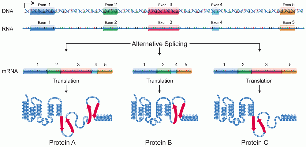
A protein isoform, or "protein variant",[1] is a member of a set of highly similar proteins that originate from a single gene and are the result of genetic differences.[2] While many perform the same or similar biological roles, some isoforms have unique functions. A set of protein isoforms may be formed from alternative splicings, variable promoter usage, or other post-transcriptional modifications of a single gene; post-translational modifications are generally not considered. (For that, see Proteoforms.) Through RNA splicing mechanisms, mRNA has the ability to select different protein-coding segments (exons) of a gene, or even different parts of exons from RNA to form different mRNA sequences. Each unique sequence produces a specific form of a protein.

The discovery of isoforms could explain the discrepancy between the small number of protein coding regions of genes revealed by the Human Genome Project and the large diversity of proteins seen in an organism: different proteins encoded by the same gene could increase the diversity of the proteome. Isoforms at the RNA level are readily characterized by cDNA transcript studies. Many human genes possess confirmed alternative splicing isoforms. It has been estimated that ~100,000 expressed sequence tags (ESTs) can be identified in humans.[1] Isoforms at the protein level can manifest in the deletion of whole domains or shorter loops, usually located on the surface of the protein.[3]

Definition

[edit]

One single gene has the ability to produce multiple proteins that differ both in structure and composition;[4][5] this process is regulated by the alternative splicing of mRNA, though it is not clear to what extent such a process affects the diversity of the human proteome, as the abundance of mRNA transcript isoforms does not necessarily correlate with the abundance of protein isoforms.[6] Three-dimensional protein structure comparisons can be used to help determine which, if any, isoforms represent functional protein products, and the structure of most isoforms in the human proteome has been predicted by AlphaFold and publicly released at isoform.io.[7] The specificity of translated isoforms is derived by the protein's structure/function, as well as the cell type and developmental stage during which they are produced.[4][5] Determining specificity becomes more complicated when a protein has multiple subunits and each subunit has multiple isoforms.

For example, the 5' AMP-activated protein kinase (AMPK), an enzyme, which performs different roles in human cells, has 3 subunits:[8]

  • α, catalytic domain, has two isoforms: α1 and α2 which are encoded from PRKAA1 and PRKAA2
  • β, regulatory domain, has two isoforms: β1 and β2 which are encoded from PRKAB1 and PRKAB2
  • γ, regulatory domain, has three isoforms: γ1, γ2, and γ3 which are encoded from PRKAG1, PRKAG2, and PRKAG3

In human skeletal muscle, the preferred form is α2β2γ1.[8] But in the human liver, the most abundant form is α1β2γ1.[8]

Mechanism

[edit]
Different mechanisms of RNA splicing

The primary mechanisms that produce protein isoforms are alternative splicing and variable promoter usage, though modifications due to genetic changes, such as mutations and polymorphisms are sometimes also considered distinct isoforms.[9]

Alternative splicing is the main post-transcriptional modification process that produces mRNA transcript isoforms, and is a major molecular mechanism that may contribute to protein diversity.[5] The spliceosome, a large ribonucleoprotein, is the molecular machine inside the nucleus responsible for RNA cleavage and ligation, removing non-protein coding segments (introns).[10]

Because splicing is a process that occurs between transcription and translation, its primary effects have mainly been studied through genomics techniques—for example, microarray analyses and RNA sequencing have been used to identify alternatively spliced transcripts and measure their abundances.[9] Transcript abundance is often used as a proxy for the abundance of protein isoforms, though proteomics experiments using gel electrophoresis and mass spectrometry have demonstrated that the correlation between transcript and protein counts is often low, and that one protein isoform is usually dominant.[11] One 2015 study states that the cause of this discrepancy likely occurs after translation, though the mechanism is essentially unknown.[12] Consequently, although alternative splicing has been implicated as an important link between variation and disease, there is no conclusive evidence that it acts primarily by producing novel protein isoforms.[11]

Alternative splicing generally describes a tightly regulated process in which alternative transcripts are intentionally generated by the splicing machinery. However, such transcripts are also produced by splicing errors in a process called "noisy splicing," and are also potentially translated into protein isoforms. Although ~95% of multi-exonic genes are thought to be alternatively spliced, one study on noisy splicing observed that most of the different low-abundance transcripts are noise, and predicts that most alternative transcript and protein isoforms present in a cell are not functionally relevant.[13]

Other transcriptional and post-transcriptional regulatory steps can also produce different protein isoforms.[14] Variable promoter usage occurs when the transcriptional machinery of a cell (RNA polymerase, transcription factors, and other enzymes) begin transcription at different promoters—the region of DNA near a gene that serves as an initial binding site—resulting in slightly modified transcripts and protein isoforms.

Characteristics

[edit]

Generally, one protein isoform is labeled as the canonical sequence based on criteria such as its prevalence and similarity to orthologous—or functionally analogous—sequences in other species.[15] Isoforms are assumed to have similar functional properties, as most have similar sequences, and share some to most exons with the canonical sequence. However, some isoforms show much greater divergence (for example, through trans-splicing), and can share few to no exons with the canonical sequence. In addition, they can have different biological effects—for example, in an extreme case, the function of one isoform can promote cell survival, while another promotes cell death—or can have similar basic functions but differ in their sub-cellular localization.[16] A 2016 study, however, functionally characterized all the isoforms of 1,492 genes and determined that most isoforms behave as "functional alloforms." The authors came to the conclusion that isoforms behave like distinct proteins after observing that the functional of most isoforms did not overlap.[17] Because the study was conducted on cells in vitro, it is not known if the isoforms in the expressed human proteome share these characteristics. Additionally, because the function of each isoform must generally be determined separately, most identified and predicted isoforms still have unknown functions.

Types

[edit]

Isoforms can be categorized based on the nature of their differences into structural isoforms and sequence isoforms. Structural isoforms arise from alternative splicing events that result in different exon compositions, including exon skipping/inclusion, alternative 5' or 3' splice sites, and intron retention. These mechanisms produce transcripts and proteins with distinct domain architectures - for example, the inclusion or exclusion of entire functional domains, or the use of alternative donor/acceptor sites that add or remove partial exon sequences. In contrast, sequence isoforms typically result from single nucleotide variations, insertions, deletions, or post-translational modifications that alter the amino acid sequence without changing the overall exon structure [18].

Alternative splicing is the main post-transcriptional modification process that produces mRNA transcript isoforms, while isoforms can result in different functions, activities, or expression patterns [19]. The distinction is functionally important: structural isoforms often exhibit dramatically different properties due to the presence or absence of entire protein domains, whereas sequence isoforms may show more subtle functional variations. Both mechanisms contribute significantly to proteome diversity, with structural variation through alternative splicing being particularly prevalent in higher eukaryotes where it affects the majority of multi-exon genes.

[edit]

Glycoform

[edit]

A glycoform is an isoform of a protein that differs only with respect to the number or type of attached glycan. Glycoproteins often consist of a number of different glycoforms, with alterations in the attached saccharide or oligosaccharide. These modifications may result from differences in biosynthesis during the process of glycosylation, or due to the action of glycosidases or glycosyltransferases. Glycoforms may be detected through detailed chemical analysis of separated glycoforms, but more conveniently detected through differential reaction with lectins, as in lectin affinity chromatography and lectin affinity electrophoresis. Typical examples of glycoproteins consisting of glycoforms are the blood proteins as orosomucoid, antitrypsin, and haptoglobin. An unusual glycoform variation is seen in neuronal cell adhesion molecule, NCAM involving polysialic acids, PSA.

Examples

[edit]
  • G-actin: despite its conserved nature, it has a varying number of isoforms (at least six in mammals).
  • Creatine kinase, the presence of which in the blood can be used as an aid in the diagnosis of myocardial infarction, exists in 3 isoforms.
  • Hyaluronan synthase, the enzyme responsible for the production of hyaluronan, has three isoforms in mammalian cells.
  • UDP-glucuronosyltransferase, an enzyme superfamily responsible for the detoxification pathway of many drugs, environmental pollutants, and toxic endogenous compounds has 16 known isoforms encoded in the human genome.[20]
  • G6PDA: normal ratio of active isoforms in cells of any tissue is 1:1 shared with G6PDG. This is precisely the normal isoform ratio in hyperplasia. Only one of these isoforms is found during neoplasia.[21]

Monoamine oxidase, a family of enzymes that catalyze the oxidation of monoamines, exists in two isoforms, MAO-A and MAO-B.

See also

[edit]

References

[edit]
[edit]
Revisions and contributorsEdit on WikipediaRead on Wikipedia
from Grokipedia
A protein isoform is a variant of a protein encoded by the same as other isoforms, differing primarily in sequence due to mechanisms such as of pre-mRNA, which generates multiple mRNA transcripts from a single . These variants often share high sequence similarity but can exhibit distinct structural features, subcellular localizations, and functional properties, enabling fine-tuned regulation of cellular processes. While is the predominant mechanism—estimated to affect 35–95% of human multiexon —isoforms may also arise from alternative promoter usage, alternative polyadenylation, or genetic polymorphisms, though post-translational modifications are sometimes broadly included despite producing sequence-identical forms. Protein isoforms play crucial roles in biological diversity and , particularly in complex multicellular organisms, where they expand the functional repertoire of the without requiring new genes. For instance, isoforms of (VEGF) regulate differently, with specific splice variants promoting or inhibiting formation, influencing processes like tumor growth and . Similarly, isoforms such as β-actin and γ-actin, differing by just four , have non-redundant functions: β-actin is essential for cell survival and due to regulatory elements in its mRNA, while γ-actin supports cytoskeletal stability in specific tissues. In disease contexts, aberrant isoform expression contributes to pathologies; for example, truncated androgen receptor isoform AR-V7 drives resistance to therapies, highlighting isoforms as potential biomarkers and therapeutic targets. The study of protein isoforms has advanced with techniques like top-down , which enables intact protein analysis to distinguish subtle sequence differences, though challenges persist due to their low abundance and high homology. not only diversifies protein interactions—often resulting in less than 50% overlap between isoform pairs—but also underlies evolutionary innovations, allowing organisms to adapt to environmental stresses or developmental needs. Overall, isoforms underscore the complexity of , bridging and in health and disease.

Fundamentals

Definition

Protein isoforms are variants of a protein produced from a single locus, differing in their amino acid sequences or post-translational modifications while sharing the same genomic origin. These variants arise primarily through processes such as of pre-mRNA, which allows a single to generate multiple mature mRNA transcripts, or through modifications after translation, such as or , that alter the protein's structure or function without changing the underlying sequence. Unlike alleles, which represent genetic sequence variations at the same locus across individuals or populations, or paralogs, which are homologous proteins encoded by duplicated genes at different loci, protein isoforms are non-allelic products from one specific , enabling functional diversity within a single genetic unit. The term "isoform" emerged alongside early studies of tissue-specific protein variants, such as distinct heavy chain forms in different muscle types, highlighting how isoforms contribute to specialized cellular functions. By the mid-1970s, such investigations had established isoforms as key to understanding tissue-specific protein expression. In the , as of 2023, approximately 19,000–20,000 protein-coding generate over 100,000 distinct isoforms, primarily through , which vastly expands the proteome's complexity from a relatively compact set of . This prevalence underscores the role of isoforms in enabling adaptive responses, such as tissue-specific expression or developmental regulation, where a single can produce multiple proteins tailored to cellular needs. , as a primary mechanism, briefly exemplifies how exons are combinatorially assembled to yield isoform diversity, though detailed processes are explored elsewhere.

Nomenclature

Protein isoforms are named and classified using standardized conventions to facilitate unambiguous scientific communication, primarily guided by international protein nomenclature guidelines that emphasize consistent formatting and descriptive accuracy for protein entries. These guidelines, developed collaboratively by major databases, recommend using gene symbols in uppercase for vertebrates and avoiding ambiguous or overly general terms in protein descriptions. In , the primary resource for protein sequence and annotation, isoforms are denoted by appending a dash followed by a sequential number to the primary accession, such as P12345-1 for the isoform and P12345-2 for an alternative one. For splice variants and other alternative products, descriptive suffixes like -201 are used to indicate specific origins, such as or promoter usage, ensuring traceability to the generating mechanism while maintaining a hierarchical structure within the entry. This convention allows for isoform-specific annotations, including sequence differences and functional notes, all centralized in a single entry where possible. For human genes, the (HGNC) provides approved symbols and names but does not routinely assign unique identifiers to isoforms; instead, it endorses linking protein isoforms to transcript-level identifiers from collaborative resources. Transcript IDs from Ensembl, such as ENST00000380152, are commonly used to specify the mRNA variant, which is then mapped to corresponding protein accessions like P12345-1 in , enabling precise cross-referencing across genomic and proteomic datasets. Databases like Ensembl and play a crucial role in assigning unique, stable identifiers to isoforms, mitigating confusion in genes producing multiple forms. Ensembl employs versioned transcript IDs (e.g., ENST000003) to catalog splice isoforms based on genomic alignment and expression data, while uses distinct accession prefixes (e.g., NP_ for proteins) with version numbers to represent curated isoforms, often selecting a representative "select" transcript per in collaboration with Ensembl via the MANE project. These systems ensure interoperability and reduce redundancy in multi-isoform analyses. Nomenclature challenges persist, including ambiguities from overlapping terms like "," which often implies mutational changes, versus "isoform," denoting regulated alternative products from the same , leading to inconsistent usage in . Early in the relied on ad-hoc naming, such as spot numbers from 2D gels, which lacked standardization and scalability. Post-2000, the advent of database-driven systems like (established in 2002) shifted toward systematic, identifier-based approaches, improving resolution but highlighting ongoing needs for unified terminology in proteoform descriptions.

Generation Mechanisms

Transcriptional and Splicing Variants

Protein isoforms arise at the RNA level through transcriptional and splicing variants, which generate diversity by processing pre-mRNA in multiple ways. , a primary mechanism, involves the selective inclusion or exclusion of s during mRNA maturation, allowing a single to produce multiple transcript variants. Key modes include , where an and its flanking s are omitted from the mature mRNA; mutually exclusive exons, in which one of two exons is included while the other is excluded; retention, where an remains in the transcript; alternative splice site usage, which selects different boundaries for exons; alternative promoter utilization, leading to transcripts with varying 5' untranslated regions or first exons; and alternative polyadenylation sites, which alter the 3' end and potentially the coding sequence. These processes are tightly regulated by cis-acting elements and trans-acting factors. Exonic and intronic splicing enhancers (ESEs, ISEs) promote exon inclusion, often by recruiting serine/arginine-rich (SR) proteins, while splicing silencers (ESSs, ISSs) repress it, typically via heterogeneous nuclear ribonucleoproteins (hnRNPs). SR proteins, such as SRSF1, bind enhancers to facilitate spliceosome assembly, whereas hnRNPs like hnRNP A1 antagonize this by binding silencers and blocking exon recognition. Tissue-specific expression of these regulators contributes to isoform diversity; for instance, varying levels of SR and hnRNP family members across cell types dictate exon choices, enabling context-dependent transcript variants essential for cellular specialization. In humans, approximately 95% of multi-exon genes undergo , generating an average of four isoforms per gene and vastly expanding the from a limited . Isoform diversity can be modeled combinatorially, where the total number of potential isoforms approximates the product of choices at each independent splicing event: Total isoforms(exon choices per event)\text{Total isoforms} \approx \prod (\text{exon choices per event}) This multiplicative framework underscores how even a few alternative events per transcript can yield exponential variety, though actual expression is constrained by regulatory networks. Splicing patterns exhibit evolutionary conservation across vertebrates, with core splice site motifs and many exon-intron structures preserved from to mammals, reflecting functional importance. However, isoform usage— the relative abundance and tissue-specific of variants—shows greater variability, allowing to diverse physiological demands while maintaining essential splicing machinery.

Post-Translational Modifications

Post-translational modifications (PTMs) generate protein isoforms through covalent alterations occurring after , diversifying protein function and distinct from sequence-based variants. These modifications introduce chemical groups or cleave segments, creating structurally and functionally distinct forms that respond to cellular needs. Key PTMs contributing to isoform diversity include phosphorylation, which attaches a to serine, , or residues, imparting a negative charge that modulates electrostatic interactions and conformational changes; glycosylation, featuring N-linked attachments at residues in the Asn-X-Ser/Thr or O-linked additions at serine/, which influence folding, stability, and intercellular recognition; ubiquitination, involving conjugation with chains that signal proteasomal degradation or alter localization; and proteolytic cleavage, where site-specific endoproteases excise domains to yield mature, active isoforms from precursors. PTMs exhibit dynamic reversibility and context specificity, enabling rapid isoform switching; for example, is balanced by opposing actions of kinases and phosphatases, while pathways activate cascades that propagate modifications across protein networks. Mass spectrometry analyses reveal extensive PTM prevalence, with extrapolations indicating that over 70% of proteins undergo and similar proportions experience ubiquitination or , resulting in multiple coexisting isoforms per protein. Modification kinetics follow enzymatic models such as Michaelis-Menten for rate-limiting steps in PTM enzymes like kinases: v=Vmax[S]Km+[S]v = \frac{V_{\max} [S]}{K_m + [S]} where vv represents the modification rate, VmaxV_{\max} the maximum velocity, [S][S] the substrate concentration, and KmK_m the Michaelis constant reflecting enzyme-substrate affinity. Computational tools facilitate PTM site prediction, including NetPhos, which uses ensembles to forecast serine, , and motifs with reported accuracies of approximately 80-90% and error rates of 10-20% on benchmark datasets.

Structural and Functional Characteristics

Structural Features

Protein isoforms exhibit sequence variations primarily arising from alternative splicing, which introduces insertions, deletions, or exon shuffling that can alter secondary structural elements such as alpha-helices and beta-sheets. These changes often manifest as localized disruptions in hydrogen bonding networks, potentially stabilizing or destabilizing helical segments. Similarly, post-translational modifications (PTMs) like phosphorylation can induce conformational shifts by introducing negative charges that repel nearby residues, promoting loop formations or helix destabilization in affected regions. Biophysical properties of isoforms differ notably in their isoelectric points (pI), with causing a downward shift due to the introduction of a dianionic charge at physiological . This pI alteration affects electrophoretic mobility and can influence isoform separation in gels by 0.5-2 units depending on the protein's baseline pI and modification site. and thermal stability also vary among isoforms. Domain architecture in isoforms often involves the retention, loss, or rearrangement of functional modules. For example, in the β (PKCβ) family, splice variants PKCβI and PKCβII differ in their C-terminal regions due to , leading to distinct regulatory properties and folds. Computational 3D modeling with has elucidated these isoform-specific folds, predicting unique tertiary arrangements for over 3,400 human isoforms, including disordered regions that differ in confidence scores (pLDDT) between splice variants of the same . Experimental validation of isoform structures relies on techniques like and (NMR) spectroscopy, which have resolved atomic-level details for a limited number of variants, as splice and modification isoforms represent a small fraction of the (PDB) entries. Recent advances, including predictions and cryo-EM, are expanding coverage of isoform structures as of 2025. These methods underscore the prevalence of modular structural diversity in isoforms generated via splicing or PTMs.

Functional Implications

Protein isoforms often exhibit modulated enzymatic activities due to structural alterations introduced by or post-translational modifications (PTMs) that affect critical functional sites. For instance, in the case of α-galactosidase A, an event results in an isoform retaining only approximately 10% of the wild-type enzyme's activity, representing a 90% reduction in catalytic efficiency owing to disruptions in the . Such changes can fine-tune metabolic pathways or render isoforms partially inactive, thereby regulating overall cellular response without complete . Similarly, splice variants lacking key catalytic residues, as observed in certain isoforms of enzymes like NEIL3, are enzymatically inactive and may serve regulatory roles by competing for substrates. PTM-based isoforms significantly influence subcellular localization and protein-protein interactions, enabling diverse functional roles within the cell. Myristoylation, a lipid PTM, directs isoforms to specific compartments; for example, the sperm-specific 1 isoform (HK1S), generated by , acquires a unique N-terminal residue that permits myristoylation, anchoring it to the plasma membrane and for localized glycolytic activity in spermatozoa. In terms of interactions, domain swaps via splicing can alter binding interfaces; the isoform containing the extra domain A (EDA), produced by inclusion of an alternative exon, enhances interactions with (TLR4) and , promoting inflammatory signaling and distinct from the EDA-excluded variant. Isoforms can provide functional or specialization, with some acting as non-functional decoys to buffer signaling pathways while others exhibit tissue-specific enhancements. For example, certain splice variants of the corticotropin-releasing factor receptor 1 (CRF1) lack signaling capability and function as decoys, sequestering ligands to attenuate receptor activation and modulate stress responses. In contrast, hyper-specialized isoforms like the muscle-specific M1 versus the embryonic M2 variant demonstrate tissue-restricted activities, with M2 supporting aerobic in proliferating cells through altered . Isoform in binding or activation can be modeled kinetically using the Hill equation, where the fractional occupancy θ is given by θ=[L]nKd+[L]n\theta = \frac{[L]^n}{K_d + [L]^n} with [L] as concentration, n as the Hill reflecting , and K_d as the ; such models illustrate how isoform-specific n values enhance phenotypic robustness in enzymatic networks. approaches, such as isobaric tags for relative and absolute quantification (iTRAQ), reveal how isoform abundance ratios correlate with functional outcomes, often showing 2- to 10-fold expression differences across cellular states. These ratios, derived from labeling and , highlight dynamic shifts in isoform dominance that drive functional diversification, as seen in proteome-wide analyses of impacts.

Classification and Types

Splice Isoforms

Splice isoforms arise from of pre-mRNA, generating protein variants with distinct sequences due to the inclusion, exclusion, or modification of exons. These isoforms are classified based on their impact on the and . Frame-preserving isoforms maintain the original , resulting in full-length variants with insertions, deletions, or substitutions that do not alter the overall length significantly, often leading to modular changes in protein domains. Frame-shifting isoforms introduce changes in the through events like alternative splice site usage, causing N-terminal or C-terminal alterations that can extend or truncate specific regions while preserving core functional motifs. Truncated isoforms result from premature stop codons, typically via retention or , yielding shorter proteins that may lack essential domains or act as regulators. Genomic studies from the indicate that a significant proportion (around 25%) of splice isoforms are detected in large-scale datasets across human tissues, suggesting functionality for this subset and highlighting their role in diversity rather than mere transcriptional noise. A striking example of splicing complexity is the DSCAM gene in , which generates over 38,000 isoforms through mutually exclusive selection, enabling neuronal self-avoidance and wiring specificity. Among functional subtypes, dominant-negative splice isoforms inhibit the activity of wild-type counterparts by forming non-functional complexes or competing for binding partners, as seen in variants that disrupt signaling pathways. Neomorphic isoforms confer novel functions unrelated to the canonical protein, such as altered subcellular localization or interaction profiles, expanding cellular capabilities beyond the original . Databases like annotate splice isoforms using flags for events, with approximately 10,000 human protein entries featuring such variants (as of 2025), facilitating systematic classification and functional prediction.

Modification-Based Isoforms

Modification-based isoforms arise from post-translational modifications (PTMs), which introduce chemical diversity to proteins without altering their , thereby generating functional variants that respond dynamically to cellular signals. These isoforms differ from splice variants by being reversible and context-dependent, often modulating protein activity, localization, stability, or interactions through enzyme-mediated additions or removals of functional groups. Phospho-isoforms represent a prominent class, where phosphorylation at multiple serine, threonine, or tyrosine residues creates distinct states that regulate signaling cascades. Approximately 70% of proteins undergo at least once, with multi-site enabling combinatorial regulation; for instance, motifs such as RSXpSXP bind 14-3-3 proteins, which stabilize or sequester targets like Raf-1 kinase to control MAPK pathway activation. These phospho-states can switch protein conformations, as seen in where hierarchical by GSK3 toggles enzymatic activity. Glyco-isoforms emerge from variations in N- or O-linked glycosylation, particularly in branching patterns that influence , stability, and . High-mannose glycans, rich in residues, predominate in early endoplasmic reticulum processing and confer rapid clearance compared to complex types with branched antennae of , , and , which enhance serum stability by shielding proteolytic sites. Sialylation variants exemplify this, as in serum proteins like , where differing content (0-2 per branch) alters charge and circulation time, with hypersialylated forms resisting hepatic uptake. Other PTM types further diversify isoforms, including on residues of tails, which neutralizes positive charges to loosen structure and promote , generating acetyl-isoforms like H3K9ac that recruit readers. Sumoylation conjugates small ubiquitin-like modifiers to s, often enhancing nuclear localization; for example, sumoylated ATF7 accumulates in the nucleus to repress target genes, while desumoylation facilitates export. Proteolytic cleavage also yields isoforms, as in the maturation of proinsulin to insulin, where endopeptidases excise the , activating the for glucose regulation. The combinatorial complexity of PTMs amplifies isoform diversity, where a protein with five independent modifiable sites can theoretically produce 25=322^5 = 32 variants, each potentially eliciting unique responses. This underpins the PTM codes hypothesis, positing that specific modification patterns encode signaling specificity, as in transcription factors where phospho-acetyl combos dictate coactivator binding over simple single-site effects.

Biological and Evolutionary Roles

Cellular and Physiological Functions

Protein isoforms play critical roles in cellular signaling pathways by enabling fine-tuned responses through . For instance, in the (MAPK) cascade, splice variants of components such as MEK1b and ERK1c form an independent signaling axis that regulates mitotic Golgi fragmentation, distinct from the canonical MEK1/2-ERK1/2 pathway, thereby modulating the duration and specificity of signaling outputs during . Similarly, alternative splicing of JNK isoforms influences their stability and interaction with scaffold proteins like JIP1, altering the persistence of stress-activated signaling in cellular processes such as and proliferation. In developmental contexts, protein isoforms contribute to key cellular events like in the . Differential expression of isoforms, such as the ε isoform intensely localized in the hippocampal mossy fiber region postnatally, supports neuronal maturation and formation in development. Recent single-cell sequencing studies have revealed extensive isoform diversity in the developing human , with over 214,000 distinct isoforms identified across excitatory neurons, where switches in isoform usage regulate binding and protein structures essential for and cellular identity establishment. At the physiological level, tissue-specific isoforms enable adaptive contractility in muscle tissues. In the human heart, atrial cells predominantly express the α-myosin heavy chain isoform, which supports rapid contraction with a higher activity (k_cat 18 s⁻¹) and shortening velocity (0.45 µm/s), while ventricular cells rely on the β-isoform for sustained force generation with greater ATP economy (tension cost 2.4 mmol kN⁻¹ m⁻¹ s⁻¹). This isoform distribution optimizes atrial refilling and ventricular ejection, illustrating how structural variants adapt physiological performance to organ-specific demands. In homeostasis, splice variants maintain balanced conductance; for example, inclusion of exon 37a in the CaV2.2 significantly increases in nociceptive neurons, enhancing excitability without altering voltage dependence, thus regulating synaptic transmission and cellular signaling fidelity. Post-2020 single-cell analyses have further elucidated isoform gradients in embryonic development, showing cell-type-specific patterns that drive over 70% novel isoform detection in human neocortical progenitors, contributing significantly to variance in cell fate decisions during . In gastrula embryos, such profiling identifies stripe-specific isoform usage along the anterior-posterior axis, with plasma membrane-related isoforms distinguishing germ layers and influencing early lineage commitment. These findings underscore how isoform ratios preserve physiological balance across tissues and developmental stages.

Evolutionary Aspects

Alternative splicing, a mechanism generating protein isoforms from single genes, emerged early in eukaryotic evolution in the common ancestor of eukaryotes through the development of spliceosomal introns and initial splicing errors that enabled regulated inclusion. shuffling played a pivotal role as a driver of isoform diversity, facilitating the recombination of protein domains across genes and contributing to the structural novelty observed in metazoan lineages. This process allowed for the rapid evolution of multifunctional proteins without relying solely on , enhancing genetic flexibility in response to environmental pressures. Protein isoforms confer adaptive advantages by promoting , enabling organisms to produce diverse functional variants from the same genomic locus without necessitating sequence mutations, thereby accelerating adaptation. In vertebrates, has driven significant expansion, significantly expanding the beyond the count and supporting like tissue-specific functions and behavioral repertoires. Comparative studies highlight how this mechanism amplifies proteomic output, particularly in neural and developmental contexts, fostering evolutionary innovation. Recent analyses indicate that rates have steadily increased over the past 1.4 billion years, particularly within the metazoan lineage, coinciding with rising organismal . Conservation patterns across species reveal that core, constitutively expressed isoforms maintain high sequence identity, often exceeding 90% across vertebrates, underscoring their essential roles under strong purifying selection. In contrast, alternative isoforms exhibit greater divergence, with indicating faster evolutionary rates for splicing patterns in non-core exons. Evolutionary pressures shape these dynamics, including positive selection on splice sites in immune genes to enable rapid isoform switching against pathogens. Neutral drift predominates in non-coding regions flanking splice sites, allowing accumulation of neutral variations that subtly modulate isoform prevalence without fitness costs. Adapted selection models, such as per-isoform dN/dS ratios (ω = dN/dS), quantify these forces, revealing elevated rates in alternative variants indicative of relaxed constraints or adaptive divergence.

Applications and Study Methods

Detection and Analysis Techniques

Protein isoforms, arising from , post-translational modifications (PTMs), or other mechanisms, require specialized techniques for detection and characterization at both transcript and protein levels. serves as a cornerstone for identifying transcript isoforms that encode proteins, with short-read platforms like Illumina providing high-depth coverage but facing limitations in resolving complex splicing patterns due to read fragmentation. Long-read sequencing methods, such as Iso-Seq, overcome these by generating full-length transcripts, achieving high splice junction resolution accuracy and enabling precise isoform assembly without reliance on reference genomes. At the protein level, liquid chromatography-tandem (LC-MS/MS) is essential for detecting PTM-based isoforms, such as phosphorylated or ubiquitinated variants, by fragmenting peptides and matching spectra to databases. Modern LC-MS/MS systems offer high throughput, identifying over 10,000 peptides per hour while distinguishing isoform-specific sequences through bottom-up or top-down approaches that preserve PTM information. Complementary molecular methods include isoform-specific (PCR), which employs primers designed to unique junctions or variable regions to amplify and quantify individual isoforms from reverse-transcribed , providing validation for sequencing data. Computational tools enhance isoform analysis by processing raw data into interpretable models. StringTie, a widely used assembler, employs network flow algorithms for de novo transcriptome reconstruction from RNA-seq alignments, outperforming earlier methods in recovering full-length isoforms and estimating abundances with reduced fragmentation bias. For PTM isoforms, databases like PhosphoSitePlus curate over 330,000 modification sites across mammalian proteomes, facilitating mapping of experimental mass spectrometry data to specific isoform variants and integrating motifs for regulatory insights. These tools often integrate with pipelines like IsoQuant for long-read data, improving accuracy in novel isoform discovery. Recent advances in the have introduced CRISPR-based editing for isoform-specific manipulation, such as splice-site targeting to generate mutant isoforms in cell lines, allowing functional dissection without affecting the full locus. models, including frameworks for isoform function prediction, leverage sequence and structural to achieve accuracies above 85%, aiding in prioritizing candidates for experimental validation. Despite these progresses, challenges persist in detecting low-abundance isoforms, which constitute less than 1% of total protein content and often evade capture due to dynamic range limitations in sequencing and . Short-read exacerbates quantification errors through ambiguous multi-mapping of reads across similar isoforms, leading to up to 20-30% inaccuracies in abundance estimates that long-read methods partially mitigate but do not fully resolve.

Role in Disease and Therapeutics

Dysregulation of protein isoforms plays a critical role in various diseases, particularly through aberrant and post-translational modifications (PTMs). In cancer, mutations in splicing factors are recurrent and drive isoform imbalances that promote oncogenesis; for instance, such mutations affect approximately 50% of hematologic malignancies, including myelodysplastic syndromes (MDS) and (CMML), leading to aberrant splice isoforms that enhance tumor proliferation and survival. In neurodegenerative disorders like (AD), PTM isoforms of , such as hyperphosphorylated forms, aggregate into neurofibrillary tangles, a hallmark that disrupts neuronal function and contributes to cognitive decline. Therapeutic interventions increasingly target specific protein isoforms to correct these dysregulations. Antisense () that modulate splicing have shown clinical success; , an ASO approved by the FDA in 2016, restores full-length SMN2 protein isoform expression in (SMA) by blocking an inhibitory splice site, improving motor function in patients across age groups. Additionally, isoform-specific small-molecule inhibitors are in development for kinases, such as () isoforms; drugs like isoform-selective PI3Kα inhibitors (e.g., ) have advanced to clinical use for cancers with PIK3CA mutations, while others targeting PI3Kδ or β isoforms are in ongoing trials for hematologic and solid tumors, demonstrating reduced off-target effects compared to pan-inhibitors. Recent advancements as of 2025 include the integration of (AI) in designing isoform-selective therapeutics, with companies like preparing to initiate human clinical trials for AI-generated small-molecule drugs targeting specific protein conformations relevant to and . Broader efforts encompass numerous clinical trials focused on isoform-targeted approaches, such as degraders and inhibitors, with promising phase I outcomes in subsets of patients, including PSA30 response rates up to 55% in trials using (AR) degraders. Isoform profiles also hold prognostic value as biomarkers; proteomics-based analysis of tau PTM isoforms, for example, identifies patient heterogeneity in AD and predicts disease progression, supporting personalized therapeutic decisions with implications for outcome forecasting in neurodegeneration.

Examples

Immunoglobulin Isoforms

Immunoglobulin isoforms, particularly those of the μ heavy chain in IgM, are generated through and of the primary transcript from the locus. The μ heavy chain gene features two polyadenylation sites: a proximal site downstream of the Cμ4 exon, which produces the secreted isoform (μs), and a distal site after the membrane-specific exons M1 and M2, which yields the membrane-bound isoform (μm). In resting or immature s, splicing typically joins the Cμ4 exon directly to the M1 exon, excluding the secreted polyadenylation signal, while polyadenylation occurs at the distal site to form the membrane-bound mRNA. Upon B cell activation and differentiation into plasma cells, increased levels of the cleavage stimulation factor CstF-64 promote usage of the proximal polyadenylation site, coupled with splicing that excludes the M1 and M2 exons, favoring the secreted form. The membrane-bound μ isoform functions as part of the (BCR) complex, facilitating recognition and intracellular signaling essential for activation and survival. In contrast, the secreted μ isoform is released as pentameric or hexameric IgM antibodies, enabling complement activation and neutralization in . During differentiation, the ratio of membrane-bound to secreted μ transcripts shifts dramatically in favor of secreted forms in plasma cells, reflecting the transition from -sensing to antibody production. Structurally, the membrane-bound isoform incorporates a and a short cytoplasmic encoded by the M1 and M2 exons, which anchor the BCR to the plasma membrane and mediate signaling through interactions with Ig-α and Ig-β chains. This exon inclusion adds a hydrophobic α-helix spanning the , absent in the secreted isoform, which terminates after the Cμ4 exon with a hydrophilic for . Evolutionarily, the dual production of membrane-bound and secreted IgM isoforms is conserved across jawed vertebrates, underpinning adaptive immunity by allowing B cells to both survey antigens via surface receptors and deploy soluble effectors, with variations in RNA processing pathways observed in basal lineages like teleost fish. Mutations in the μ heavy chain gene can disrupt isoform balance, leading to immunodeficiencies resembling (XLA). For instance, splice-site mutations, such as a G-to-A substitution at 1831, inhibit production of the membrane-bound μ isoform while altering the secreted form, blocking development at the pre-B stage and causing profound with recurrent infections. Similarly, deletions encompassing the membrane exons prevent μm expression, underscoring the essential role of the membrane isoform in maturation.

Troponin Isoforms

Troponin T (TnT) is a key subunit of the that regulates in by conferring calcium sensitivity to the thin filaments. In vertebrates, three homologous genes encode distinct TnT isoforms tailored to specific muscle types: TNNT1 produces the slow isoform (TnT1), TNNT3 encodes the fast isoform (TnT3), and TNNT2 generates the cardiac-specific isoform, which is unique to heart muscle and differs significantly in its N-terminal region to support continuous contractile demands. These isoforms exhibit tissue-specific expression, with TnT1 predominant in type I slow-twitch fibers for endurance activities, TnT3 in type II fast-twitch fibers for rapid force generation, and cardiac TnT optimized for rhythmic . Regulation of TnT isoforms involves alternative splicing, particularly during developmental stages, where the cardiac TNNT2 gene undergoes exon skipping to produce fetal-specific variants that transition to adult forms postnatally. For instance, in the developing heart, early isoforms include exon 5, which confers lower calcium sensitivity and greater flexibility for embryonic contractility; this exon is predominantly excluded in adult cardiac TnT, along with variable inclusion of exon 4, resulting in higher calcium affinity. Additionally, post-translational modifications such as phosphorylation modulate function; protein kinase C phosphorylates cardiac TnT at Ser194, reducing the calcium sensitivity of force development and actomyosin ATPase activity, thereby fine-tuning relaxation and preventing excessive contraction. This site-specific phosphorylation alters troponin-tropomyosin interactions, decreasing maximal force by influencing the inhibitory state of the thin filament. In , isoform switching occurs with re-expression of fetal cardiac TnT variants, such as those including 5, which exhibit lower calcium sensitivity compared to isoforms, contributing to diminished contractility as an adaptive response to stress but ultimately impairing systolic function. Studies in failing myocardium show this shift correlates with reduced peak force generation, with functional assays indicating reduced contractile performance due to altered thin filament . These changes are commonly detected using analysis with isoform-specific antibodies, which reveal shifts in band patterns corresponding to spliced variants in diseased tissue samples. Evolutionarily, TnT isoform diversification is vertebrate-specific, arising from gene duplication events that enabled specialization for distinct muscle physiologies, such as sustained cardiac beating versus phasic skeletal movements, enhancing overall locomotor and circulatory efficiency in higher vertebrates.

Proteoforms

A proteoform is defined as all of the different molecular forms in which the protein product of a single gene can be found, including those arising from genetic variations, alternative splicing of RNA transcripts, and post-translational modifications (PTMs). This terminology was proposed by the Consortium for Top-Down Proteomics in 2013 to provide a unified descriptor for protein complexity, addressing ambiguities in prior terms like "isoform" or "protein species." Unlike narrower definitions, proteoform encompasses the full spectrum of variants from a single genomic locus, emphasizing the atomic-level resolution of sequence and compositional differences. Protein isoforms, which arise primarily from alternative splicing or allelic variations, represent only a subset of proteoforms, as the latter also include myriad combinations of isoforms with site-specific PTMs such as , , or ubiquitination. For instance, a given splice isoform may exist as multiple proteoforms depending on the number and of PTMs, which can alter function, localization, or stability. Estimates suggest that the approximately 20,000 genes give rise to over 1 million distinct proteoforms, highlighting the vast expansion of proteomic diversity beyond the genome. The study of proteoforms necessitates approaches that preserve and analyze intact protein molecules, with top-down (MS) emerging as a key method for their identification and characterization. In top-down MS, whole proteoforms are ionized, separated by mass-to-charge ratio, and fragmented to reveal precise sequences and modification patterns, enabling differentiation of subtle variants. This differs from , which involves enzymatic digestion into peptides prior to MS analysis, often inferring proteoform identity indirectly and missing combinatorial PTM information on individual molecules. The Human Proteoform Project, launched in 2021, aims to comprehensively map human proteoforms using advanced technologies. As of 2025, it is progressing toward developing proteoform atlases to advance precision medicine. Focusing on proteoforms offers significant advantages over isoform-centric analyses by capturing the complete heterogeneity of the , which is essential for understanding context-dependent protein behaviors in cellular processes and states. Such comprehensive profiling reveals functional nuances that isoform studies alone overlook, facilitating advances in and discovery.

Glycoforms

Glycoforms represent a subset of protein isoforms arising from variations in , a where chains (glycans) are covalently attached to specific residues, primarily (N-linked) or serine/ (O-linked), resulting in proteins with differing glycan structures such as biantennary (two-branched) versus triantennary (three-branched) complex N-glycans. These structural differences in glycan composition, branching, and terminal modifications (e.g., sialylation or fucosylation) produce microheterogeneity at individual glycosylation sites, leading to distinct glycoforms of the same polypeptide backbone. Glycoforms are prevalent, with more than 50% of proteins undergoing glycosylation, and a single protein can exhibit over 100 distinct glycoforms due to combinatorial glycan diversity. Glycoforms are generated primarily in the Golgi apparatus through sequential action of glycosyltransferases, enzymes that add monosaccharides to nascent glycoproteins transiting from the , with variations arising from differences in enzyme expression, substrate availability, and compartmental localization. This process is highly cell-type specific; for instance, liver cells predominantly produce biantennary glycans with high sialylation for serum proteins, while brain tissue favors more complex triantennary or poly-sialylated structures on neural glycoproteins, reflecting tissue-specific glycosyltransferase profiles such as elevated expression of N-acetylglucosaminyltransferase-IX in neurons. These variations enable glycoform diversity tailored to cellular contexts, influencing protein trafficking and function without altering the core sequence. Functionally, glycoforms modulate protein interactions and stability; for example, sialic acid-capped glycoforms promote immune evasion by masking recognition sites on pathogens or host cells, preventing binding to immune lectins like siglecs and enabling self-tolerance through negative charges that repel immune effectors. Glycosylation also enhances protein stability, often extending circulatory half-life by 2- to 5-fold via shielding from proteolysis and altering pharmacokinetics, as seen in sialylated lysosomal enzymes where α2-3-linked sialic acid increases half-life threefold. Additionally, glycoform-specific glycan motifs determine binding specificity to lectins, carbohydrate-recognizing proteins that mediate cell adhesion, signaling, and pathogen clearance; for instance, triantennary glycans may preferentially engage galectins for immune modulation, while biantennary forms interact with selectins for leukocyte rolling. Analysis of glycoforms relies on specialized techniques like glycoproteomics via (Glyco-MS), which identifies site-specific glycopeptides and quantifies glycan heterogeneity through tandem MS fragmentation of intact glycoforms, enabling detection of thousands of variants from complex samples. Complementary methods include arrays, where immobilized with defined glycan-binding specificities capture and profile glycoforms via fluorescent detection, providing of structural motifs without enzymatic release of glycans. These approaches have revealed extensive glycoform diversity, underscoring glycosylation's role as a dynamic regulatory layer in protein isoform biology. Recent advances as of 2025 include workflows like GlycanDIA for high-throughput glycomic profiling and enhanced for brain N-glycoforms.

References

Add your contribution
Related Hubs
User Avatar
No comments yet.