Hubbry Logo
OsteosclerosisOsteosclerosisMain
Open search
Osteosclerosis
Community hub
Osteosclerosis
logo
8 pages, 0 posts
0 subscribers
Be the first to start a discussion here.
Osteosclerosis
Osteosclerosis
from Wikipedia
Osteosclerosis
Sclerosis of the bones of the pelvis due to prostate cancer metastases
SpecialtyMedical genetics Edit this on Wikidata

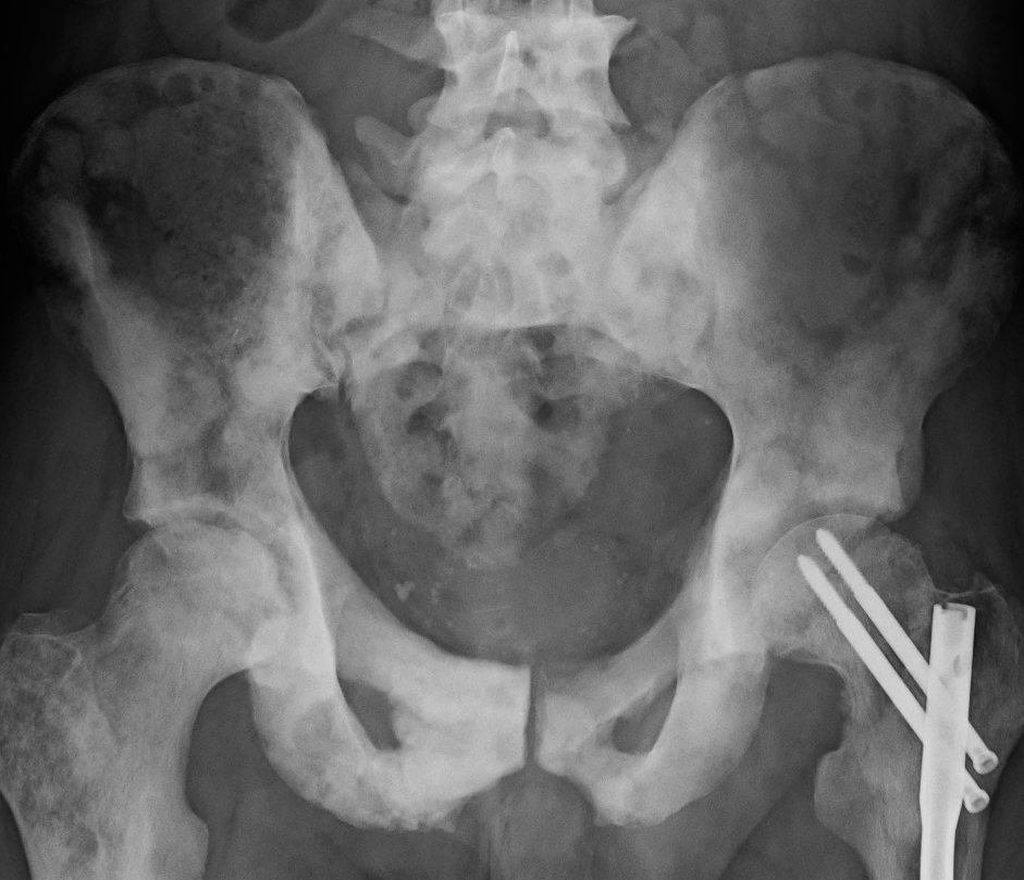
Osteosclerosis is a disorder characterized by abnormal hardening of bone and an elevation in bone density. It may predominantly affect the medullary portion and/or cortex of bone. Plain radiographs are a valuable tool for detecting and classifying osteosclerotic disorders.[1][2] It can manifest in localized or generalized osteosclerosis. Localized osteosclerosis can be caused by Legg–Calvé–Perthes disease, sickle-cell disease and osteoarthritis among others. Osteosclerosis can be classified in accordance with the causative factor into acquired and hereditary.[2][1]

Types

[edit]

Acquired osteosclerosis

[edit]

Hereditary osteosclerosis

[edit]

Diagnosis

[edit]

Osteosclerosis can be detected with a simple radiography. There are white portions of the bone which appear due to the increased number of bone trabeculae.[citation needed]

Animals

[edit]

In the animal kingdom, there also exists a non-pathological form of osteosclerosis, resulting in unusually solid bone structure with little to no marrow. It is often seen in aquatic vertebrates, especially those living in shallow waters,[8] providing ballast as an adaptation for an aquatic lifestyle. It makes bones heavier, but also more fragile. In those animal groups, osteosclerosis often occurs together with bone thickening (pachyostosis). This joint occurrence is called pachyosteosclerosis.[9]

See also

[edit]

References

[edit]
[edit]
Revisions and contributorsEdit on WikipediaRead on Wikipedia
from Grokipedia
Osteosclerosis is a disorder characterized by abnormal hardening of bone and an elevation in , often resulting from increased osteoblastic activity, reduced , or the formation of bone-like tissue by tumor cells. This condition can manifest as localized lesions or diffuse skeletal involvement, distinguishing it from normal processes. Osteosclerosis encompasses a heterogeneous group of primary genetic disorders and secondary conditions, with primary forms such as osteopetrosis—including its autosomal recessive (ARO) and autosomal dominant (ADO) variants—arising from mutations in genes affecting osteoclast function, leading to impaired bone resorption. Secondary osteosclerosis frequently stems from neoplastic processes, including osteoblastic metastases from breast or prostate cancer, or bone tumors like osteosarcoma, as well as non-neoplastic causes such as Paget's disease, chronic osteomyelitis, or healed fractures. Rare variants include intramedullary osteosclerosis (IMOS), a benign sclerosing disorder primarily affecting the diaphyses of long bones in adults, often without identifiable etiology. Clinically, osteosclerosis may be asymptomatic, particularly in benign forms like osteopoikilosis, or present with symptoms depending on the underlying cause and extent of involvement, such as , fractures, due to encroachment, cranial nerve palsies, or growth disturbances in pediatric cases. Diagnosis relies on radiographic imaging revealing increased , often with characteristic patterns like diffuse sclerosis or punctate opacities, supplemented by CT, MRI, or to differentiate from mimics such as metastases or infections; may be required for confirmation in ambiguous cases. Treatment is tailored to the etiology, ranging from supportive care and analgesics for benign conditions to for severe genetic forms such as malignant , for neoplastic causes, or surgical interventions like for symptomatic IMOS.

Definition and Overview

Definition

Osteosclerosis is a medical condition characterized by an abnormal increase in , resulting from excessive bone formation, reduced , or a combination of both, which manifests as radiographically dense on imaging studies. This hardening primarily affects the cortical or medullary portions of , leading to increased skeletal mass without necessarily altering the overall architecture in all cases. The term "osteosclerosis" is derived from the Greek roots (bone) and sklērōsis (hardening). Unlike , which involves progressive bone loss and decreased mineral density due to imbalanced remodeling favoring resorption, osteosclerosis represents the converse process of net bone gain. , by contrast, refers to a specific hereditary subtype of osteosclerosis driven by genetic defects in function, resulting in dense but fragile bones prone to fractures. Osteosclerosis is broadly classified into focal (localized to specific bone regions, such as enostoses or dense bone islands) or diffuse (generalized involvement of the skeleton), based on the extent and distribution of the density changes.

Epidemiology

Osteosclerosis is a rare skeletal disorder characterized by increased bone density, with an overall global incidence that remains low and not well-quantified due to its heterogeneous presentation. Some hereditary forms, such as dysosteosclerosis and osteosclerotic bone dysplasia, have a prevalence of less than 1 in 1,000,000 individuals and typically manifest in infancy or early childhood, affecting males and females equally across demographics; more common benign hereditary variants like osteopoikilosis have an estimated prevalence of 1 in 50,000. In contrast, acquired forms are more common, particularly in populations with underlying chronic conditions, though exact population-level incidence rates are limited by diagnostic challenges and varying definitions. Among acquired causes, osteosclerosis is notably associated with (CKD), where it arises as part of CKD-mineral bone disorder; prevalence in affected patients has been reported to range from 0% to 54% in historical cohorts, though modern management has reduced its frequency. Demographic trends show a higher occurrence in adults over 50 years, linked to age-related chronic illnesses, while younger adults and children are more likely to present with hereditary variants without bias. Risk factors include advanced age, prolonged exposure to chronic diseases like renal failure, and environmental factors such as overexposure, which elevate susceptibility without inherent or ethnic predispositions beyond exposure patterns. Geographic variations are prominent in regions with endemic fluorosis, where high groundwater fluoride levels contribute to skeletal fluorosis manifesting as osteosclerosis. In , fluorosis affects approximately 30% of the population in endemic areas, with skeletal involvement reported in up to 24.8% of exposed individuals in some studies. Similarly, in , over 1 million people suffer from (1,085,512 cases as of 2017), with notable occurrence in high-risk areas affected by coal-burning fluorosis such as . These patterns underscore environmental exposures as key drivers in specific locales, contrasting with the sporadic nature of cases elsewhere.

Pathophysiology

Bone Remodeling Mechanisms

Bone remodeling is a dynamic physiological process that maintains skeletal integrity by continuously replacing old or damaged tissue with new , ensuring mechanical strength and . This process involves a precise balance between , mediated by osteoclasts, and bone formation, carried out by osteoblasts. In healthy adults, resorption and formation are tightly coupled, resulting in no net change in bone mass under normal conditions. The fundamental unit of bone remodeling is the basic multicellular unit (BMU), which coordinates the activities of osteoclasts, osteoblasts, and osteocytes within a localized compartment. Osteoclasts, multinucleated cells derived from monocyte-macrophage lineage, initiate remodeling by attaching to the surface and secreting acids and enzymes to dissolve the mineralized matrix, creating resorption lacunae approximately 40–60 μm in depth. This resorption phase lasts about 2–4 weeks. Osteoblasts, originating from mesenchymal stem cells, follow by migrating into the lacunae, where they deposit new organic matrix (osteoid) and promote its mineralization to refill the defect, a process spanning 3–4 months. Osteocytes, terminally differentiated osteoblasts embedded within the bone matrix, act as mechanosensors, detecting microdamage or mechanical stress and signaling to recruit osteoclast precursors via factors like , thereby initiating targeted remodeling while regulating osteoblast activity through sclerostin secretion to inhibit excessive formation. Hormonal regulation fine-tunes the remodeling balance through systemic and local signaling pathways. (PTH), secreted in response to low serum calcium, exerts dual effects: intermittent pulses stimulate osteoblast survival and bone formation via Wnt/β-catenin activation, while continuous elevation promotes resorption by upregulating expression in osteoblasts and osteocytes. , in its active form 1α,25-dihydroxyvitamin D3, supports mineralization by enhancing osteoblast differentiation and matrix production, while also influencing the /OPG ratio to modulate osteoclastogenesis; it synergizes with PTH to maintain calcium . The /OPG pathway serves as a critical local regulator, where (receptor activator of nuclear factor κB ligand), produced by osteoblasts and osteocytes, binds on osteoclast precursors to drive differentiation and activation, whereas OPG (osteoprotegerin) acts as a receptor to inhibit this process, thereby preserving the resorption-formation equilibrium. Quantitatively, this balanced process renews approximately 10% of the adult annually, with the entire replaced every 10 years, varying by type—cortical turns over more slowly (about 3–5% per year) compared to trabecular (up to 25–30%).

Pathological Processes

Osteosclerosis arises from an imbalance in , where excessive osteoblast-mediated formation or inhibition of activity predominates, leading to a net accumulation of . This disruption favors production over resorption, resulting in pathological hardening and increased density without proportional improvement in quality. In conditions like , defective function impairs breakdown, while in sclerosteosis, unchecked osteoblast activity drives overproduction. Such imbalances can stem from genetic mutations or acquired factors, ultimately compromising marrow function and vascularity. At the molecular level, dysregulation of key signaling pathways contributes to this remodeling imbalance. Overactivation of the Wnt/β-catenin pathway, a central regulator of differentiation, promotes excessive bone formation by enhancing proliferation and survival while suppressing osteoclastogenesis. In hereditary forms, loss-of-function mutations in the SOST gene reduce sclerostin production, a potent Wnt secreted by osteocytes; this inhibition of sclerostin unleashes Wnt signaling, leading to unrestrained activity and generalized osteosclerosis as seen in sclerosteosis. Similarly, aberrant TGF-β signaling, particularly gain-of-function mutations in the TGFB1 gene, stimulates recruitment and matrix deposition, driving diaphyseal in disorders like Camurati-Engelmann disease. These pathways often intersect, amplifying feedback mechanisms that sustain pathological bone accrual. Histologically, osteosclerosis manifests as increased trabecular thickness, where bony spicules become densely packed and interconnected, often resembling marble-like tissue with reduced intertrabecular spaces. Marrow cavities diminish due to encroaching bone, limiting hematopoietic and adipose tissues, while cortical bone exhibits pronounced thickening from successive layers of subperiosteal and endosteal deposition. These changes, observed in bone biopsies, reflect immature or disorganized mineralization patterns, with variable osteoid seams and minimal resorption lacunae, contributing to bone fragility despite elevated density. The progression of osteosclerosis typically evolves from focal to diffuse sclerosis through iterative cycles of unbalanced remodeling. Early stages involve localized periosteal or endosteal proliferation, forming linear or nodular thickenings that expand via osteoblast-driven . In chronic states, loops—such as sustained Wnt activation or TGF-β-mediated —propagate involvement to adjacent regions, culminating in widespread marrow obliteration and skeletal . This model is evident in progressive dysplasias, where initial hyperostotic foci merge into confluent sclerotic masses over years, exacerbating mechanical stress and secondary complications.

Etiology

Acquired Forms

Acquired osteosclerosis results from non-genetic factors that imbalance , favoring excessive formation or reduced resorption, and is distinct from hereditary forms due to its potential reversibility upon addressing the trigger. Medications represent a key iatrogenic cause, with bisphosphonates inhibiting osteoclast-mediated resorption and thereby increasing density, sometimes manifesting as sclerotic changes in adults on prolonged therapy. Similarly, long-term use, which blocks to suppress osteoclastogenesis, can induce osteosclerosis through unopposed osteoblastic activity, as evidenced in clinical reports of dense bone formation. Endocrine disorders contribute via disrupted mineral homeostasis; for instance, in leads to , where elevated drives high bone turnover and focal or diffuse osteosclerosis, particularly in the . Malignancies, especially , cause acquired osteosclerosis through osteoblastic metastases that secrete factors like endothelin-1 and BMPs, stimulating aberrant bone formation and resulting in sclerotic lesions often visible on imaging. Environmental exposures, such as chronic high intake in fluorosis, promote osteosclerosis by enhancing function and periosteal bone deposition, leading to dense, brittle bones predominantly in the spine and . In contrast to hereditary variants, acquired osteosclerosis frequently improves or reverses with intervention, such as discontinuing offending medications or treating the underlying condition; for example, shows partial resolution of sclerotic changes after elimination. Mechanisms specific to these forms involve external perturbations like iatrogenic inhibition from or 's direct mitogenic effects on , often linking to secondary pathological processes such as altered remodeling in renal disease.

Hereditary Forms

Hereditary forms of osteosclerosis encompass a group of rare genetic disorders characterized by increased due to defective or excessive formation, primarily affecting function or Wnt signaling pathways. These conditions arise from mutations in genes regulating , leading to diffuse or focal skeletal sclerosis. Unlike acquired forms, hereditary osteosclerosis manifests from birth or early life, often with multisystem involvement including cranial nerve compression and fractures. Osteopetrosis represents one of the most prominent hereditary causes, with multiple subtypes distinguished by inheritance patterns and genetic defects. The autosomal recessive form (ARO), the most severe variant, is frequently caused by biallelic mutations in the TCIRG1 gene, which encodes a subunit of the vacuolar H+-ATPase essential for osteoclast acidification and resorption activity. Mutations in CLCN7, encoding a critical for lysosomal function in , also underlie ARO and can lead to intermediate or severe phenotypes with central nervous system involvement. In contrast, the autosomal dominant form (ADO), particularly type II, results from heterozygous CLCN7 mutations, producing milder skeletal changes with preserved vision but increased fracture risk. Pycnodysostosis, another key autosomal recessive condition, stems from mutations in the CTSK gene, which encodes cathepsin K, a vital for degradation in the resorption lacunae. Over 30 distinct CTSK variants have been identified, leading to , acro-osteolysis, and generalized osteosclerosis with brittle bones prone to fractures. This disorder highlights the role of lysosomal enzymes in bone homeostasis, with mutations causing incomplete degradation of bone matrix and subsequent sclerosis. Inheritance patterns in hereditary osteosclerosis vary, with autosomal recessive forms like (ARO) and typically requiring two mutated alleles and showing higher penetrance in consanguineous families, while autosomal dominant variants exhibit variable expressivity. For instance, van Buchem disease, an autosomal recessive sclerosing dysplasia, arises from a homozygous 52-kb deletion downstream of the SOST gene, disrupting sclerostin expression and causing progressive of the skull and long bones. Autosomal dominant examples include sclerosteosis type 2, caused by mutations in the LRP4 gene that enhance Wnt signaling and bone formation. Variable is common, as seen in ADO where family members may range from asymptomatic to severely affected. Rare variants further illustrate the . Endosteal , or Worth-type high bone mass, is an autosomal dominant disorder due to gain-of-function in LRP5, a co-receptor in the Wnt/β-catenin pathway that inhibits sclerostin-mediated suppression of activity, resulting in endosteal thickening without metaphyseal involvement. Camurati-Engelmann disease, also autosomal dominant, involves heterozygous in TGFB1 affecting the latency-associated peptide of beta-1, leading to dysregulated signaling that promotes diaphyseal and . These conditions underscore how pathway-specific disruptions—Wnt activation or TGF-β excess—contribute to focal sclerosis. Most hereditary forms present congenitally or in , with radiographic evidence of sclerosis evident by age 2-5 years, progressing variably based on . is often guarded due to irreversible bone overgrowth, with severe recessive types like ARO carrying high infantile mortality from , infections, or , while dominant forms allow longer survival but with chronic complications such as cranial neuropathies. Early genetic via sequencing improves monitoring, though progression remains inexorable without targeted therapies.

Clinical Features

Symptoms and Signs

Osteosclerosis can be asymptomatic, particularly in benign forms such as osteopoikilosis, where it is often discovered incidentally on imaging. In symptomatic cases, manifestations vary by type and are primarily related to the underlying cause; severe forms like osteopetrosis involve abnormal hardening and brittleness of bones, leading to increased fragility despite higher density. Common presenting symptoms include persistent bone pain, which can be dull and aching, often exacerbated by physical activity or pressure on affected areas, and a heightened risk of fractures following minimal trauma. In hereditary forms such as , cranial nerve compression due to bony overgrowth frequently results in neurological signs, including progressive , vision impairment or blindness from optic nerve involvement, and facial paralysis. Physical signs in these cases often include , frontal bossing, and skeletal deformities like or , alongside dental abnormalities such as delayed eruption or retention of primary teeth. Severe presentations may also feature and from bone marrow encroachment, leading to , , and easy bruising, as well as due to . Focal osteosclerosis, often seen in acquired conditions like metastatic , typically presents with localized and tenderness in the involved bone, such as the spine or , potentially accompanied by swelling or reduced mobility in the affected limb. In contrast, diffuse forms, including those associated with systemic disorders like chronic renal disease or certain infections, may cause generalized , , and systemic rather than isolated complaints. Age-specific manifestations vary significantly; in pediatric patients with hereditary osteosclerosis, early growth delays, frequent fractures, and developmental issues like delayed psychomotor milestones are prominent, often compounded by recurrent infections from immune compromise. Among elderly individuals with acquired osteosclerosis, such as from endocrine disorders or malignancies, symptoms more commonly involve mobility limitations, chronic back or joint pain, and gait disturbances due to cumulative fractures or osteoarthritis.

Complications

Osteosclerosis, characterized by abnormal bone densification, predisposes individuals to several major complications, primarily due to the brittle nature of the excessively sclerotic bone and impaired function. Pathological fractures represent one of the most common long-term consequences, occurring because the dense bone lacks the normal elasticity and vascular supply needed for proper remodeling and healing. In autosomal dominant type II, a hereditary form of osteosclerosis, approximately 80% of patients experience fractures, with an average of three fractures per affected individual, most frequently involving the . These fractures often result in non-union, delayed union, , or hardware failure during surgical intervention, exacerbating morbidity. The reduced vascularity within sclerotic bone also heightens the risk of , as poor blood flow impairs the delivery of immune cells and nutrients to infection sites. This complication is particularly prevalent following fractures or surgical procedures, where bacterial invasion can lead to chronic infections that are difficult to eradicate. In , mandibular is a frequent issue due to the narrowing of medullary canals, further complicating oral health. Additionally, dental problems such as abscesses, cysts, fistulas, delayed , and arise from sclerosis, increasing the susceptibility to recurrent infections and requiring specialized dental surveillance. Neurological deficits constitute another critical complication, stemming from the compression of and foramina by overgrowth of sclerotic bone. Optic atrophy leading to progressive blindness is a severe outcome in malignant infantile forms, while auditory nerve involvement causes deafness, and facial nerve compression results in . These deficits often worsen over time without intervention, contributing to significant . In autosomal dominant forms, about 5% of patients develop optic or auditory damage. Systemic effects can further compound the prognosis, particularly in specific subtypes. In forms associated with II deficiency, may exacerbate or lead to renal failure through chronic acidosis and electrolyte imbalances. replacement by sclerotic tissue induces , , and increased bleeding risk, which heighten overall morbidity and susceptibility post-surgery. Prognostic factors, such as the extent of marrow encroachment and delayed diagnosis, directly influence severity; untreated severe cases carry high mortality from , infections, or neurological deterioration by the first decade of life.

Diagnosis

Imaging Modalities

Plain serves as the initial imaging modality for detecting osteosclerosis, revealing increased radiopacity due to excessive formation, often appearing as diffuse sclerosis in the affected . In conditions like , a characteristic "bone-within-bone" appearance is observed, where endosteal formation creates layered structures visible in the , vertebrae, and long . Similarly, in , specific patterns such as the "rugger-jersey spine"—dense sclerotic bands at the vertebral endplates alternating with central lucency—highlight localized osteosclerosis. Computed tomography (CT) provides superior detail for characterizing osteosclerosis, particularly in measuring cortical thickness and identifying intramedullary involvement, which appears as dense sclerotic areas within the . CT is especially useful for focal lesions, demonstrating cortical thickening and bone expansion without significant periosteal reaction in cases like intramedullary osteosclerosis. Magnetic resonance imaging (MRI) excels in assessing bone marrow changes associated with osteosclerosis, showing low-signal-intensity medullary streaks or replacement of normal fatty marrow by sclerotic tissue on T1- and T2-weighted sequences. This modality helps differentiate osteosclerosis from other marrow pathologies by evaluating soft tissue and vascular involvement. Dual-energy X-ray absorptiometry (DEXA) quantifies bone mineral density in osteosclerosis, often revealing elevated T-scores indicative of high bone mass, aiding in confirmation and monitoring of the condition. Advanced imaging techniques like CT and MRI demonstrate higher diagnostic accuracy for focal osteosclerotic lesions compared to plain X-rays, with sensitivities exceeding 90% for MRI and around 85% for CT in differentiating sclerotic changes from mimics.

Differential Diagnosis

Osteosclerosis, characterized by increased bone density on imaging, must be differentiated from other conditions that produce similar sclerotic appearances to avoid misdiagnosis. Primary differentials include Paget's disease of bone, which often presents with a mixed lytic-sclerotic pattern and cortical thickening, distinguishable by its focal involvement and elevated serum alkaline phosphatase levels. Fibrous dysplasia mimics osteosclerosis through expansile lesions with a ground-glass matrix on radiographs, but it typically shows unilateral or polyostotic distribution without diffuse involvement. Hyperostosis frontalis interna, a benign thickening of the inner frontal bone table, can resemble localized cranial osteosclerosis and is identified by its symmetric, non-aggressive frontal predominance without systemic symptoms. Rare mimics include meningioma-induced hyperostosis, where reactive bone overgrowth occurs adjacent to the tumor, often visible as localized skull sclerosis on CT with an associated dural-based mass. Sickle cell dactylitis, or hand-foot syndrome, may produce sclerotic changes in the metacarpals and phalanges due to infarction and marrow hyperplasia, featuring a "bone-within-bone" appearance on imaging and linked to hemolytic crises. Distinguishing these conditions often requires histologic confirmation via , which reveals a pattern of lamellar and woven in Paget's or fibrous stroma with irregular trabeculae in fibrous dysplasia. Metabolic panels are essential to exclude mimics like , assessing calcium, phosphate, , and levels to identify secondary causes of sclerosis. A typical diagnostic begins with plain radiographs or CT to characterize the sclerotic pattern, followed by laboratory evaluation including metabolic and for hereditary forms, and in ambiguous cases to confirm the underlying .

Conservative Treatments

Conservative treatments for osteosclerosis primarily aim to manage symptoms, address underlying causes in reversible acquired forms, and prevent complications, while exercising caution in hereditary cases where interventions may exacerbate issues. In acquired forms, such as or those secondary to metabolic disturbances, pharmacological approaches focus on modulating to inhibit excessive activity and stabilize progression. Bisphosphonates, which suppress function, have shown some efficacy in alleviating associated symptoms like in , a localized form of acquired osteosclerosis, with studies reporting symptom improvement in treated patients compared to controls. However, bisphosphonates are contraindicated or used with extreme caution in hereditary forms like , as they can further impair already deficient activity, potentially inducing or worsening osteopetrosis-like changes and increasing fracture risk. In severe hereditary cases like malignant , subcutaneous gamma-1b (three times weekly) may be used adjunctively to stimulate and improve hematologic parameters, particularly as a bridge to HSCT or in non-transplant candidates. Supportive care emphasizes symptom and nutritional optimization to mitigate bone fragility and associated deficiencies common in both acquired and hereditary osteosclerosis. often involves nonsteroidal drugs (NSAIDs) to address musculoskeletal discomfort and , providing effective symptomatic without altering . supplementation, typically with or cholecalciferol to maintain serum 25-hydroxy vitamin D levels above 30 ng/mL, is recommended for correcting deficiencies that may accompany renal or metabolic causes of acquired osteosclerosis, supporting overall and reducing secondary complications like . In hereditary , calcium supplementation is added if dietary intake is inadequate or develops, aiding in maintaining mineral balance without promoting further sclerosis. Lifestyle interventions play a key role in preventing fractures and maintaining mobility, particularly given the brittle nature of sclerotic bone. Weight-bearing exercises, such as walking or low-impact activities under physical therapy guidance, are advised to enhance muscle strength and balance, thereby reducing fall risk and supporting bone integrity without overloading fragile sites. In endemic areas where acquired osteosclerosis results from chronic fluoride exposure (skeletal fluorosis), strict avoidance of fluoride-rich water, food, and supplements is essential to halt progression and allow partial reversibility in early stages. Activity modification, including rest and ice application for acute pain, complements these measures to promote long-term functional stability. Monitoring protocols are crucial for assessing progression and treatment efficacy, tailored to the form of osteosclerosis. Serial , such as radiographs or computed tomography, is recommended every 6-12 months to evaluate changes, lesion extent, and risk, with more frequent assessments if symptoms worsen or in pediatric hereditary cases. Laboratory evaluations, including serum calcium, phosphorus, , and levels, should accompany every 6-12 months, or quarterly if using active vitamin D analogs, to guide adjustments in supportive care and detect complications early.

Surgical Options

Surgical interventions for osteosclerosis, particularly in severe forms like infantile , are reserved for cases where conservative measures fail to address life-threatening complications such as bone marrow failure, cranial nerve compression, or skeletal deformities. (HSCT), often referred to as bone marrow transplantation, serves as the primary curative option for autosomal recessive malignant infantile osteopetrosis by replacing defective osteoclasts with donor-derived functional cells to restore . Indications include early-onset , , and , with transplantation ideally performed before 3 months of age to maximize efficacy. Success rates vary by donor type, achieving 50-70% long-term survival with HLA-matched sibling donors, though alternative donor transplants yield lower rates around 40%. For neurological complications, such as compression leading to vision loss or other cranial neuropathies, decompressive is indicated to alleviate and relieve nerve impingement caused by sclerotic overgrowth. This procedure involves careful drilling of hyperdense to access and decompress affected nerves, often the via transcranial or endoscopic approaches, with reported stabilization or modest improvement in vision in select cases. Surgical risks are heightened due to the brittle nature of osteopetrotic , necessitating specialized neurosurgical techniques to avoid injury or incomplete decompression. Orthopedic surgeries address fracture susceptibility and deformities in osteosclerosis, where bones are dense yet fragile. Osteotomies, such as valgus intertrochanteric osteotomies, correct angular deformities like or , improving mobility and preventing further joint degeneration. For fractures, particularly of the or long bones, open reduction and using specialized hardware—like locking plates or intramedullary nails—is employed, often requiring preoperative canal creation due to obliterated marrow cavities. These procedures face challenges from poor and delayed , with union rates comparable to normal bone when fixation is secure, though re-fracture risks persist in up to one-third of cases. Emerging techniques focus on hereditary forms, with trials targeting mutations in genes like CLCN7, which encodes a essential for function. As of 2023, (siRNA)-based therapies suppressing CLCN7 expression have received FDA designation for autosomal dominant , showing preclinical promise in reducing bone density without HSCT's immunological risks. /Cas9-mediated editing of CLCN7 mutations in patient-derived induced pluripotent stem cells has also demonstrated corrected differentiation , paving the way for autologous cell therapies ineligible for transplantation. These approaches remain experimental, with no widespread clinical adoption yet. Post-transplant outcomes include reduced rates of hematological complications and improved in pediatric survivors, with event-free survival exceeding 70% in optimized protocols. Adult patients, when treated, face higher risks including graft failure, infections, and persistent neurodegeneration due to advanced disease at diagnosis, though data is limited with reported successes in small cases. Surgical options complement conservative treatments like but are not substitutes for early intervention.

Veterinary Aspects

Occurrence in Animals

Osteopetrosis, a severe form of osteosclerosis characterized by increased due to defective function, occurs as an inherited autosomal recessive lethal condition in , particularly in Angus and breeds. This disorder results from a deletion in the SLC4A2 , leading to dense, brittle bones, cranial deformities, and often or early neonatal death. Affected calves exhibit overly dense yet fragile skeletal structures that impair marrow function and neural development. Fluorosis, another common cause of osteosclerosis in , arises from chronic exposure to excessive in contaminated water or feed sources, resulting in with features of osteosclerosis, , and ligamentous . This environmental condition is prevalent in grazing animals such as , sheep, and horses in endemic regions with high- . The overall prevalence of in domestic animals in affected areas of is approximately 8.5%, with higher rates in and buffaloes than in sheep and , where levels exceed safe thresholds, leading to lameness, stiffness, and reduced productivity. Species-specific manifestations include the grey-lethal mutation in mice, which causes malignant autosomal recessive and serves as a key animal model for studying human hereditary forms due to shared defects in osteoclast maturation via the Ostm1 . In cats, diffuse osteosclerosis has been reported in association with chronic renal , potentially due to inherited high bone mass disorders in young individuals. While osteosclerosis in animals shares no direct zoonotic transmission to humans, environmental factors like contamination in shared water sources pose parallel exposure risks for and human populations in endemic areas.

Diagnostic and Therapeutic Approaches in

In , the diagnosis of osteosclerosis relies primarily on , which reveals increased , periosteal exostoses, and cortical thickening characteristic of conditions like fluorosis-induced skeletal changes in . serves as a complementary tool in large animals, such as and , to assess involvement, including secondary lameness or effusions associated with deformities, though it is not directly diagnostic for the osseous alterations themselves. Genetic testing is available for hereditary forms like in breeds including Angus and , identifying SLC4A2 gene deletions in carriers to prevent affected offspring, but remains largely confined to research models for other species due to limited clinical applicability. Therapeutic approaches emphasize supportive care tailored to the underlying cause, with no curative options for established bone changes. In fluorosis cases among , management involves immediate removal from fluoride-contaminated sources and administration of intravenously for acute exposure to bind free ions, alongside dietary supplementation with calcium to mitigate absorption in chronic scenarios. For severe hereditary in calves, which often presents with non-viable neonates exhibiting extreme and impaired mobility, is the standard humane intervention to alleviate suffering. Species-specific strategies highlight adaptations to clinical contexts. In dogs with cancer-induced osteosclerosis, such as from , bisphosphonates like zoledronate or pamidronate are employed to inhibit activity, reduce , and slow tumor-related resorption, often in combination with palliative . For zoo elephants affected by fluorosis-related osteosclerosis, focuses on sourcing low-fluoride feeds and water, with monitoring of mineral intake to prevent exacerbation through environmental contaminants. Veterinary challenges include economic barriers in agricultural settings, where radiographic and costs can outweigh benefits for low-value , leading to reliance on clinical history and basic . Additionally, much emphasizes animal models, such as mouse knockout studies, to translate findings on defects to therapeutics, indirectly informing veterinary protocols.

References

Add your contribution
Related Hubs
User Avatar
No comments yet.