Hubbry Logo
ThioflavinThioflavinMain
Open search
Thioflavin
Community hub
Thioflavin
logo
8 pages, 0 posts
0 subscribers
Be the first to start a discussion here.
Thioflavin
Thioflavin
from Wikipedia
Thioflavin T
Names
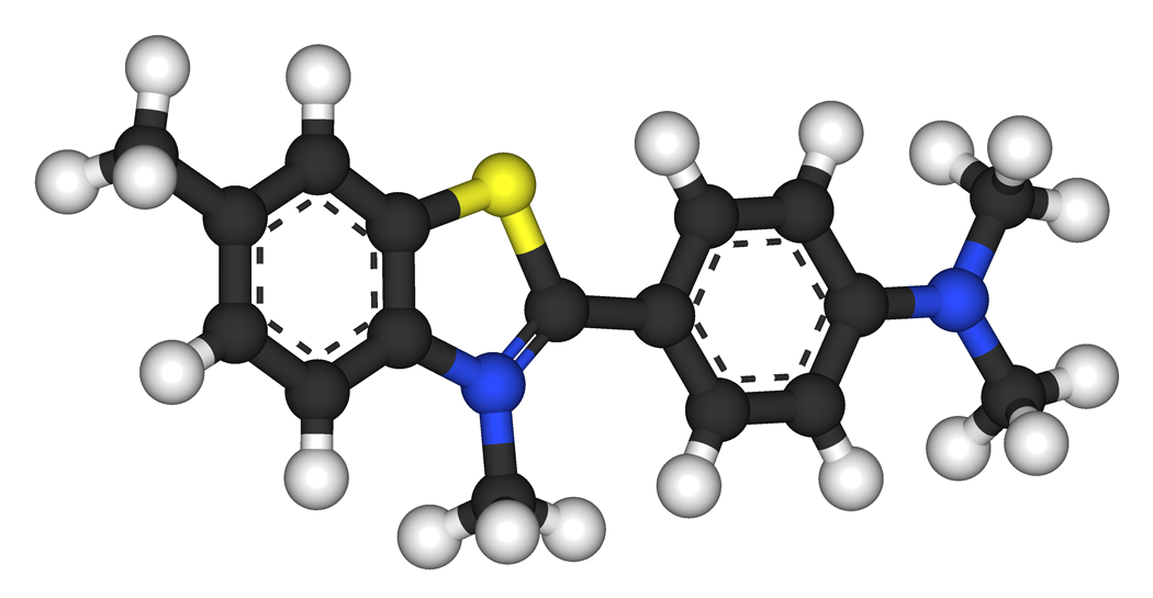
Preferred IUPAC name
2-[4-(Dimethylamino)phenyl]-3,6-dimethyl-1,3-benzothiazol-3-ium chloride
Identifiers
3D model (JSmol)
ChEBI
ChEMBL
ChemSpider
ECHA InfoCard 100.017.482 Edit this at Wikidata
UNII
  • InChI=1S/C17H19N2S.ClH/c1-12-5-10-15-16(11-12)20-17(19(15)4)13-6-8-14(9-7-13)18(2)3;/h5-11H,1-4H3;1H/q+1;/p-1 checkY
    Key: JADVWWSKYZXRGX-UHFFFAOYSA-M checkY
  • InChI=1/C17H19N2S.ClH/c1-12-5-10-15-16(11-12)20-17(19(15)4)13-6-8-14(9-7-13)18(2)3;/h5-11H,1-4H3;1H/q+1;/p-1
    Key: JADVWWSKYZXRGX-REWHXWOFAC
  • [Cl-].s2c1cc(ccc1[n+](c2c3ccc(N(C)C)cc3)C)C
Properties
C17H19ClN2S
Molar mass 318.86 g/mol
Density 1.301 g/cm3
Melting point 137.9 °C (decomp.)
Except where otherwise noted, data are given for materials in their standard state (at 25 °C [77 °F], 100 kPa).
☒N verify (what is checkY☒N ?)

Thioflavins are fluorescent dyes that are available as at least two compounds, namely Thioflavin T and Thioflavin S. Both are used for histology staining and biophysical studies of protein aggregation.[1] In particular, these dyes have been used since 1959 to investigate amyloid formation.[2][3] They are also used in biophysical studies of the electrophysiology of bacteria.[4] Thioflavins are corrosive, irritant, and acutely toxic, causing serious eye damage.[5] Thioflavin T has been used in research into Alzheimer's disease and other neurodegenerative diseases.

Thioflavin T

[edit]

Thioflavin T (Basic Yellow 1, Methylene yellow, CI 49005, or ThT) is a benzothiazole salt obtained by the methylation of dehydrothiotoluidine with methanol in the presence of hydrochloric acid. The dye is widely used to visualize and quantify the presence of misfolded protein aggregates called amyloid, both in vitro and in vivo (e.g., plaques composed of amyloid beta found in the brains of Alzheimer's disease patients).[1]

When it binds to beta sheet-rich structures, such as those in amyloid aggregates, the dye displays enhanced fluorescence and a characteristic red shift of its emission spectrum.[6][7] Additional studies also consider fluorescence changes as result of the interaction with double stranded DNA.[8] This change in fluorescent behavior can be caused by many factors that affect the excited state charge distribution of thioflavin T, including binding to a rigid, highly-ordered nanopocket, and specific chemical interactions between thioflavin T and the nanopocket.[9][10]

Prior to binding to an amyloid fibril, thioflavin T emits weakly around 427 nm. Quenching effects of the nearby excitation peak at 450 nm is suspected to play a role in minimizing emissions.

When excited at 450 nm, thioflavin T produces a strong fluorescence signal at approximately 482 nm upon binding to amyloids. Thioflavin T molecule consists of a phenylamine and a benzothiazole ring connected through a carbon-carbon bond. These two rings can rotate freely when the molecule is in solution. The free rotation of these rings results in quenching of any excited state generated by photon excitation. However, when thioflavin T binds to amyloid fibrils, the two rotational planes of the two rings become immobilized and therefore, this molecule can maintain its excited state.[1]

Thioflavin T fluorescence is often used as a diagnostic of amyloid structure, but it is not perfectly specific for amyloid. Depending on the particular protein and experimental conditions, thioflavin T may[9] or may not[11] undergo a spectroscopic change upon binding to precursor monomers, small oligomers, unaggregated material with a high beta sheet content, or even alpha helix-rich proteins. Conversely, some amyloid fibers do not affect thioflavin T fluorescence,[12] raising the prospect of false negative results.

X-ray crystal structure of thioflavin T bound to an amyloid-like oligomer of β2 microglobulin
Structure of thioflavin T bound to an amyloid-like oligomer of β2 microglobulin (in gray), in a complex that displays enhanced and red shifted fluorescence. Many factors that shift the excited state charge from the dimethylaminobenzyl portion of thioflavin T (in blue) to the benzothiazole portion (in red), including binding to rigid, highly-ordered amyloid aggregates, can produce this 'positive' thioflavin T signal.[9]


Thioflavin S stain (left in green) and amyloid-Beta antibody immunocytochemistry (right) on adjacent sections of the hippocampus of a patient suffering from Alzheimer's disease. Thioflavin S binds both senile plaques (SP) and neurofibrillary tangles (NFT), the two characteristic cortical lesions of Alzheimer's. Amyloid beta is a peptide derived from the amyloid precursor protein which is only found in senile plaques, and so only plaques are visible in the right hand image. The left image also has a red signal which exactly superimposes the green signal in lipofuscin granules (LP), which are autofluorescent inclusions derived from lysosomes which accumulate in the human brain during normal aging.

In adult C. elegans, exposure to thioflavin T results "in a profoundly extended lifespan and slowed aging" at some levels, but decreased lifespan at higher levels.[13]

Thioflavin S

[edit]

Thioflavin S is a homogenous mixture of compounds that results from the methylation of dehydrothiotoluidine with sulfonic acid. It is also used to stain amyloid plaques. Like thioflavin T it binds to amyloid fibrils but not monomers and gives a distinct increase in fluorescence emission. However unlike thioflavin T, it does not produce a characteristic shift in the excitation or emission spectra.[6] This latter characteristic of thioflavin S results in high background fluorescence, making it unable to be used in quantitative measurements of fibril solutions.[6] Another dye that is used to identify amyloid structure is Congo red.

See also

[edit]

References

[edit]
Revisions and contributorsEdit on WikipediaRead on Wikipedia
from Grokipedia
Thioflavin refers to a class of synthetic fluorescent dyes, most notably Thioflavin T (ThT) and Thioflavin S (ThS), that exhibit dramatically enhanced upon binding to the beta-sheet-rich structures of fibrils, making them essential tools in and biochemistry for detecting protein aggregates associated with diseases like Alzheimer's. Thioflavin T, chemically known as 2-[4-(dimethylamino)phenyl]-3,6-dimethyl-1,3-benzothiazol-3-ium chloride (CAS 2390-54-7), is a cell-permeable cationic with a molecular formula of C₁₇H₁₉ClN₂S and a molecular weight of 318.86 g/mol. In aqueous solutions, ThT displays weak due to free around its central carbon-carbon bond, which quenches emission; however, binding to restricts this , shifting its excitation maximum to approximately 450 nm and emission to 482 nm, resulting in a increase of over 1000-fold. This property arises from ThT's interaction with hydrophobic grooves on fibril surfaces, often involving aromatic stacking with residues in beta-sheets. Thioflavin S, a sulfonated derivative, shares similar spectral properties but is less commonly used for quantitative assays due to its heterogeneity. As the "gold standard" for amyloid detection, ThT is routinely employed to monitor fibril formation kinetics in vitro through real-time fluorescence assays, typically at concentrations of 10–50 μM, and for histological staining of amyloid plaques in tissue sections from neurodegenerative disease models. Its specificity for amyloid structures has facilitated extensive research into protein misfolding pathologies, including amyloid-beta and tau aggregates in Alzheimer's disease, alpha-synuclein in Parkinson's, and prion proteins. Beyond amyloids, ThT has emerging applications as a probe for mitochondrial membrane potential at low concentrations (under 1 μM) and as a nucleolar stain in neuronal cells, highlighting its versatility in cellular imaging. Despite its widespread use, optimal staining protocols emphasize avoiding high dye concentrations to prevent non-specific binding and photobleaching.

History and Development

Discovery and Early Use

Thioflavin T, a cationic derivative also known as Basic Yellow 1, was originally developed as a fluorescent for applications. This industrial origin positioned thioflavin dyes as versatile staining agents before their adaptation for scientific purposes. The transition to biological use occurred in 1959 when Paul S. Vassar and C. F. A. Culling introduced thioflavin T as a selective fluorescent stain for deposits in histological sections, particularly in cases of . Their pioneering work demonstrated enhanced yellow-green under light when thioflavin T bound to , offering greater sensitivity and specificity compared to earlier dyes like . This marked the shift from an industrial tool to a key method in for detecting extracellular protein aggregates. The seminal paper, published in the Archives of Pathology, highlighted its utility in connective tissues and , establishing thioflavin T as a standard for qualitative visualization in fixed tissues. In the , early studies expanded thioflavin to amyloid deposits in neurodegenerative and systemic diseases, including and . Thioflavin S, a related variant introduced earlier as a in the early , began gaining use in amyloid research during this period. Researchers like and colleagues applied thioflavin S in the 1970s for fluorescent light of senile plaques in Alzheimer's presenile dementia, revealing amyloid cores within plaques. Similarly, Martin Roth and associates correlated senile plaque counts with dementia severity in elderly populations in the , using silver methods to quantify deposits in brain tissues. These investigations underscored the role of thioflavin dyes in identifying β-pleated sheet structures characteristic of amyloid in both cerebral plaques and peripheral tissues affected by , laying groundwork for their routine use in .

Evolution in Research Applications

In 1989, thioflavin T (ThT) was introduced as a fluorometric probe for the quantitative determination of , enabling precise monitoring of fibril kinetics through enhanced upon binding. This approach revolutionized the study of aggregation by providing a sensitive, real-time method to track formation rates, surpassing earlier qualitative staining techniques. The method's reliability quickly positioned ThT as the gold standard for detection, facilitating reproducible quantification across diverse protein systems. During the 1990s, biophysical investigations deepened understanding of ThT's binding specificity to amyloid structures, revealing its preferential interaction with β-sheet-rich and minimal response to non- aggregates. These studies, including detailed spectroscopic analyses, confirmed ThT's utility in distinguishing amyloid conformations and laid the groundwork for its broader application in protein misfolding research related to neurodegenerative diseases. By the 2000s, ThT assays were adapted for of amyloid inhibitors, accelerating efforts against aggregation in conditions like through automated readouts of inhibition efficacy. Post-2017 advancements have integrated ThT with techniques, such as stochastic optical reconstruction microscopy (), to visualize morphology at the nanoscale. This combination has enabled detailed mapping of ultrastructure and polymorphism in real time, enhancing insights into aggregation pathways and therapeutic targeting in neurodegeneration. These developments underscore ThT's evolution from a basic kinetic probe to a versatile tool in advanced imaging and biophysical assays as of 2025.

Chemical Structure and Properties

Molecular Composition and Synthesis

Thioflavins constitute a class of cationic benzothiazole dyes featuring a dimethylamino-substituted phenyl group at the 2-position of the benzothiazole ring, which imparts their characteristic fluorescence properties upon binding to specific structures. The core molecular composition for the prototypical Thioflavin T (ThT) is represented by the formula C17_{17}H19_{19}ClN2_{2}S, corresponding to 2-[4-(dimethylamino)phenyl]-3,6-dimethylbenzothiazol-3-ium chloride (SMILES: CN(C)C1=CC=C(C=C1)C2=N+C3=C(S2)C=C(C=C3)C.[Cl-]), which represents the cation with the chloride counterion. This structure includes a positively charged nitrogen in the thiazole ring, balanced by a chloride counterion, along with methyl substituents at the 3- and 6-positions. The molar mass of ThT is 318.86 g/mol. The chloride salt form enhances solubility in polar solvents such as water (up to approximately 25 mM) and alcohols like methanol and ethanol, facilitating its use in aqueous biological assays. The synthesis of thioflavin dyes generally proceeds via the construction of the heterocycle followed by N-quaternization to introduce the cationic character. The ring formation typically involves an acid- or base-catalyzed between a substituted o-aminothiophenol and a para-substituted derivative, which cyclizes under oxidative conditions to yield the neutral 2-aryl intermediate. For ThT specifically, this step employs 2-amino-5-methylbenzenethiol (providing the 6-methyl group) and 4-(dimethylamino), often conducted in or polyphosphoric acid at elevated temperatures (around 100–200 °C) to promote dehydration and ring closure. The resulting intermediate, known historically as dehydrothiotoluidine, is then quaternized at the nitrogen. Quaternization for ThT is commonly achieved by treatment with methyl in an inert solvent to form the iodide salt, which can be exchanged to the form, or more traditionally, by reaction with and under pressure (e.g., in an at 150–180 °C for several hours). This step yields the final cationic in high purity after recrystallization from or . For the sulfonated variant Thioflavin S (ThS), the synthesis diverges in the quaternization phase, where dehydrothiotoluidine is instead reacted with or a equivalent, resulting in a heterogeneous of sulfonated products rather than a single chloride salt. These routes allow for modular variations by altering substituents on the starting materials, enabling the preparation of thioflavin analogs with tuned or binding affinities.

Physical and Spectroscopic Characteristics

Thioflavin T, the primary variant of thioflavin dyes, appears as a yellow to orange-brown powder at room temperature. It has a melting point of 137.9 °C, at which point it decomposes rather than fully melting. The compound exhibits solubility in water of approximately 5–25 mM at room temperature, though lower (≈0.33 g/L or 1 mM using ethanol co-solvent) in neutral buffers like PBS (pH 7.2) at 20 °C; solubility increases significantly in hot water up to 100 mg/mL. In , thioflavin T displays an absorption maximum at approximately 412 nm, corresponding to its characteristic color. The free emits weakly at around 440 nm upon excitation, with severely quenched due to rotational motion in the , specifically intramolecular torsion around the benzothiazole-phenyl bond. This non-radiative decay pathway results in an extremely low of about 0.0001 in at . Thioflavin T is light-sensitive and prone to under prolonged exposure to light, necessitating storage in dark conditions. It also shows reduced stability in strong acidic or basic environments, where both absorption intensity in the visible region and residual decrease markedly, accompanied by pH-dependent shifts in color. These properties contrast with the dramatic enhancement in and spectral shifts observed upon binding to rigid structures like amyloid fibrils.

Mechanism of Action

Binding to Amyloid Structures

Thioflavin T (ThT), the most commonly studied variant of thioflavin, primarily binds to through intercalation into the grooves formed by the cross-β sheet architecture. This binding occurs via a combination of hydrophobic interactions between the and moieties of ThT and the side chains of β-strands, particularly in regions rich in aromatic residues such as and , alongside electrostatic interactions involving the positively charged quaternary ammonium group of ThT and negatively charged surfaces. The aligns parallel to the 's long axis, occupying channels created by cross-strand side-chain ladders, which stabilizes the association without disrupting the 's overall structure. The specificity of ThT for amyloid structures stems from its preference for the rigid, periodic cross-β architecture, where β-strands are stacked perpendicular to the axis. This contrasts with weaker or negligible binding to non- aggregates, such as those featuring α-helical conformations or amorphous protein clusters, due to the absence of suitable groove geometries for stable intercalation. Studies on diverse systems, including Aβ and proteins, confirm that ThT exhibits minimal affinity for α-helical peptides or disordered aggregates, highlighting its utility as a selective probe for β-sheet-rich . Binding stoichiometry varies by fibril type and binding mode, typically ranging from 0.1 to 0.9 ThT molecules per , reflecting both high- and low-affinity sites. For instance, in Sup35p , one mode yields about 0.9 ThT per at lower affinity sites, while a higher-affinity mode binds roughly 0.1 ThT per ; similarly, show 0.11–0.24 ThT per across binding modes. This variable binding reflects saturation of specific grooves rather than uniform coverage, ensuring minimal perturbation to integrity. Factors such as and significantly influence binding affinity, with dissociation constants (Kd) for ThT typically ranging from 1–10 μM under physiological conditions. Higher enhances affinity by shielding electrostatic repulsions between the positively charged ThT and similarly charged regions, increasing binding capacity up to several-fold in systems like insulin and fibrils. Acidic reduces affinity due to increased electrostatic repulsion, though elevated can mitigate this effect, as observed in engineered β-sheet mimics with Kd ≈ 2 μM.

Fluorescence Enhancement Process

The fluorescence enhancement of Thioflavin T (ThT) upon binding to amyloid fibrils primarily arises from the restriction of intramolecular torsional motion, which locks the dye molecule into a planar conformation and suppresses non-radiative decay pathways. In its unbound state, ThT exhibits rapid rotation around the central C-C bond connecting the and rings, leading to a twisted intramolecular charge transfer (TICT) state that dissipates excitation energy as heat, resulting in a low of approximately 0.0001 in . Upon binding to the rigid β-sheet structures of amyloid fibrils, this torsional freedom is sterically hindered, stabilizing the locally excited (LE) state and promoting radiative decay, which elevates the quantum yield to around 0.28–0.30. This binding-induced conformational change also induces significant spectral shifts in ThT's absorption and emission profiles, enhancing detectability. The excitation maximum red-shifts from approximately 412 nm (or 385 nm in some reports) in the free state to 440–450 nm when bound, while the emission maximum shifts from 427–445 nm to 482 nm, accompanied by a dramatic increase in intensity—often by three orders of magnitude (up to 1000-fold). These alterations stem from the altered electronic environment in the bound planar form, which modifies the dye's dipole moment and vibronic coupling. For quantitative assays, the fluorescence intensity can be modeled using a simplified binding equation: I=I0(1+K[F])I = I_0 (1 + K [F]), where II is the observed intensity, I0I_0 is the baseline intensity of unbound ThT, KK is the (typically on the order of 10410^410510^5 M1^{-1} for amyloid ), and [F][F] represents the concentration; this approximation assumes low fibril occupancy and is useful for initial design but requires validation against saturation binding data. Despite its utility, the enhancement process has limitations, including potential self-quenching at high ThT concentrations (>50 μM), where formation reduces signal intensity, and non-specific binding to other structured proteins or nucleic acids that can introduce background or variable enhancement. Optimal conditions thus involve low dye concentrations (10–20 μM) to maximize signal-to-noise ratios while minimizing these artifacts.

Biological and Medical Applications

Detection of Amyloid Fibrils

Thioflavin T (ThT) is widely employed in standard assays to quantify formation through its enhanced upon binding. A typical protocol involves preparing protein samples at concentrations conducive to fibrillation, such as 10-100 μM for peptides like Aβ, and adding ThT at 10-50 μM in a buffered solution, often with gentle agitation to promote aggregation. is measured using excitation at 440 nm and emission at 485 nm, with the signal showing a linear response to concentration up to approximately 100 μM, enabling reliable quantification of content. For histological detection of amyloid plaques in tissue sections, such as those from models, a common protocol entails fixing and sectioning brain tissue, followed by incubation in 0.1% ThT dissolved in (PBS) for 5-10 minutes at . Sections are then rinsed multiple times in PBS to remove unbound dye and mounted for imaging, where amyloid deposits appear as bright fluorescent structures under fluorescence microscopy with excitation at 440 nm and emission at 485 nm, facilitating visualization of plaques and vascular s. Quantitative analysis of ThT fluorescence often involves normalization to total protein content to account for variations in sample loading or tissue heterogeneity, ensuring accurate comparison of amyloid burden across experiments. In kinetic studies, ThT assays track the fibrillation process in real-time by plotting fluorescence intensity over time, revealing characteristic sigmoidal curves with a lag phase (nucleation-dominated), an exponential growth phase (fibril elongation), and a plateau phase (saturation), from which parameters like lag time and apparent growth rate are derived. The primary advantages of ThT-based detection include its compatibility with real-time, high-throughput monitoring of amyloid formation without disrupting kinetics at optimal concentrations. However, disadvantages encompass potential false positives arising from non-specific binding to aggregates like DNA or micelles, or self-fluorescence at higher dye levels, necessitating careful controls and complementary validation methods.

Emerging Uses in Neuroscience and Diagnostics

Recent studies have demonstrated the utility of Thioflavin T (ThT) as a for neuronal bodies and nucleoli in live cells, expanding its role beyond detection. In 2024, researchers reported that ThT provides rapid and specific labeling of nucleolar structures and synaptic elements in neuronal cultures, with enhanced by low-intensity blue light exposure, enabling high-resolution without significant at optimized concentrations. This application leverages ThT's affinity for RNA-rich nucleoli and protein aggregates in synaptic regions, offering a simple alternative to traditional nuclear stains for studying cellular architecture in models. Thioflavin derivatives, such as Pittsburgh Compound B (PiB), have been integrated into (PET) imaging for detection of in , with ongoing clinical trials as of 2025 validating their diagnostic accuracy. PiB, a radiolabeled analog of ThT, exhibits high binding affinity to β-sheet-rich , allowing quantification of plaque burden in brain regions affected by neurodegeneration. Updated appropriate use criteria from 2025 emphasize PiB-PET's role in early diagnosis and monitoring treatment response in Alzheimer's trials, showing concordance rates exceeding 90% with biomarkers. High-throughput screening platforms employing ThT fluorescence have accelerated for proteinopathies, particularly , by monitoring α-synuclein aggregation inhibition. In a 2018 assay, ThT-based detection identified potent inhibitors like SynuClean-D from libraries of over 14,000 compounds, reducing formation by up to 80% . Subsequent 2022 studies refined two-step ThT protocols for α-synuclein, enabling scalable screening of aggregation modulators with improved reproducibility and sensitivity to early oligomeric states. ThT has also found applications in detecting bacterial and aggregates, with derivatives enhancing specificity in these contexts. In studies, ThT stains amyloid-like structures in aggregates, confirming their role in microbial adhesion and resistance, as shown in 2024 analyses of components. For prions, (RT-QuIC) assays using ThT provide ultrasensitive detection of seeding activity, quantifying protein misfolding with limits of detection below 10 attomolar.

Specific Variants

Thioflavin T

Thioflavin T (ThT), chemically known as 2-[4-(dimethylamino)phenyl]-3,6-dimethyl-1,3-benzothiazol-3-ium chloride (CAS 2390-54-7), is synthesized through the methylation of dehydrothiotoluidine using methanol in the presence of hydrochloric acid, resulting in a homogeneous product suitable for consistent analytical applications. This method, developed in the early 20th century, produces a single benzothiazole derivative that exhibits enhanced solubility and stability compared to heterogeneous mixtures in related dyes. ThT demonstrates high specificity for β-sheet structures in , with a binding affinity characterized by an equilibrium (Kd) of approximately 1 μM, enabling sensitive detection of fibrillar aggregates. Its preference in quantitative assays stems from minimal background in unbound states, allowing for reliable measurement of formation kinetics via enhancement upon binding. ThT is the most widely used variant in research, serving as the gold standard probe in the majority of studies due to its reproducibility and ease of integration into spectroscopic setups. In applications, ThT dominates amyloid investigations, including real-time tracking of α-synuclein aggregation in models through plate-reader assays. These assays typically involve incubating protein samples with 10-50 μM ThT and exciting at 440 nm to observe emission shifts from 485 nm, providing kinetic data on lag, growth, and plateau phases of fibrillation. Despite its utility, ThT exhibits photobleaching during extended imaging sessions, where prolonged excitation leads to a gradual loss of intensity, limiting its use in long-term live-cell or . For qualitative assessments requiring broader staining profiles, alternatives such as Thioflavin S may be considered, though ThT remains preferred for precise quantification.

Thioflavin S

Thioflavin S (ThS), chemically known as a mixture of sulfonated derivatives (CAS 1326-12-1), is synthesized through the sulfonic acid-mediated of dehydrothiotoluidine, yielding a heterogeneous mixture of sulfonated isomers that are incompletely characterized. This process introduces variability in the dye's composition, distinguishing it from more uniform variants like Thioflavin T. Upon binding to structures, ThS exhibits enhanced without a shift in its excitation or emission spectra, resulting in a peak emission around 455 nm under typical conditions. However, this lack of spectral change contributes to high non-specific background , particularly at elevated concentrations, which complicates precise quantification and limits its utility compared to Thioflavin T's quantitative superiority in kinetic assays. ThS finds primary application in qualitative histological staining of amyloid deposits within fixed tissues, such as brain sections from models, where it highlights plaques and threads under . Its use is less common in real-time kinetic studies of formation due to the dye's compositional heterogeneity and associated variability. Historically, ThS has played a key role in early diagnostics, with protocols optimized for sensitivity in detecting diffuse . Relative to Thioflavin T, ThS offers advantages in certain tissue staining protocols, including improved penetration into fixed specimens owing to its polar groups, facilitating broader application in .

Safety and Regulatory Considerations

Toxicity Profile

Thioflavin T exhibits acute oral toxicity in rats, with an LD50 value of 200 mg/kg ( Test Guideline 423), classifying it under GHS as toxic if swallowed (Category 3, H301). It is a severe irritant to eyes, causing serious damage (Category 1, H318), and may induce allergic skin reactions (Category 1, H317). Thioflavin S shows similar irritant properties, including potential irritation (Category 3, H335), with limited oral toxicity data available and an intraperitoneal LD50 of 400 mg/kg in mice. Chronic exposure data for thioflavins are limited, with safety assessments indicating no established repeated-dose target organ toxicity or long-term adverse health effects based on available animal models. The core structure in thioflavin T is negative for mutagenicity ( Test Guideline 471), though some related benzothiazoles have shown positive results in bacterial assays. Respiratory issues may arise from inhalation of the powder form during laboratory handling, though specific chronic respiratory links remain undocumented. Eyes are a primary target organ for thioflavin T, where exposure can lead to severe irritation and potential corneal damage due to its corrosive nature. Inhalation risks are heightened in the fine powder form of both variants, potentially causing upper irritation upon exposure. As of 2025, thioflavins are classified under the Globally Harmonized System (GHS) as hazardous substances due to , serious eye damage, skin sensitization, and environmental hazards, but they lack specific listings as carcinogens or reproductive toxicants. Thioflavin T is registered under the U.S. TSCA inventory.

Handling and Environmental Impact

When handling Thioflavin T in laboratory settings, appropriate (PPE) such as gloves, tightly fitting safety goggles, and face protection must be worn to prevent skin, eye, and respiratory exposure. Work should be conducted in a well-ventilated to minimize of dust or aerosols, and thorough handwashing is required after use to avoid accidental or absorption. For storage, Thioflavin T should be kept in a tightly closed in a cool, dry, dark, and well-ventilated place at to maintain stability and prevent degradation from light or moisture exposure. Under these conditions, the powder form has a of approximately 2-3 years. Thioflavin T exhibits high aquatic toxicity, classified as very toxic to aquatic life with long-lasting effects, and releases to the environment should be strictly avoided to prevent entry into waterways or drains. Key metrics include an of 0.0298 mg/L for growth inhibition in (Pseudokirchneriella subcapitata) over 72 hours, indicating sensitivity in algal populations. It is not readily biodegradable according to Test Guideline 301B, and its cationic contributes to persistence in sediments, though potential is low with no significant data reported (log Pow = 3.02). Waste management for Thioflavin T follows 2025 EPA guidelines under the (RCRA) for , requiring disposal as a toxic solid in approved facilities rather than sewers or household trash. Contaminated containers must be handled similarly, with no neutralization typically needed unless specified by local regulations, and no notable risks are associated with proper disposal.

References

Add your contribution
Related Hubs
User Avatar
No comments yet.