
Community hub
Recent from talks
Contribute something
Nothing was collected or created yet.
Fluorophore
View on WikipediaThis article may contain unverified or indiscriminate information in embedded lists. (February 2016) |

A fluorophore (or fluorochrome, similarly to a chromophore) is a fluorescent chemical compound that can re-emit light upon light excitation. Fluorophores typically contain several combined aromatic groups, or planar or cyclic molecules with several π bonds.[1]
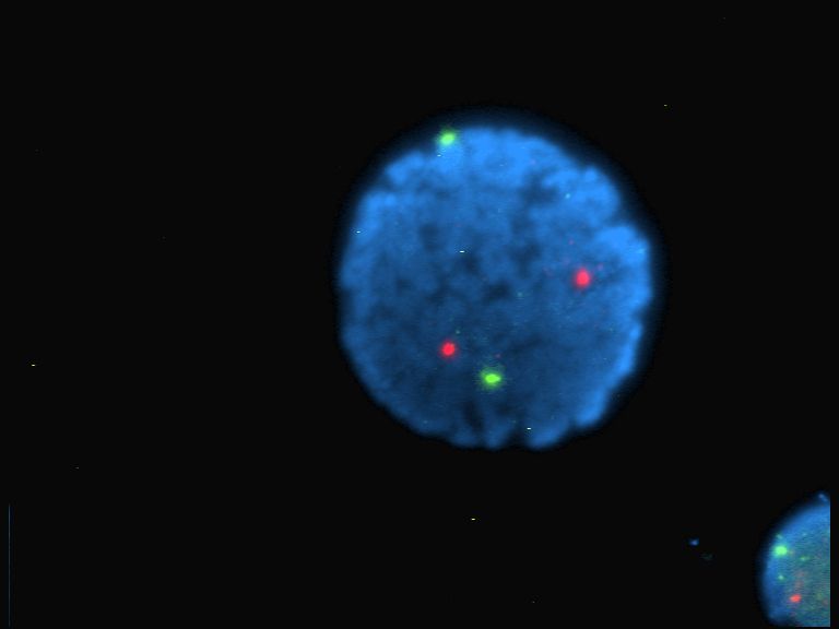
Fluorophores are sometimes used alone, as a tracer in fluids, as a dye for staining of certain structures, as a substrate of enzymes, or as a probe or indicator (when its fluorescence is affected by environmental aspects such as polarity or ions). More generally they are covalently bonded to macromolecules, serving as a markers (or dyes, or tags, or reporters) for affine or bioactive reagents (antibodies, peptides, nucleic acids). Fluorophores are notably used to stain tissues, cells, or materials in a variety of analytical methods, such as fluorescent imaging and spectroscopy.
Fluorescein, via its amine-reactive isothiocyanate derivative fluorescein isothiocyanate (FITC), has been one of the most popular fluorophores. From antibody labeling, the applications have spread to nucleic acids thanks to carboxyfluorescein. Other historically common fluorophores are derivatives of rhodamine (TRITC), coumarin, and cyanine.[2] Newer generations of fluorophores, many of which are proprietary, often perform better, being more photostable, brighter, or less pH-sensitive than traditional dyes with comparable excitation and emission.[3][4]
Fluorescence
[edit]The fluorophore absorbs light energy of a specific wavelength and re-emits light at a longer wavelength. The absorbed wavelengths, energy transfer efficiency, and time before emission depend on both the fluorophore structure and its chemical environment, since the molecule in its excited state interacts with surrounding molecules. Wavelengths of maximum absorption (≈ excitation) and emission (for example, Absorption/Emission = 485 nm/517 nm) are the typical terms used to refer to a given fluorophore, but the whole spectrum may be important to consider. The excitation spectrum may be a very narrow or broader band, or it may be all beyond a cutoff level. The emission spectrum is usually sharper than the excitation spectrum, and it is of a longer wavelength and correspondingly lower energy. Excitation energies range from ultraviolet through the visible spectrum, and emission energies may continue from visible light into the near infrared region.
The main characteristics of fluorophores are:
- Maximum excitation and emission wavelength (expressed in nanometers (nm)): corresponds to the peak in the excitation and emission spectra (usually one peak each).
- Molar absorption coefficient (in mol−1cm−1): links the quantity of absorbed light, at a given wavelength, to the concentration of fluorophore in solution.
- Quantum yield: efficiency of the energy transferred from incident light to emitted fluorescence (the number of emitted photons per absorbed photons).
- Lifetime (in picoseconds): duration of the excited state of a fluorophore before returning to its ground state. It refers to the time taken for a population of excited fluorophores to decay to 1/e (≈0.368) of the original amount.
- Stokes shift: the difference between the maximum excitation and maximum emission wavelengths.
- Dark fraction: the proportion of the molecules not active in fluorescence emission. For quantum dots, prolonged single-molecule microscopy showed that 20-90% of all particles never emit fluorescence.[5] On the other hand, conjugated polymer nanoparticles (Pdots) show almost no dark fraction in their fluorescence.[6] Fluorescent proteins can have a dark fraction from protein misfolding or defective chromophore formation.[7]
These characteristics drive other properties, including photobleaching or photoresistance (loss of fluorescence upon continuous light excitation). Other parameters should be considered, as the polarity of the fluorophore molecule, the fluorophore size and shape (i.e. for polarization fluorescence pattern), and other factors can change the behavior of fluorophores.
Fluorophores can also be used to quench the fluorescence of other fluorescent dyes or to relay their fluorescence at even longer wavelengths.
Size (molecular weight)
[edit]Most fluorophores are organic small molecules of 20–100 atoms (200–1000 Dalton; the molecular weight may be higher depending on grafted modifications and conjugated molecules), but there are also much larger natural fluorophores that are proteins: green fluorescent protein (GFP) is 27 kDa, and several phycobiliproteins (PE, APC...) are ≈240kDa. As of 2020, the smallest known fluorophore was claimed to be 3-hydroxyisonicotinaldehyde, a compound of 14 atoms and only 123 Da.[8]
Fluorescence particles like quantum dots (2–10 nm diameter, 100–100,000 atoms) are also considered fluorophores.[9]
The size of the fluorophore might sterically hinder the tagged molecule and affect the fluorescence polarity.
Families
[edit]
Fluorophore molecules could be either utilized alone, or serve as a fluorescent motif of a functional system. Based on molecular complexity and synthetic methods, fluorophore molecules could be generally classified into four categories: proteins and peptides, small organic compounds, synthetic oligomers and polymers, and multi-component systems.[10][11]
Fluorescent proteins GFP, YFP, and RFP (green, yellow, and red, respectively) can be attached to other specific proteins to form a fusion protein, synthesized in cells after transfection of a suitable plasmid carrier.
Non-protein organic fluorophores belong to following major chemical families:
- Xanthene derivatives: fluorescein, rhodamine, Oregon green, eosin, and Texas red
- Cyanine derivatives: cyanine, indocarbocyanine, oxacarbocyanine, thiacarbocyanine, and merocyanine
- Squaraine derivatives and ring-substituted squaraines, including Seta and Square dyes
- Squaraine rotaxane derivatives: See Tau dyes
- Naphthalene derivatives (dansyl and prodan derivatives)
- Coumarin derivatives
- Oxadiazole derivatives: pyridyloxazole, nitrobenzoxadiazole, and benzoxadiazole
- Anthracene derivatives: anthraquinones, including DRAQ5, DRAQ7, and CyTRAK Orange
- Pyrene derivatives: cascade blue, etc.
- Oxazine derivatives: Nile red, Nile blue, cresyl violet, oxazine 170, etc.
- Acridine derivatives: proflavin, acridine orange, acridine yellow, etc.
- Arylmethine derivatives: auramine, crystal violet, malachite green
- Tetrapyrrole derivatives: porphin, phthalocyanine, bilirubin
- Dipyrromethene derivatives: BODIPY, aza-BODIPY
These fluorophores fluoresce due to delocalized electrons which can jump a band and stabilize the energy absorbed. For example, benzene, one of the simplest aromatic hydrocarbons, is excited at 254 nm and emits at 300 nm.[12] This discriminates fluorophores from quantum dots, which are fluorescent semiconductor nanoparticles.
They can be attached to proteins to specific functional groups, such as amino groups (active ester, carboxylate, isothiocyanate, hydrazine), carboxyl groups (carbodiimide), thiol (maleimide, acetyl bromide), and organic azide (via click chemistry or non-specifically (glutaraldehyde)).
Additionally, various functional groups can be present to alter their properties, such as solubility, or confer special properties, such as boronic acid which binds to sugars or multiple carboxyl groups to bind to certain cations. When the dye contains an electron-donating and an electron-accepting group at opposite ends of the aromatic system, this dye will probably be sensitive to the environment's polarity (solvatochromic), hence called environment-sensitive. Often dyes are used inside cells, which are impermeable to charged molecules; as a result of this, the carboxyl groups are converted into an ester, which is removed by esterases inside the cells, e.g., fura-2AM and fluorescein-diacetate.
The following dye families are trademark groups, and do not necessarily share structural similarities.
- CF dye (Biotium)
- DRAQ and CyTRAK probes (BioStatus)
- BODIPY (Invitrogen)
- EverFluor (Setareh Biotech)
- Alexa Fluor (Invitrogen)
- Bella Fluor (Setareh Biotech)
- DyLight Fluor (Thermo Scientific, Pierce)
- Atto and Tracy (Sigma Aldrich)
- FluoProbes (Interchim)
- Abberior Dyes (Abberior)
- DY and MegaStokes Dyes (Dyomics)
- Sulfo Cy dyes (Cyandye)
- HiLyte Fluor (AnaSpec)
- Seta, SeTau and Square Dyes (SETA BioMedicals)
- Quasar and Cal Fluor dyes (Biosearch Technologies)
- SureLight Dyes (APC, RPEPerCP, Phycobilisomes) (Columbia Biosciences)
- APC, APCXL, RPE, BPE (Phyco-Biotech, Greensea, Prozyme, Flogen)
- Vio Dyes (Miltenyi Biotec)
Examples of frequently encountered fluorophores
[edit]Reactive and conjugated dyes
[edit]| Dye | Ex (nm) | Em (nm) | MW | Notes |
|---|---|---|---|---|
| Hydroxycoumarin | 325 | 386 | 331 | Succinimidyl ester |
| Aminocoumarin | 350 | 445 | 330 | Succinimidyl ester |
| Methoxycoumarin | 360 | 410 | 317 | Succinimidyl ester |
| Cascade Blue | (375);401 | 423 | 596 | Hydrazide |
| Pacific Blue | 403 | 455 | 406 | Maleimide |
| Pacific Orange | 403 | 551 | ||
| 3-Hydroxyisonicotinaldehyde | 385 | 525 | 123 | QY 0.15; pH sensitive |
| Lucifer yellow | 425 | 528 | ||
| NBD | 466 | 539 | 294 | NBD-X |
| R-Phycoerythrin (PE) | 480;565 | 578 | 240 k | |
| PE-Cy5 conjugates | 480;565;650 | 670 | aka Cychrome, R670, Tri-Color, Quantum Red | |
| PE-Cy7 conjugates | 480;565;743 | 767 | ||
| Red 613 | 480;565 | 613 | PE-Texas Red | |
| PerCP | 490 | 675 | 35kDa | Peridinin chlorophyll protein |
| TruRed | 490,675 | 695 | PerCP-Cy5.5 conjugate | |
| FluorX | 494 | 520 | 587 | (GE Healthcare) |
| Fluorescein | 495 | 519 | 389 | FITC; pH sensitive |
| BODIPY-FL | 503 | 512 | ||
| G-Dye100 | 498 | 524 | suitable for protein labeling and electrophoresis | |
| G-Dye200 | 554 | 575 | suitable for protein labeling and electrophoresis | |
| G-Dye300 | 648 | 663 | suitable for protein labeling and electrophoresis | |
| G-Dye400 | 736 | 760 | suitable for protein labeling and electrophoresis | |
| Cy2 | 489 | 506 | 714 | QY 0.12 |
| Cy3 | (512);550 | 570;(615) | 767 | QY 0.15 |
| Cy3B | 558 | 572;(620) | 658 | QY 0.67 |
| Cy3.5 | 581 | 594;(640) | 1102 | QY 0.15 |
| Cy5 | (625);650 | 670 | 792 | QY 0.28 |
| Cy5.5 | 675 | 694 | 1272 | QY 0.23 |
| Cy7 | 743 | 767 | 818 | QY 0.28 |
| TRITC | 547 | 572 | 444 | TRITC |
| X-Rhodamine | 570 | 576 | 548 | XRITC |
| Lissamine Rhodamine B | 570 | 590 | ||
| Texas Red | 589 | 615 | 625 | Sulfonyl chloride |
| Allophycocyanin (APC) | 650 | 660 | 104 k | |
| APC-Cy7 conjugates | 650;755 | 767 | Far Red |
Abbreviations:
- Ex (nm): Excitation wavelength in nanometers
- Em (nm): Emission wavelength in nanometers
- MW: Molecular weight
- QY: Quantum yield
Nucleic acid dyes
[edit]| Dye | Ex (nm) | Em (nm) | MW | Notes |
|---|---|---|---|---|
| Hoechst 33342 | 343 | 483 | 616 | AT-selective |
| DAPI | 345 | 455 | AT-selective | |
| Hoechst 33258 | 345 | 478 | 624 | AT-selective |
| SYTOX Blue | 431 | 480 | ~400 | DNA |
| Chromomycin A3 | 445 | 575 | CG-selective | |
| Mithramycin | 445 | 575 | ||
| YOYO-1 | 491 | 509 | 1271 | |
| Ethidium Bromide | 210;285 | 605 | 394 | in aqueous solution |
| GelRed | 290;520 | 595 | 1239 | Non-toxic substitute for Ethidium Bromide |
| Acridine Orange | 503 | 530/640 | DNA/RNA | |
| SYTOX Green | 504 | 523 | ~600 | DNA |
| TOTO-1, TO-PRO-1 | 509 | 533 | Vital stain, TOTO: Cyanine Dimer | |
| TO-PRO: Cyanine Monomer | ||||
| Thiazole Orange | 510 | 530 | ||
| CyTRAK Orange | 520 | 615 | - | (Biostatus) (red excitation dark) |
| Propidium Iodide (PI) | 536 | 617 | 668.4 | |
| LDS 751 | 543;590 | 712;607 | 472 | DNA (543ex/712em), RNA (590ex/607em) |
| 7-AAD | 546 | 647 | 7-aminoactinomycin D, CG-selective | |
| SYTOX Orange | 547 | 570 | ~500 | DNA |
| TOTO-3, TO-PRO-3 | 642 | 661 | ||
| DRAQ5 | 600/647 | 697 | 413 | (Biostatus) (usable excitation down to 488) |
| DRAQ7 | 599/644 | 694 | ~700 | (Biostatus) (usable excitation down to 488) |
Cell function dyes
[edit]| Dye | Ex (nm) | Em (nm) | MW | Notes |
|---|---|---|---|---|
| Indo-1 | 361/330 | 490/405 | 1010 | AM ester, low/high calcium (Ca2+) |
| Fluo-3 | 506 | 526 | 855 | AM ester. pH > 6 |
| Fluo-4 | 491/494 | 516 | 1097 | AM ester. pH 7.2 |
| DCFH | 505 | 535 | 529 | 2'7'Dichorodihydrofluorescein, oxidized form |
| DHR | 505 | 534 | 346 | Dihydrorhodamine 123, oxidized form, light catalyzes oxidation |
| SNARF | 548/579 | 587/635 | pH 6/9 |
Fluorescent proteins
[edit]| Dye | Ex (nm) | Em (nm) | MW | QY | BR | PS | Notes |
|---|---|---|---|---|---|---|---|
| GFP (Y66H mutation) | 360 | 442 | |||||
| GFP (Y66F mutation) | 360 | 508 | |||||
| EBFP | 380 | 440 | 0.18 | 0.27 | monomer | ||
| EBFP2 | 383 | 448 | 20 | monomer | |||
| Azurite | 383 | 447 | 15 | monomer | |||
| GFPuv | 385 | 508 | |||||
| T-Sapphire | 399 | 511 | 0.60 | 26 | 25 | weak dimer | |
| Cerulean | 433 | 475 | 0.62 | 27 | 36 | weak dimer | |
| mCFP | 433 | 475 | 0.40 | 13 | 64 | monomer | |
| mTurquoise2 | 434 | 474 | 0.93 | 28 | monomer | ||
| ECFP | 434 | 477 | 0.15 | 3 | |||
| CyPet | 435 | 477 | 0.51 | 18 | 59 | weak dimer | |
| GFP (Y66W mutation) | 436 | 485 | |||||
| mKeima-Red | 440 | 620 | 0.24 | 3 | monomer (MBL) | ||
| TagCFP | 458 | 480 | 29 | dimer (Evrogen) | |||
| AmCyan1 | 458 | 489 | 0.75 | 29 | tetramer, (Clontech) | ||
| mTFP1 | 462 | 492 | 54 | dimer | |||
| GFP (S65A mutation) | 471 | 504 | |||||
| Midoriishi Cyan | 472 | 495 | 0.9 | 25 | dimer (MBL) | ||
| Wild Type GFP | 396,475 | 508 | 26k | 0.77 | |||
| GFP (S65C mutation) | 479 | 507 | |||||
| TurboGFP | 482 | 502 | 26 k | 0.53 | 37 | dimer, (Evrogen) | |
| TagGFP | 482 | 505 | 34 | monomer (Evrogen) | |||
| GFP (S65L mutation) | 484 | 510 | |||||
| Emerald | 487 | 509 | 0.68 | 39 | 0.69 | weak dimer, (Invitrogen) | |
| GFP (S65T mutation) | 488 | 511 | |||||
| EGFP | 488 | 507 | 26k | 0.60 | 34 | 174 | weak dimer, (Clontech) |
| Azami Green | 492 | 505 | 0.74 | 41 | monomer (MBL) | ||
| ZsGreen1 | 493 | 505 | 105k | 0.91 | 40 | tetramer, (Clontech) | |
| TagYFP | 508 | 524 | 47 | monomer (Evrogen) | |||
| EYFP | 514 | 527 | 26k | 0.61 | 51 | 60 | weak dimer, (Clontech) |
| Topaz | 514 | 527 | 57 | monomer | |||
| Venus | 515 | 528 | 0.57 | 53 | 15 | weak dimer | |
| mCitrine | 516 | 529 | 0.76 | 59 | 49 | monomer | |
| YPet | 517 | 530 | 0.77 | 80 | 49 | weak dimer | |
| TurboYFP | 525 | 538 | 26 k | 0.53 | 55.7 | dimer, (Evrogen) | |
| ZsYellow1 | 529 | 539 | 0.65 | 13 | tetramer, (Clontech) | ||
| Kusabira Orange | 548 | 559 | 0.60 | 31 | monomer (MBL) | ||
| mOrange | 548 | 562 | 0.69 | 49 | 9 | monomer | |
| Allophycocyanin (APC) | 652 | 657.5 | 105 kDa | 0.68 | heterodimer, crosslinked[13] | ||
| mKO | 548 | 559 | 0.60 | 31 | 122 | monomer | |
| TurboRFP | 553 | 574 | 26 k | 0.67 | 62 | dimer, (Evrogen) | |
| tdTomato | 554 | 581 | 0.69 | 95 | 98 | tandem dimer | |
| TagRFP | 555 | 584 | 50 | monomer (Evrogen) | |||
| DsRed monomer | 556 | 586 | ~28k | 0.1 | 3.5 | 16 | monomer, (Clontech) |
| DsRed2 ("RFP") | 563 | 582 | ~110k | 0.55 | 24 | (Clontech) | |
| mStrawberry | 574 | 596 | 0.29 | 26 | 15 | monomer | |
| TurboFP602 | 574 | 602 | 26 k | 0.35 | 26 | dimer, (Evrogen) | |
| AsRed2 | 576 | 592 | ~110k | 0.21 | 13 | tetramer, (Clontech) | |
| mRFP1 | 584 | 607 | ~30k | 0.25 | monomer, (Tsien lab) | ||
| J-Red | 584 | 610 | 0.20 | 8.8 | 13 | dimer | |
| R-phycoerythrin (RPE) | 565 >498 | 573 | 250 kDa | 0.84 | heterotrimer[13] | ||
| B-phycoerythrin (BPE) | 545 | 572 | 240 kDa | 0.98 | heterotrimer[13] | ||
| mCherry | 587 | 610 | 0.22 | 16 | 96 | monomer | |
| HcRed1 | 588 | 618 | ~52k | 0.03 | 0.6 | dimer, (Clontech) | |
| Katusha | 588 | 635 | 23 | dimer | |||
| P3 Archived 2024-08-21 at the Wayback Machine | 614 | 662 | ~10,000 kDa | phycobilisome complex[13] | |||
| Peridinin Chlorophyll (PerCP) | 483 | 676 | 35 kDa | trimer[13] | |||
| mKate (TagFP635) | 588 | 635 | 15 | monomer (Evrogen) | |||
| TurboFP635 | 588 | 635 | 26 k | 0.34 | 22 | dimer, (Evrogen) | |
| mPlum | 590 | 649 | 51.4 k | 0.10 | 4.1 | 53 | |
| mRaspberry | 598 | 625 | 0.15 | 13 | monomer, faster photobleach than mPlum | ||
| mScarlet | 569 | 594 | 0.70 | 71 | 277 | monomer[14] |
Advanced fluorescent proteins
[edit]StayGold and mStayGold are advanced fluorescent proteins that have significantly contributed to the field of live-cell imaging. StayGold, known for its high photostability and brightness, was originally designed as a dimeric fluorescent protein, which, while effective, posed challenges related to the aggregation and labelling accuracy.[15] To address these limitations, mStayGold was engineered as a monomeric variant, enhancing its utility in precise protein labeling. mStayGold exhibits superior photostability, maintaining fluorescence under high irradiance conditions and demonstrates increased brightness compared to its former variant StayGold. Additionally, it matures faster, allowing for quicker imaging post-transfection. These advancements make mStayGold a versatile tool for a variety of applications, including single molecule tracking and high resolution imaging of dynamic cellular processes, thereby expanding the capabilities of fluorescent protein in biological research.[16]
Abbreviations:
- Ex (nm): Excitation wavelength in nanometers
- Em (nm): Emission wavelength in nanometers
- MW: Molecular weight
- QY: Quantum yield
- BR: Brightness: Molar absorption coefficient * quantum yield / 1000
- PS: Photostability: time [sec] to reduce brightness by 50%
Applications
[edit]Fluorophores have particular importance in the field of biochemistry and protein studies, for example, in immunofluorescence, cell analysis,[17] immunohistochemistry,[3][18] and small molecule sensors.[19][20]
Uses outside the life sciences
[edit]
Fluorescent dyes find a wide use in industry, going under the name of "neon colors", such as:
- Multi-ton scale usages in textile dyeing and optical brighteners in laundry detergents
- Advanced cosmetic formulations
- Safety equipment and clothing
- Organic light-emitting diodes (OLEDs)
- Fine arts and design (posters and paintings)
- Synergists for insecticides and experimental drugs
- Dyes in highlighters to give off a glow-like effect
- Solar panels to collect more light / wavelengths
- Fluorescent sea dye is used to help airborne search and rescue teams locate objects in the water
See also
[edit]- Category:Fluorescent dyes
- Fluorescence in the life sciences
- Quenching of fluorescence
- Fluorescence recovery after photobleaching (FRAP) - an application for quantifying mobility of molecules in lipid bilayers.
References
[edit]- ^ Juan Carlos Stockert, Alfonso Blázquez-Castro (2017). "Chapter 3 Dyes and Fluorochromes". Fluorescence Microscopy in Life Sciences. Bentham Science Publishers. pp. 61–95. ISBN 978-1-68108-519-7. Retrieved 24 December 2017.
- ^ Rietdorf J (2005). Microscopic Techniques. Advances in Biochemical Engineering / Biotechnology. Berlin: Springer. pp. 246–9. ISBN 3-540-23698-8. Retrieved 2008-12-13.
- ^ a b Tsien RY; Waggoner A (1995). "Fluorophores for confocal microscopy". In Pawley JB (ed.). Handbook of biological confocal microscopy. New York: Plenum Press. pp. 267–74. ISBN 0-306-44826-2. Retrieved 2008-12-13.
- ^ Lakowicz, JR (2006). Principles of fluorescence spectroscopy (3rd ed.). Springer. p. 954. ISBN 978-0-387-31278-1.
- ^ Pons T, Medintz IL, Farrell D, Wang X, Grimes AF, English DS, Berti L, Mattoussi H (2011). "Single-molecule colocalization studies shed light on the idea of fully emitting versus dark single quantum dots". Small. 7 (14): 2101–2108. doi:10.1002/smll.201100802. PMID 21710484.
- ^ Koner AL, Krndija D, Hou Q, Sherratt DJ, Howarth M (2013). "Hydroxy-terminated conjugated polymer nanoparticles have near-unity bright fraction and reveal cholesterol-dependence of IGF1R nanodomains". ACS Nano. 7 (2): 1137–1144. doi:10.1021/nn3042122. PMC 3584654. PMID 23330847.
- ^ Garcia-Parajo MF, Segers-Nolten GM, Veerman JA, Greve J, van Hulst NF (2000). "Real-time light-driven dynamics of the fluorescence emission in single green fluorescent protein molecules". PNAS. 97 (13): 7237–7242. Bibcode:2000PNAS...97.7237G. doi:10.1073/pnas.97.13.7237. PMC 16529. PMID 10860989.
- ^ Cozens, Tom (2020-12-16). "Fluorescent molecule breaks size record for green-emitting dyes". chemistryworld.com. Retrieved 2021-12-03.
- ^ Li Z, Zhao X, Huang C, Gong X (2019). "Recent advances in green fabrication of luminescent solar concentrators using nontoxic quantum dots as fluorophores". J. Mater. Chem. C. 7 (40): 12373–12387. doi:10.1039/C9TC03520F. S2CID 203003761.
- ^ Liu, J.; Liu, C.; He, W. (2013), "Fluorophores and Their Applications as Molecular Probes in Living Cells", Curr. Org. Chem., 17 (6): 564–579, doi:10.2174/1385272811317060003
- ^ Juan Carlos Stockert, Alfonso Blázquez-Castro (2017). "Chapter 4 Fluorescent Labels". Fluorescence Microscopy in Life Sciences. Bentham Science Publishers. pp. 96–134. ISBN 978-1-68108-519-7. Retrieved 24 December 2017.
- ^ Omlc.ogi.edu
- ^ a b c d e "Columbia Biosciences". Archived from the original on 2024-08-21. Retrieved 2014-06-07.
- ^ Bindels, Daphne S.; Haarbosch, Lindsay; van Weeren, Laura; Postma, Marten; Wiese, Katrin E.; Mastop, Marieke; Aumonier, Sylvain; Gotthard, Guillaume; Royant, Antoine; Hink, Mark A.; Gadella, Theodorus W. J. (January 2017). "mScarlet: a bright monomeric red fluorescent protein for cellular imaging". Nature Methods. 14 (1): 53–56. doi:10.1038/nmeth.4074. ISSN 1548-7105. PMID 27869816. S2CID 3539874.
- ^ Hirano, Masahiko; Ando, Ryoko; Shimozono, Satoshi; Sugiyama, Mayu; Takeda, Noriyo; Kurokawa, Hiroshi; Deguchi, Ryusaku; Endo, Kazuki; Haga, Kei; Takai-Todaka, Reiko; Inaura, Shunsuke; Matsumura, Yuta; Hama, Hiroshi; Okada, Yasushi; Fujiwara, Takahiro (July 2022). "A highly photostable and bright green fluorescent protein". Nature Biotechnology. 40 (7): 1132–1142. doi:10.1038/s41587-022-01278-2. ISSN 1546-1696. PMC 9287174. PMID 35468954.
- ^ Ando, Ryoko; Shimozono, Satoshi; Ago, Hideo; Takagi, Masatoshi; Sugiyama, Mayu; Kurokawa, Hiroshi; Hirano, Masahiko; Niino, Yusuke; Ueno, Go; Ishidate, Fumiyoshi; Fujiwara, Takahiro; Okada, Yasushi; Yamamoto, Masaki; Miyawaki, Atsushi (April 2024). "StayGold variants for molecular fusion and membrane-targeting applications". Nature Methods. 21 (4): 648–656. doi:10.1038/s41592-023-02085-6. ISSN 1548-7105. PMC 11009113. PMID 38036853.
- ^ Sirbu, Dumitru; Luli, Saimir; Leslie, Jack; Oakley, Fiona; Benniston, Andrew C. (2019). "Enhanced in vivo Optical Imaging of the Inflammatory Response to Acute Liver Injury in C57BL/6 Mice Using a Highly Bright Near-Infrared BODIPY Dye". ChemMedChem. 14 (10): 995–999. doi:10.1002/cmdc.201900181. ISSN 1860-7187. PMID 30920173. S2CID 85544665.
- ^ Taki, Masayasu (2013). "Chapter 5. Imaging and sensing of cadmium in cells". In Astrid Sigel; Helmut Sigel; Roland K. O. Sigel (eds.). Cadmium: From Toxicology to Essentiality. Metal Ions in Life Sciences. Vol. 11. Springer. pp. 99–115. doi:10.1007/978-94-007-5179-8_5. PMID 23430772.
- ^ Sirbu, Dumitru; Butcher, John B.; Waddell, Paul G.; Andras, Peter; Benniston, Andrew C. (2017-09-18). "Locally Excited State-Charge Transfer State Coupled Dyes as Optically Responsive Neuron Firing Probes" (PDF). Chemistry - A European Journal. 23 (58): 14639–14649. doi:10.1002/chem.201703366. ISSN 0947-6539. PMID 28833695.
- ^ Jiang, Xiqian; Wang, Lingfei; Carroll, Shaina L.; Chen, Jianwei; Wang, Meng C.; Wang, Jin (2018-08-20). "Challenges and Opportunities for Small-Molecule Fluorescent Probes in Redox Biology Applications". Antioxidants & Redox Signaling. 29 (6): 518–540. doi:10.1089/ars.2017.7491. ISSN 1523-0864. PMC 6056262. PMID 29320869.
External links
[edit]- The Database of fluorescent dyes
- Table of fluorochromes
- The Molecular Probes Handbook - a comprehensive resource for fluorescence technology and its applications.