Hubbry Logo
Factor XFactor XMain
Open search
Factor X
Community hub
Factor X
logo
8 pages, 0 posts
0 subscribers
Be the first to start a discussion here.
Factor X
Factor X
from Wikipedia

F10
Available structures
PDBOrtholog search: PDBe RCSB
Identifiers
AliasesF10, FX, FXA, coagulation factor X
External IDsOMIM: 613872; MGI: 103107; HomoloGene: 30976; GeneCards: F10; OMA:F10 - orthologs
Orthologs
SpeciesHumanMouse
Entrez
Ensembl
UniProt
RefSeq (mRNA)

NM_000504
NM_001312674
NM_001312675

NM_001242368
NM_007972

RefSeq (protein)

NP_000495
NP_001299603
NP_001299604

NP_001229297
NP_031998

Location (UCSC)Chr 13: 113.12 – 113.15 MbChr 8: 13.09 – 13.11 Mb
PubMed search[3][4]
Wikidata
View/Edit HumanView/Edit Mouse

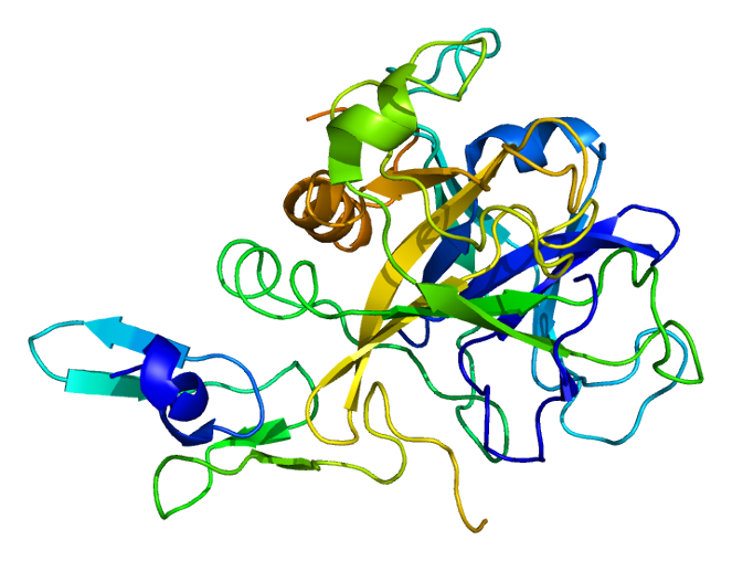
Coagulation factor X (EC 3.4.21.6), or Stuart factor, is an enzyme of the coagulation cascade, encoded in humans by F10 gene.[5] It is a serine endopeptidase (protease group S1, PA clan). Factor X is synthesized in the liver and requires vitamin K for its synthesis.

Factor X is activated, by hydrolysis, into factor Xa by both factor IX with its cofactor, factor VIII in a complex known as intrinsic pathway; and factor VII with its cofactor, tissue factor in a complex known as extrinsic pathway.[6] It is therefore the first member of the final common pathway or thrombin pathway.

It acts by cleaving prothrombin in two places (an Arg-Thr and then an Arg-Ile bond), which yields the active thrombin. This process is optimized when factor Xa is complexed with activated co-factor V in the prothrombinase complex.

Factor Xa is inactivated by protein Z-dependent protease inhibitor (ZPI), a serine protease inhibitor (serpin). The affinity of this protein for factor Xa is increased 1000-fold by the presence of protein Z, while it does not require protein Z for inactivation of factor XI. Defects in protein Z lead to increased factor Xa activity and a propensity for thrombosis. The half life of factor X is 40–45 hours.

Structure

[edit]

The first crystal structure of human factor Xa was deposited in May 1993. To date, 191 crystal structures of factor Xa with various inhibitors have been deposited in the protein data bank. The active site of factor Xa is divided into four subpockets as S1, S2, S3 and S4. The S1 subpocket determines the major component of selectivity and binding. The S2 sub-pocket is small, shallow and not well defined. It merges with the S4 subpocket. The S3 sub-pocket is located on the rim of the S1 pocket and is quite exposed to solvent. The S4 sub-pocket has three ligand binding domains: the "hydrophobic box", the "cationic hole" and the water site. Factor Xa inhibitors generally bind in an L-shaped conformation, where one group of the ligand occupies the anionic S1 pocket lined by residues Asp189, Ser195, and Tyr228, and another group of the ligand occupies the aromatic S4 pocket lined by residues Tyr99, Phe174, and Trp215. Typically, a fairly rigid linker group bridges these two interaction sites.[7]

Genetics

[edit]

The human factor X gene is located on chromosome 13 (13q34).

Role in disease

[edit]

Inborn deficiency of factor X is very rare (1:1,000,000), and may present with epistaxis (nosebleeds), hemarthrosis (bleeding into joints) and gastrointestinal blood loss. Apart from congenital deficiency, low factor X levels may occur occasionally in a number of disease states. For example, factor X deficiency may be seen in amyloidosis, where factor X is adsorbed to the amyloid fibrils in the vasculature.

Deficiency of vitamin K or antagonism by warfarin (or similar medication) leads to the production of an inactive factor X. In warfarin therapy, this is desirable to prevent thrombosis. As of late 2007, four out of five emerging anti-coagulation therapeutics targeted this enzyme.[8]

Inhibiting Factor Xa would offer an alternate method for anticoagulation. Direct Xa inhibitors are popular anticoagulants.

Polymorphisms in Factor X have been associated with an increased prevalence in bacterial infections, suggesting a possible role directly regulating the immune response to bacterial pathogens.[9]

Therapeutic use

[edit]

Factor X is part of fresh frozen plasma and the prothrombinase complex. There are two commercially available Factor X concentrates: "Factor X P Behring" manufactured by CSL Behring,[10] and high purity Factor X Coagadex produced by Bio Products Laboratory and approved for use in the United States by the FDA in October 2015, and in the EU in March 2016, after earlier acceptance by CHMP and COMP.[11][12][13][14]

Kcentra, manufactured by CSL Behring, is a concentrate containing coagulation Factors II, VII, IX and X, and antithrombotic Proteins C and S.[15]

Use in biochemistry

[edit]

The factor Xa protease can be used in biochemistry to cleave off protein tags that improve expression or purification of a protein of interest. Its preferred cleavage site (after the arginine in the sequence Ile-Glu/Asp-Gly-Arg, IEGR or IDGR) can easily be engineered between a tag sequence and the protein of interest. After expression and purification, the tag is then proteolytically removed by factor Xa.

Factor Xa

[edit]
Blood coagulation pathways in vivo showing the central role played by thrombin

Factor Xa is the activated form of the coagulation factor X, also known as thrombokinase. Factor X is an enzyme, a serine endopeptidase, which plays a key role at several stages of the coagulation system. Factor X is synthesized in the liver. The most commonly used anticoagulants in clinical practice, warfarin and the heparin series of anticoagulants and fondaparinux, act to inhibit the action of Factor Xa in various degrees.

Traditional models of coagulation developed in the 1960s envisaged two separate cascades, the extrinsic (tissue factor (TF)) pathway and the intrinsic pathway. These pathways converge to a common point, the formation of the Factor Xa/Va complex which together with calcium and bound on a phospholipids surface, generate thrombin (Factor IIa) from prothrombin (Factor II).

A new model, the cell-based model of anticoagulation appears to explain more fully the steps in coagulation. This model has three stages: 1) initiation of coagulation on TF-bearing cells, 2) amplification of the procoagulant signal by thrombin generated on the TF-bearing cell and 3) propagation of thrombin generation on the platelet surface. Factor Xa plays a key role in all three of these stages.[16]

In stage 1, Factor VII binds to the transmembrane protein TF on the surface of cells and is converted to Factor VIIa. The result is a Factor VIIa/TF complex, which catalyzes the activation of Factor X and Factor IX. Factor Xa formed on the surface of the TF-bearing cell interacts with Factor Va to form the prothrombinase complex which generates small amounts of thrombin on the surface of TF-bearing cells.

In stage 2, the amplification stage, if enough thrombin has been generated, then activation of platelets and platelet-associated cofactors occurs.

In stage 3, thrombin generation, Factor XIa activates free Factor IX on the surface of activated platelets. The activated Factor IXa with Factor VIIIa forms the "tenase" complex. This "tenase" complex activates more Factor X, which in turn forms new prothrombinase complexes with Factor Va. Factor Xa is the prime component of the prothrombinase complex which converts large amounts of prothrombin—the "thrombin burst". Each molecule of Factor Xa can generate 1000 molecules of thrombin. This large burst of thrombin is responsible for fibrin polymerization to form a thrombus.

Factor Xa also plays a role in other biological processes that are not directly related to coagulation, like wound healing, tissue remodelling, inflammation, angiogenesis and atherosclerosis.

Inhibition of the synthesis or activity of Factor X is the mechanism of action for many anticoagulants in use today. Warfarin, a synthetic derivative of coumarin, is the most widely used oral anticoagulant in the US. In some European countries, other coumarin derivatives (phenprocoumon and acenocoumarol) are used. These agents known as vitamin K antagonists (VKA), inhibit the vitamin K-dependent carboxylation of Factors II (prothrombin), VII, IX, X in the hepatocyte. This carboxylation after the translation is essential for the physiological activity.[17]

Heparin (unfractionated heparin) and its derivatives low molecular weight heparin (LMWH) bind to a plasma cofactor, antithrombin (AT) to inactivate several coagulation factors IIa, Xa, XIa and XIIa. The affinity of unfractionated heparin and the various LMWHs for Factor Xa varies considerably. The efficacy of heparin-based anticoagulants increases as selectivity for Factor Xa increases. LMWH shows increased inactivation of Factor Xa compared to unfractionated heparin, and fondaparinux, an agent based on the critical pentasacharide sequence of heparin, shows more selectivity than LMWH. This inactivation of Factor Xa by heparins is termed "indirect" since it relies on the presence of AT and not a direct interaction with Factor Xa.

Recently a new series of specific, direct acting inhibitors of Factor Xa has been developed. These include the drugs rivaroxaban, apixaban, betrixaban, LY517717, darexaban (YM150), edoxaban and 813893. These agents have several theoretical advantages over current therapy. They may be given orally. They have rapid onset of action. And they may be more effective against Factor Xa in that they inhibit both free Factor Xa and Factor Xa in the prothrombinase complex.[18]

History

[edit]

American and British scientists described deficiency of factor X independently in 1953 and 1956, respectively. As with some other coagulation factors, the factor was initially named after these patients, a Mr Rufus Stuart (1921) and a Miss Audrey Prower (1934). At that time, those investigators could not know that the human genetic defect they had identified would be found in the previously characterized enzyme called thrombokinase.

Thrombokinase was the name coined by Paul Morawitz in 1904 to describe the substance that converted prothrombin to thrombin and caused blood to clot.[19] That name embodied an important new concept in understanding blood coagulation – that an enzyme was critically important in the activation of prothrombin. Morawitz believed that his enzyme came from cells such as platelets yet, in keeping with the state of knowledge about enzymes at that time, he had no clear idea about the chemical nature of his thrombokinase or its mechanism of action. Those uncertainties led to decades during which the terms thrombokinase and thromboplastin were both used to describe the activator of prothrombin and led to controversy about its chemical nature and origin.[20]

In 1947, J Haskell Milstone isolated a proenzyme from bovine plasma which, when activated, converted prothrombin to thrombin. Following Morawitz’s designation, he called it prothrombokinase [21] and by 1951 had purified the active enzyme, thrombokinase. Over the next several years he showed that thrombokinase was a proteolytic enzyme that, by itself, could activate prothrombin. Its activity was greatly enhanced by addition of calcium, other serum factors, and tissue extracts,[22] which represented the thromboplastins that promoted the conversion of prothrombin to thrombin by their interaction with thrombokinase. In 1964 Milstone summarized his work and that of others: “There are many chemical reactions which are so slow that they would not be of physiological use if they were not accelerated by enzymes. We are now confronted with a reaction, catalyzed by an enzyme, which is still too slow unless aided by accessory factors.” [23]

Interactions

[edit]

Factor X has been shown to interact with Tissue factor pathway inhibitor.[24]

References

[edit]

Further reading

[edit]
[edit]
Revisions and contributorsEdit on WikipediaRead on Wikipedia
from Grokipedia
Factor X, also known as Stuart-Prower factor, is a vitamin K-dependent glycoprotein synthesized primarily in the liver that circulates in human plasma at concentrations of approximately 10 μg/mL and serves as a central convergence point in the blood cascade. It exists as an inactive precursor that is activated to the Factor Xa through limited by either the extrinsic pathway (via the tissue factor-Factor VIIa complex) or the intrinsic pathway (via the Factor IXa-Factor VIIIa complex), thereby linking the two initiation arms of . Once activated, Factor Xa assembles with its cofactor Factor Va, calcium ions, and anionic phospholipids on cell surfaces to form the prothrombinase complex, which catalyzes the rapid conversion of prothrombin to , a key that amplifies by activating platelets, fibrinogen, and other downstream factors. This process is essential for generation and clot formation, making Factor X indispensable for normal . Structurally, Factor X comprises a heterodimeric molecule of about 59 kDa, consisting of a light chain (17 kDa) with a γ-carboxyglutamic acid (Gla)-rich domain for phospholipid binding, two epidermal growth factor (EGF)-like domains, and a heavy chain (32 kDa) containing the catalytic serine protease domain, all connected by disulfide bridges; the Gla domain undergoes post-translational γ-carboxylation dependent on vitamin K to enable calcium-mediated membrane association. The gene encoding Factor X, F10, is located on chromosome 13q34 and spans about 22 kb, producing a protein that is secreted into plasma after processing to remove the signal peptide. Inherited deficiencies in Factor X, resulting from mutations in F10, are rare autosomal disorders (prevalence ~1:1,000,000) that manifest as moderate to severe bleeding tendencies, including mucosal hemorrhage, hemarthroses, and postoperative bleeding, often requiring prophylactic replacement therapy. Acquired reductions in Factor X levels can occur in conditions like amyloidosis, liver disease, or vitamin K deficiency, further highlighting its clinical significance. Beyond coagulation, Factor Xa exhibits signaling functions, such as (PAR) activation on vascular cells, influencing , , and tumor progression, which has expanded its therapeutic relevance. Direct oral anticoagulants (DOACs) like , , and specifically inhibit Factor Xa, providing effective prophylaxis and treatment for thromboembolic disorders with a lower bleeding risk compared to vitamin K antagonists like , revolutionizing antithrombotic therapy since their approval in the early 2010s. Ongoing research explores Factor Xa's role in , , and cancer, underscoring its multifaceted contributions to and .

Structure and Synthesis

Protein Structure

Factor X is a vitamin K-dependent glycoprotein synthesized in the liver as a single-chain precursor, which is processed into a mature two-chain form with a molecular weight of approximately 59 . The light chain, comprising approximately 17 , includes the N-terminal followed by two epidermal growth factor-like (EGF-like) domains, while the heavy chain (approximately 32 ) consists of an activation peptide and the C-terminal domain; these chains are covalently linked by a bond between Cys56 and Cys74 (in mature numbering). Post-translational modifications are critical to its structure and function. The Gla domain undergoes vitamin K-dependent γ-carboxylation at 11 residues within the Gla domain (positions 6, 7, 14, 16, 19, 20, 25, 26, 29, 32, and 39 in mature numbering), forming γ-carboxyglutamates that coordinate calcium ions. Additionally, β-hydroxylation modifies an residue (Asp63) in the first EGF-like domain, and the protein features four N-linked sites (Asn36, Asn78, Asn188, Asn333) and O-linked sites, including on Thr17 and Thr29 of the activation peptide, contributing to its stability and solubility. Although no high-resolution exists for the full , homology models derived from the 2.0 Å of active Factor Xa (PDB ID: 1XKA) depict an extended, inactive conformation in which the domain's —His57, Asp102, and Ser195 ( numbering)—is distorted, with the activation loop preventing proper alignment for .

Genetic Encoding and Expression

The F10 gene, which encodes factor X, is located on the long arm of human chromosome 13 at position 13q34 and spans approximately 27 kb with eight exons. The F10 gene is primarily expressed in hepatocytes of the liver, where it is transcribed into mRNA that is translated into a single-chain precursor protein, known as prepro-factor X, consisting of 488 including a and propeptide. This precursor undergoes proteolytic processing in the Golgi apparatus to remove the and propeptide, yielding the mature two-chain form linked by bonds, prior to secretion into the bloodstream. Expression of the F10 gene is regulated by promoter regions containing binding sites for hepatocyte nuclear factors, particularly HNF4α, which acts as a key in maintaining liver-specific expression of factors. Additionally, the K-dependent γ-glutamyl carboxylase (GGCX), encoded by the GGCX gene on chromosome 2p12, interacts with the propeptide of the precursor to catalyze post-translational γ-carboxylation of residues, essential for calcium binding and functional activity. Rare genetic variants in the F10 gene, such as missense point mutations, can lead to structural anomalies in the encoded protein by altering critical residues in functional domains. For example, the Ala275Val substitution disrupts the stability of the epidermal growth factor-like domain, impairing proper folding and secretion. Similarly, mutations like Asp413Asn affect the domain, compromising catalytic efficiency without abolishing expression entirely.

Activation and Mechanism

Conversion to Factor Xa

Factor X, a in the cascade, is activated to its form, Factor Xa, through proteolytic cleavage primarily at the Arg194-Ile195 bond in the heavy chain. This cleavage releases a 52-residue activation from the of the heavy chain, resulting in a disulfide-linked two-chain consisting of a light chain (residues 1-139) and a heavy chain (residues 195-448). The process occurs via two main pathways: the extrinsic pathway, mediated by the tissue factor-Factor VIIa complex on phospholipid surfaces, and the intrinsic pathway, driven by the Factor IXa-Factor VIIIa tenase complex, also assembled on anionic phospholipid membranes. In both cases, the γ-carboxyglutamic acid ()-rich domain of Factor X facilitates binding to these surfaces, promoting efficient complex assembly. Upon cleavage, the newly formed (Ile195) inserts into the activation pocket of the heavy chain, forming a with Asp378, which repositions the (His236, Asp282, Ser379) into an active conformation characteristic of serine proteases. This structural rearrangement transforms the inactive into the enzymatically active Factor Xa, enabling its subsequent roles in the cascade. A secondary cleavage at Lys435-Ser436 may occur, yielding the β form of Factor Xa, though the α form predominates under physiological conditions. The kinetics of activation vary by pathway and surface dependence. For the extrinsic pathway, Factor VIIa-tissue factor complex exhibits a Km of 205 nM and kcat of 70 min⁻¹ for Factor X cleavage. In the intrinsic pathway, the membrane-bound tenase complex achieves high , with apparent Km values around 23-190 nM and kcat up to 1740 min⁻¹ depending on phospholipid composition, yielding catalytic efficiencies (kcat/Km) enhanced by up to 10⁶-fold over solution-phase reactions. Regulation of Factor X activation prevents excessive . (TFPI) binds directly to Factor Xa, forming a complex with tissue factor-Factor VIIa that inhibits further Factor X activation in the extrinsic pathway. III, accelerated by , inhibits Factor VIIa and Factor IXa, thereby suppressing activation in both pathways, with rate enhancements of several thousand-fold in the presence of .

Catalytic Activity

Factor Xa functions as a that catalyzes the of specific in its substrates through a classical mechanism. The employs a consisting of , aspartate 282, and (in mature Factor X numbering), where the serine residue acts as a to attack the carbonyl carbon of the scissile , facilitated by the histidine-aspartate pair that enhances its reactivity. This mechanism is highly specific for substrates with an residue at the P1 position, cleaving after Arg271-Thr272 and Arg320-Ile321 in prothrombin to generate active , with the S2 subsite preferring small residues like or adjacent to the arginine. The catalytic activity of Factor Xa is dramatically enhanced when it assembles into the prothrombinase complex on phospholipid membranes in the presence of calcium ions and its cofactor, Factor Va. This complex reduces the Michaelis constant (Km) for prothrombin from approximately 131 μM (for free Factor Xa) to about 0.2 μM, while increasing the maximum velocity (Vmax) to around 1900 nmol /min/nmol Factor Xa, resulting in an overall enhancement of prothrombin activation by approximately 300,000-fold compared to Factor Xa alone. These kinetic parameters follow Michaelis-Menten kinetics, where the cofactor and membrane assembly optimize substrate binding and turnover by aligning prothrombin optimally with the . Factor Xa activity is regulated by natural inhibitors to prevent uncontrolled . (TFPI) directly inhibits Factor Xa by forming a quaternary complex with and Factor VIIa, thereby blocking further prothrombin activation in the extrinsic pathway. Additionally, protein Z-dependent protease inhibitor (ZPI), in complex with protein Z, potently inhibits Factor Xa on surfaces and can also target the Factor X, with inhibition rates enhanced over 1,000-fold in the presence of protein Z and calcium.

Role in Hemostasis

Position in Coagulation Cascade

Factor X occupies a pivotal position in the cascade as the convergence point of the intrinsic and extrinsic pathways, marking the start of the common pathway that culminates in clot formation. In the extrinsic pathway, exposure of to triggers the activation of Factor VII to VIIa, which then complexes with to cleave and activate Factor X to its enzymatic form, Factor Xa. Concurrently, the intrinsic pathway, initiated by contact activation of , propagates through sequential activations of Factors XI, IX, and VIII, culminating in the intrinsic tenase complex (Factors IXa and VIIIa on surfaces with calcium) that also generates Factor Xa. This dual activation ensures robust initiation of regardless of the triggering mechanism, with both pathways converging efficiently at Factor X to amplify the response. Following activation, Factor Xa assembles into the prothrombinase complex with Factor Va, calcium ions, and phospholipid membranes (often provided by activated platelets), which potently converts prothrombin (Factor II) to (Factor IIa). , in turn, proteolytically cleaves fibrinogen (Factor I) into monomers that spontaneously polymerize into a protofibril network; this clot is then covalently stabilized by activated Factor XIII (cross-linked by ), ensuring mechanical strength and resistance to . This sequential progression from Factor X activation through generation and formation represents the core of the common pathway, bridging upstream pathway initiation to downstream hemostatic plug consolidation. The process involving Factor X is further amplified by loops mediated by , which activates Factors V and VIII to their cofactor forms (Va and VIIIa), thereby enhancing the efficiency of both the tenase and prothrombinase complexes and accelerating Factor Xa and production. also plays a critical role in platelet activation by proteolytically cleaving G protein-coupled protease-activated receptors (PARs), primarily PAR1 and PAR4 on platelet surfaces, leading to shape change, granule release, and aggregation that provide additional catalytic surfaces for the cascade. These mechanisms create an autocatalytic amplification to rapidly generate sufficient for effective . In human plasma, Factor X is present at a concentration of approximately 10 μg/mL, supporting its readiness for rapid activation in response to vascular . Its biological half-life is about 40 hours, allowing sustained circulating levels under normal conditions.

Interactions with Other Factors

Factor Xa forms a calcium-dependent complex with activated Factor V (Factor Va) on the surface of phospholipid membranes, constituting the prothrombinase complex that efficiently converts prothrombin to . This assembly enhances the catalytic efficiency of Factor Xa by several orders of magnitude through allosteric modulation and substrate presentation. Similarly, in the extrinsic pathway, Factor X is activated by the tissue factor (TF)-Factor VIIa complex, known as the extrinsic tenase, which binds Factor X and cleaves it at specific arginine-isoleucine bonds to generate Factor Xa. The interaction between Factor Xa and Factor Va exhibits high binding affinity, with apparent dissociation constants (Kd) typically in the range of 0.5–1 nM, facilitating rapid complex formation on procoagulant surfaces. The (EGF)-like domains in Factor X, particularly the EGF1 and EGF2 domains, contribute to interaction specificity; for instance, the EGF2 domain mediates recognition by the TF-Factor VIIa complex during activation, while the Gla domain anchors both zymogen and activated forms to membranes. These domains ensure selective docking amid the complexity of plasma proteins, preventing off-target activations. Factor Xa is regulated by (TFPI), which binds directly to Factor Xa via its Kunitz-2 domain, forming a stable inhibitory complex that blocks further substrate access and prevents excessive propagation. In the anticoagulant pathway, Factor Xa interacts with , which binds Factor Xa with a Kd of approximately 18 nM and inhibits its amidolytic and prothrombinase activities independently of activated . also enhances the anticoagulant effects of activated by facilitating the inactivation of downstream cofactors, indirectly modulating Factor Xa-driven generation. Beyond , Factor Xa exerts a limited role in through (PAR) signaling, primarily activating PAR-1 and PAR-2 on endothelial and immune cells to induce expression such as IL-6 and TNF-α, though this is context-dependent and often overshadowed by its procoagulant functions. These signaling events contribute to vascular responses in inflammatory settings but do not dominate Factor Xa's physiological profile.

Pathophysiology

Factor X Deficiency

Factor X deficiency is a rare bleeding disorder characterized by insufficient functional levels of factor X, a critical component of the coagulation cascade. The inherited form is autosomal recessive, resulting from biallelic mutations in the F10 gene, with an estimated prevalence of 1 in 1,000,000 individuals worldwide. Acquired factor X deficiency, which is more common than the congenital type, arises from secondary causes such as (particularly affecting up to 8-14% of cases), , , or use of anticoagulant medications like . Normal plasma factor X levels range from 70-130% of standard activity, and deficiencies below 50% typically manifest clinically. Symptoms primarily involve abnormal bleeding due to impaired thrombin generation, with manifestations varying by severity and type of deficiency. Common presentations include mucocutaneous bleeding such as epistaxis, gingival hemorrhage, easy bruising, and menorrhagia, alongside gastrointestinal or genitourinary bleeding. In severe cases, hemarthroses, muscle hematomas, and life-threatening events like occur, particularly in neonates or during trauma. Disease severity correlates with residual factor X activity: levels below 1% indicate severe deficiency with spontaneous bleeding, while 6-10% activity results in moderate symptoms often triggered by injury or ; milder forms (above 10%) may be asymptomatic until challenged. Acquired deficiencies often present later in life and may be associated with underlying conditions like , leading to similar hemorrhagic complications. Diagnosis begins with laboratory evaluation showing prolonged prothrombin time (PT) and activated partial thromboplastin time (aPTT), reflecting factor X's role in both extrinsic and intrinsic pathways. Confirmation requires a specific factor X activity , typically using one-stage clotting methods, which quantifies functional levels. Genetic testing for F10 mutations is essential for inherited cases, with over 180 pathogenic variants identified to date, including missense, nonsense, and splice-site alterations predominantly affecting the catalytic or domains. In acquired forms, resolution of upon addressing the underlying cause (e.g., supplementation) supports the . Recent studies from 2023 to 2025 have expanded understanding of , identifying novel in diverse populations and enhancing precision. For instance, case reports from and the described severe congenital deficiencies linked to previously unreported variants, underscoring the need for global databases to improve carrier screening and prenatal in underrepresented regions. Multicenter analyses have refined variant classification, aiding in phenotype-genotype correlations and supporting targeted . As of 2025, additional novel variants such as p.F139L have been reported, associated with mild phenotypes in heterozygotes.

Contribution to Thrombotic Disorders

Factor X plays a pivotal role in the prothrombinase complex, where activated Factor X (Factor Xa) assembles with Factor Va, calcium, and phospholipids to convert prothrombin to , amplifying . In pathological states such as , dysregulated prothrombinase activity on damaged endothelial surfaces or plaque rupture sites promotes excessive generation, contributing to arterial formation. Similarly, in , upregulates expression, leading to unchecked Factor X activation and prothrombinase assembly on activated cells, which exacerbates microvascular and . Genetic polymorphisms in the F10 gene can enhance Factor X activity, increasing thrombotic propensity. Subjects with Factor X levels above the 90th percentile (≥126 U/dL) exhibit a 1.6-fold increased VTE risk. Elevated Factor X levels are linked to both venous and arterial thrombotic events. In VTE cohorts, high Factor X activity correlates with incident deep vein thrombosis and , independent of other vitamin K-dependent factors. Arterial thrombosis, including acute coronary syndromes, involves heightened Factor Xa generation at atherosclerotic lesions, fostering platelet-rich clot stabilization. During the (2020-2025), Factor Xa contributed to by cleaving the , enhancing viral entry and promoting , which amplified thrombotic complications in severe cases. The interplay of Factor X dysregulation with other prothrombotic factors heightens VTE risk. High Factor X levels (above the 90th percentile, ≥126 U/dL) are associated with approximately 1.6-fold increased VTE risk in population studies. In (APS), Factor X activity assays provide diagnostic utility for monitoring anticoagulation efficacy, as can artifactually prolong ; chromogenic Factor X levels help calibrate dosing to mitigate thrombotic events without over-anticoagulation.

Clinical Applications

Therapeutic Replacement

Therapeutic replacement for Factor X deficiency focuses on replenishing the deficient clotting factor to manage or prevent bleeding episodes, particularly in congenital cases where baseline levels are low. Plasma-derived concentrates are the cornerstone of treatment, including high-purity Factor X products like Coagadex, which is approved for routine prophylaxis, perioperative management, and on-demand treatment of bleeding in patients aged 12 years and older with hereditary Factor X deficiency. Prothrombin complex concentrates (PCCs), which contain Factors II, VII, IX, and X, serve as an alternative, especially when single-factor products are unavailable; these are commonly used for acute bleeding control. For bleeding episodes, initial dosing with Coagadex is typically 25 IU/kg for patients 12 years and older or 30 IU/kg for those under 12, with adjustments based on clinical response, while PCCs are dosed at 20-30 IU/kg to achieve a Factor X activity increase of 40-60 IU/dL. For congenital Factor X deficiency with frequent bleeding, prophylactic regimens aim to maintain steady-state levels and reduce episode frequency. Coagadex prophylaxis involves twice-weekly infusions at 25 IU/kg, though some protocols adapt to weekly dosing based on individual and bleeding history; (FFP) remains a viable alternative, administered at 10-20 mL/kg loading followed by 3-6 mL/kg every 12-24 hours to sustain trough levels above 10-20%. These approaches have demonstrated efficacy in preventing spontaneous bleeds, such as hemarthroses or mucosal hemorrhages, though FFP carries higher fluid volume risks. Recombinant Factor X options are currently limited, with no approved products available, though preclinical studies have explored recombinant expression for potential future use. Post-infusion monitoring is essential to ensure therapeutic , targeting Factor X activity levels of 10-40% of normal (or 10-40 IU/dL) to achieve without excessive risk of ; trough levels for prophylaxis are often maintained at or above 5 IU/dL. Complications from plasma-derived products include potential transmission of infectious agents like viruses, though this risk has been minimized since the through donor screening, viral inactivation processes (e.g., solvent-detergent treatment and nanofiltration in Coagadex), and advanced , resulting in no reported transmissions in clinical use. Emerging research into , drawing from phase I/II trials in hemophilia for AAV-based factor delivery, holds promise for adaptable long-term correction of Factor X deficiency, but remains investigational as of 2025 with no ongoing human trials specifically for this disorder.

Anticoagulant Inhibitors

Anticoagulant inhibitors targeting Factor X or Factor Xa represent a major advancement in thrombosis prevention and treatment, primarily through direct or indirect mechanisms that interrupt the coagulation cascade at this critical amplification step. These agents are widely used to mitigate risks of venous thromboembolism (VTE) and in conditions like (AF), offering advantages over traditional antagonists such as , including predictable without routine monitoring. Direct oral anticoagulants (DOACs) that selectively inhibit Factor Xa include , , and , which bind to the active site of Factor Xa, preventing the conversion of prothrombin to without affecting other serine proteases. Rivaroxaban has a of 5-9 hours in healthy individuals, reaching peak plasma concentrations 2-4 hours post-ingestion. Apixaban exhibits a of approximately 12 hours (range 8-15 hours), with renal clearance accounting for about 27% of elimination. Edoxaban, similarly, has a of 10-14 hours, with roughly 50% renal excretion. These agents are administered orally, typically once or twice daily, and their use has been established in large randomized trials demonstrating noninferiority or superiority to for prevention in nonvalvular AF. Indirect inhibitors, such as , exert their effect by binding to III, inducing a conformational change that enhances the inhibition of Factor Xa by over 300-fold, without directly affecting . is administered subcutaneously once daily, with standard dosing of 2.5 mg for VTE prophylaxis in most surgical settings and 7.5 mg (adjusted to 5 mg for creatinine clearance 20-50 mL/min) for acute VTE treatment. Unlike unfractionated , it does not require anti-Xa monitoring due to its predictable response. Reversal of Factor Xa inhibitors can be achieved with , a recombinant modified Factor Xa decoy protein approved by the FDA in 2018 for life-threatening bleeding associated with and . In clinical practice, Factor Xa inhibitors are indicated for VTE prevention and treatment following or acute events, as well as for stroke prevention in nonvalvular . The 2023 ACC/AHA/ACCP/HRS Guideline for the Diagnosis and Management of states that DOACs are reasonable (class 2a) over in patients with (BMI ≥40 kg/m²), and are preferred in those with moderate renal impairment ( clearance 30-50 mL/min), provided dose adjustments are made, due to favorable efficacy and safety profiles in these populations. Meta-analyses of pivotal trials indicate that DOACs reduce the of or systemic by 20-30% compared to in patients, with consistent benefits across , , and . risks are generally lower for (reduced by up to 50%) but may be elevated for with and, to a lesser extent, ; management involves withholding the agent, supportive care, and specific reversal with for severe cases to rapidly restore .

Laboratory Uses

Factor X plays a central role in laboratory assays designed to evaluate pathways and monitor therapies. In the (PT) assay, which assesses the extrinsic and common pathways of , patient plasma containing Factor X is mixed with reagent—a preparation of , phospholipids, and calcium—to initiate clotting and measure the time to formation, thereby detecting deficiencies or inhibitors affecting Factor X activity. This test is particularly sensitive to reductions in Factor X levels, as the common pathway relies on Factor X activation to Factor Xa for subsequent prothrombin conversion. The time (RVVT), often performed in a dilute form (dRVVT), specifically activates Factor X through enzymes in the , bypassing upstream factors in the intrinsic and extrinsic pathways, making it a targeted tool for detecting lupus anticoagulants—autoantibodies that prolong clotting times by interfering with phospholipid-dependent reactions. The assay involves adding dilute to patient plasma, with confirmation steps using phospholipid-rich reagents to distinguish true lupus anticoagulant from other inhibitors; a prolonged screen-to-confirm ratio greater than 1.20 indicates the presence of the anticoagulant. This method's specificity for Factor X activation ensures it is unaffected by deficiencies in factors VIII, IX, XI, or XII, enhancing its utility in diagnostics. Chromogenic assays provide a quantitative measure of Factor Xa activity by employing synthetic substrates that release a upon , allowing spectrophotometric detection without relying on clot formation. The substrate S-2765 (Z-D-Arg-Gly-Arg-pNA·2HCl), for instance, is highly specific for Factor Xa and is widely used in anti-Factor Xa assays calibrated to direct oral anticoagulants (DOACs) like or , enabling precise monitoring of drug levels in plasma by quantifying inhibition of exogenously added Factor Xa. These assays are insensitive to lupus anticoagulants and fibrinogen abnormalities, offering advantages over clotting-based tests for in patients on FXa inhibitors. In research settings, recombinant Factor Xa serves as a standardized tool for studying prothrombin activation and the prothrombinase complex, where it cleaves prothrombin to generate in controlled enzymatic reactions, facilitating investigations into kinetics and inhibitor mechanisms. Recent advancements as of 2025 include point-of-care biosensors, such as electrochemical immunosensors, that detect DOAC activity by indirectly assessing Factor Xa inhibition in , providing rapid, calibration-free quantification for bedside management. These portable devices integrate microfluidic elements and signal amplification to achieve sensitivities comparable to laboratory chromogenic assays, supporting timely adjustments in antithrombotic therapy.

History

Discovery

The discovery of Factor X, initially known by separate eponyms, stemmed from investigations into rare bleeding disorders in the mid-1950s. In 1956, British hematologists Trevor P. Telfer, K.W. Denson, and Donald R. Wright identified a novel coagulation defect in a 22-year-old woman named Audrey Prower, who exhibited prolonged prothrombin times and bleeding tendencies not corrected by known factors like V or VII. This "Prower factor" was characterized through plasma mixing studies and family pedigree analysis in the UK, revealing an autosomal recessive inheritance pattern associated with severe hemorrhagic episodes. Independently, in 1957, American researchers Cecil Hougie, Emily M. Barrow, and J. Brantley Graham at the University of North Carolina described the "Stuart factor" in a patient named Rufus Stuart from a North Carolina pedigree, initially misdiagnosed as factor VII (SPCA) deficiency. Their work utilized the thromboplastin generation test (TGT) to demonstrate that Stuart plasma lacked a distinct component essential for intermediate stages of thromboplastin formation, distinguishing it from other deficiencies and confirming its role in a similar bleeding diathesis. Further studies quickly revealed that the Stuart and Prower factors were identical, based on their shared functional properties in clotting assays and comparable deficiencies in affected pedigrees. This realization prompted collaborative efforts among coagulation experts, including Oscar D. Ratnoff, who contributed to early characterizations linking the factor to the common pathway of coagulation by showing its activation downstream of both intrinsic and extrinsic routes. In 1959, the International Committee on Haemostasis and Thrombosis (now ISTH), under the auspices of the World Health Organization, unified the nomenclature during a meeting in Montreux, Switzerland, officially designating it as Factor X to reflect its position as the tenth identified clotting factor in the evolving cascade model. This standardization resolved debates over alternative Roman numeral assignments (such as VI) and facilitated global research consistency. Early characterization relied on rudimentary but innovative assays developed in the 1950s. The TGT, introduced by Rosemary Biggs and A.S. Douglas in 1953, was pivotal for detecting the defect by assessing plasma's ability to generate thromboplastin in mixtures, revealing the factor's necessity for both pathways. Complementary one-stage clotting tests, such as the prothrombin time assay using rabbit brain thromboplastin extracts, quantified Factor X activity by measuring the time to fibrin clot formation in deficient versus normal plasmas, with corrections achieved by adding adsorbed bovine plasma. These methods, though qualitative at first, enabled the initial segregation of Factor X from prothrombin and other precursors, laying the groundwork for its placement in the coagulation cascade.

Milestones in Research

In the and , significant progress was made in the purification of Factor X from and bovine plasma, enabling its isolation in larger quantities through refined chromatographic and precipitation techniques. This allowed for detailed biochemical characterization, including the determination of its in 1975 for the bovine form, which revealed a two-chain structure linked by disulfide bonds. The sequencing also identified key functional residues in the heavy chain, confirming Factor X as a upon activation to Factor Xa. During the and , advancements accelerated understanding of Factor X. The human Factor X cDNA was cloned in 1986, providing insights into its genomic organization across eight exons and confirming its vitamin K-dependent gamma-carboxylation for calcium binding and membrane interaction. This cloning facilitated the development of prothrombin complex concentrates enriched with vitamin K-dependent factors, including Factor X, which were introduced in the late and refined in the for safer plasma-derived therapy in bleeding disorders. In the 2000s, advanced with the determination of multiple crystal structures of Factor Xa, including complexes with inhibitors, which elucidated its geometry and substrate interactions critical for . The approvals of direct oral anticoagulants (DOACs) targeting Factor Xa marked a clinical milestone: received European approval in 2008 and U.S. approval in 2011 for venous thromboembolism prevention, followed by in 2012, revolutionizing anticoagulation by offering predictable without routine monitoring. From the onward, research has focused on innovative therapeutics and diagnostics for Factor X-related disorders. Preclinical approaches, such as platelet-targeted delivery of activated Factor X, demonstrated efficacy in hemophilia models by bypassing upstream deficiencies. Recombinant Factor X production advanced with optimized expression in HEK293 cells, yielding biologically active protein for potential therapeutic use in congenital deficiencies. In 2024, studies highlighted Factor Xa's role in cancer-associated , showing that inhibitors reduced venous risk in patients, though with elevated bleeding concerns, informing tailored prophylaxis strategies. Additionally, AI-driven tools emerged for predicting pathogenicity of Factor X variants, enhancing genotype-phenotype correlations in disorders through analysis of sequence and structural data.

References

Add your contribution
Related Hubs
User Avatar
No comments yet.