Hubbry Logo
Lateralization of brain functionLateralization of brain functionMain
Open search
Lateralization of brain function
Community hub
Lateralization of brain function
logo
8 pages, 0 posts
0 subscribers
Be the first to start a discussion here.
Lateralization of brain function
from Wikipedia

Diagram of the human brain.
The human brain is divided into two hemispheres–left and right. Scientists continue to explore how some cognitive functions tend to be dominated by one side or the other; that is, how they are lateralized.
  Right cerebral hemisphere
  Left cerebral hemisphere

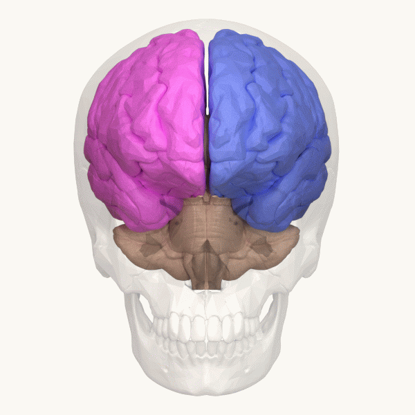
The lateralization of brain function (or hemispheric dominance[1][2]/ lateralization[3][4]) is the tendency for some neural functions or cognitive processes to be specialized to one side of the brain or the other. The median longitudinal fissure separates the human brain into two distinct cerebral hemispheres connected by the corpus callosum. Both hemispheres exhibit brain asymmetries in both structure and neuronal network composition associated with specialized function.

Lateralization of brain structures has been studied using both healthy and split-brain patients. However, there are numerous counterexamples to each generalization and each human's brain develops differently, leading to unique lateralization in individuals. This is different from specialization, as lateralization refers only to the function of one structure divided between two hemispheres. Specialization is much easier to observe as a trend, since it has a stronger anthropological history.[5]

The best example of an established lateralization is that of Broca's and Wernicke's areas, where both are often found exclusively on the left hemisphere. Function lateralization, such as semantics, intonation, accentuation, and prosody, has since been called into question and largely been found to have a neuronal basis in both hemispheres.[6] Another example is that each hemisphere in the brain tends to represent one side of the body. In the cerebellum, this is the ipsilateral side, but in the forebrain this is predominantly the contralateral side.

Lateralized functions

[edit]

Language and speech

[edit]

Language functions are lateralized to the left hemisphere in 96% of right-handers and 60% of left-handers.[7][8][9]

Meaning of words, called lexicon, is processed bilaterally which has been tested through the word superiority effect. This finding is consistent with the distributed memory and knowledge systems required for lexical entries; however, each hemisphere's lexicon is considered unique since it may be organized and accessed differently.[8] For example, the right hemisphere lacks letter recognition, and cannot judge lexical relationships such as superordinate words or antonyms.[8]

The permitted organization of words, called grammar, is lateralized in only one hemisphere, typically the left one. These functions include "understanding verbs, pluralizations, the possessive, and active-passive differences" and understanding changes in meaning due to word order.[8] However, the right hemisphere is able to judge when a sentence is grammatically correct, which may indicate that patterns of speech are learned by rote rather than applied through understanding rules.[8]

Speech production and language comprehension are specialized in Broca's and Wernicke's areas respectively, which are located in the left hemisphere for 96% of right-handers and 70% of left-handers.[8][10] However, there are some cases in which speech is produced in both hemispheres in split-brain patients, also lateralization can shift due to plasticity over time.[8] The emotional content of language, called emotional prosody, is right-lateralized.[8]

In writing, studies attempting to isolate the linguistic component of written language in terms of brain lateralization could not provide enough evidence of a difference in the relative activation of the brain hemispheres between left-handed and right-handed adults.[11]

Sensory processing

[edit]

Sensory processing for the left and right sides of the body is often lateralized to the contralateral hemisphere due to nerve fiber decussation.

Because of the functional division of the left and right sides of the body, the processing of information in the sensory cortices is essentially identical. That is, the processing of visual and auditory stimuli, spatial manipulation, facial perception, and artistic ability are represented bilaterally.[9] Numerical estimation, comparison and online calculation depend on bilateral parietal regions[12][13] while exact calculation and fact retrieval are associated with left parietal regions, perhaps due to their ties to linguistic processing.[12][13]

Vision

[edit]
Lateralization of the left and right visual hemifields due to decussation.

In vision, retinal ganglion cells undergo partial decussation at the optic chiasm, where axons from the nasal retinas cross to the opposite hemisphere, while axons from the temporal retinas remain on the ipsilateral side.[14][15] As a result, visual input from the left visual hemifields are processed by the right hemisphere's visual cortex, while input from the right visual hemifields are processed by the left hemisphere's visual cortex.[15]

Hearing

[edit]

In hearing, spiral ganglion neurons in the vestibulocochlear nerve project to the ipsilateral cochlear nuclei in the medulla.[15][16] However, second-order axons from the ventral cochlear nucleus branch to both the ipsilateral and contralateral superior olivary complexes.[15][16] Consequently, hearing is strongly lateralized only at the ipsilateral cochlear nuclei, while further processing in the inferior colliculi, the medial geniculate nucleus of the thalamus, and the auditory cortex occurs bilaterally with a slight contralateral dominance.[15][16] This lateralization explains why damage to one cochlear nucleus causes deafness in the ipsilateral ear, whereas damage above the cochlear nucleus typically results in only slight hearing loss.[15]

When tasked to repeat words in a dichotic listening task, individuals tend to say words played in their right ear, a phenomenon called right-ear advantage.[8] Since hearing is slightly contralateral dominant, this effect is consistent with the left hemisphere lateralization of language.[8] When tasked to recall melodies in a dichotic listening task, people instead tend to have a left-ear advantage.[8]

Touch

[edit]

In the somatosensory system, sensations of touch, vibration, pressure, pain, and temperature are primarily processed in the contralateral somatosensory cortex of the brain. Mechanoreceptors responsible for touch and vibration transmit signals through the dorsal column-medial lemniscal pathway, where they decussate at the dorsal column nuclei in the medulla before ascending.[15] Touch from the face and top of the head follows the trigeminal touch pathway, where second-order neurons decussate at the trigeminal nucleus.[15]

Pain and temperature signals from nociceptors travel a different pathway, the spinothalamic pathway, where second-order neurons decussate earlier in the spinal cord.[15] For pain and temperature in the face and top of the head, second-order neurons decussate at the spinal trigeminal nucleus of the brainstem.[15] The earlier decussation of pain signals compared to touch explains Brown-Séquard syndrome, a condition in which damage to one half of the spinal cord leads to ipsilateral insensitivity to touch but contralateral insensitivity to pain and temperature.[15]

Motor system

[edit]

Voluntary movement is lateralized to the contralateral motor cortex, so the right hemisphere controls the left side of the body, while the left hemisphere controls the right side.

In the two lateral pathways, the corticospinal tract is responsible for control of distal muscles and begins at the contralateral motor cortex or contralateral somatosensory areas, and decussates between the medulla and spinal cord.[15] The rubrospinal tract responsible for distal muscle and posture begins at the contralateral red nucleus and quickly decussates in the pons.[15]

In the four ventromedial pathways, the vestibulospinal tract responsible for head balance begins at the ipsilateral vestibular nucleus of the medulla and splits into a bilateral and ipsilateral path. The bilateral path controls neck and back muscles for head balance, while the ipsilateral path maintains upright posture of the legs.[15] The tectospinal tract responsible for orienting the head toward sensory stimuli begins at the contralateral superior colliculus and quickly decussates at the red nucleus.[15] The reticulospinal tracts responsible for controlling muscles against gravity begin at the ipsilateral reticular formation and do not decussate.

Value systems

[edit]

Rather than just being a series of places where different brain modules occur, there are running similarities in the kind of function seen in each side, for instance how right-side impairment of drawing ability making patients draw the parts of the subject matter with wholly incoherent relationships, or where the kind of left-side damage seen in language impairment not damaging the patient's ability to catch the significance of intonation in speech.[17] This has led British psychiatrist Iain McGilchrist to view the two hemispheres as having different value systems, where the left hemisphere tends to reduce complex matters such as ethics to rules and measures, and the right hemisphere is disposed to the holistic and metaphorical.[18]

Clinical significance

[edit]

Depression is linked with a hyperactive right hemisphere, with evidence of selective involvement in "processing negative emotions, pessimistic thoughts and unconstructive thinking styles", as well as vigilance, arousal and self-reflection, and a relatively hypoactive left hemisphere, "specifically involved in processing pleasurable experiences" and "relatively more involved in decision-making processes".[19] Additionally, "left hemisphere lesions result in an omissive response bias or error pattern whereas right hemisphere lesions result in a commissive response bias or error pattern."[20] The delusional misidentification syndromes, reduplicative paramnesia and Capgras delusion are also often the result of right hemisphere lesions.[21]

Lateral view of the Brain

Hemisphere damage

[edit]

Damage to either the right or left hemisphere, and its resulting deficits provide insight into the function of the damaged area. There is truth to the idea that some brain functions reside more on one side of the brain than the other. We know this in part from what is lost when a stroke affects a particular part of the brain. Left hemisphere damage has many effects on language production and perception. Damage or lesions to the right hemisphere can result in a lack of emotional prosody[22] or intonation when speaking.[23] The left hemisphere is often involved with dealing of detail-oriented perception while the right hemisphere deals mostly with wholeness or an overall concept of things.[23]

Right hemisphere damage also has grave effects on understanding discourse. People with damage to the right hemisphere have a reduced ability to generate inferences, comprehend and produce main concepts, and a reduced ability to manage alternative meanings. Furthermore, people with right hemisphere damage often exhibit discourse that is abrupt and perfunctory or verbose and excessive. They can also have pragmatic deficits in situations of turn taking, topic maintenance and shared knowledge. .[23] Although both sides of the hemisphere has different responsibilities and tasks, they both complete each other and create a bigger picture.[23] Lateral brain damage can also affect visual perceptual spatial resolution. People with left hemisphere damage may have impaired perception of high resolution, or detailed, aspects of an image. People with right hemisphere damage may have impaired perception of low resolution, or big picture, aspects of an image.

Plasticity

[edit]

If a specific region of the brain, or even an entire hemisphere, is injured or destroyed, its functions can sometimes be assumed by a neighboring region in the same hemisphere or the corresponding region in the other hemisphere, depending upon the area damaged and the patient's age.[24] When injury interferes with pathways from one area to another, alternative (indirect) connections may develop to communicate information with detached areas, despite the inefficiencies.

Broca's aphasia

[edit]

Broca's aphasia is a specific type of expressive aphasia and is so named due to the aphasia that results from damage or lesions to the Broca's area of the brain, that exists most commonly in the left inferior frontal hemisphere. Thus, the aphasia that develops from the lack of functioning of the Broca's area is an expressive and non-fluent aphasia. It is called 'non-fluent' due to the issues that arise because Broca's area is critical for language pronunciation and production. The area controls some motor aspects of speech production and articulation of thoughts to words and as such lesions to the area result in specific non-fluent aphasia.[25]

Wernicke's aphasia

[edit]

Wernicke's aphasia is the result of damage to the area of the brain that is commonly in the left hemisphere above the Sylvian fissure. Damage to this area causes primarily a deficit in language comprehension. While the ability to speak fluently with normal melodic intonation is spared, the language produced by a person with Wernicke's aphasia is riddled with semantic errors and may sound nonsensical to the listener. Wernicke's aphasia is characterized by phonemic paraphasias, neologism or jargon. Another characteristic of a person with Wernicke's aphasia is that they are unconcerned by the mistakes that they are making.

Society and culture

[edit]

Possible misapplication

[edit]
Gross oversimplification of hemisphericity
Oversimplification of hemisphericity

The concept of "right-brained" or "left-brained" individuals is considered a widespread myth which oversimplifies the true nature of the brain's cerebral hemispheres. Proof leading to the "mythbuster" of the left-/right-brained concept is increasing as more and more studies are brought to light. Harvard Health Publishing includes a study from the University of Utah in 2013, that exhibited brain scans revealing similarity on both sides of the brain, personality and environmental factors aside.[26] Although certain functions show a degree of lateralization in the brain—with language predominantly processed in the left hemisphere, and spatial and nonverbal reasoning in the right—these functions are not exclusively tied to one hemisphere.[27]

Terence Hines states that the research on brain lateralization is valid as a research program, though commercial promoters have applied it to promote subjects and products far outside the implications of the research.[28] For example, the implications of the research have no bearing on psychological interventions such as eye movement desensitization and reprocessing (EMDR) and neurolinguistic programming,[29][30] brain-training equipment, or management training.[31]

[edit]
Oversimplification of lateralization in pop psychology. This belief was widely held even in the scientific community for some years.

Some popularizations oversimplify the science about lateralization, by presenting the functional differences between hemispheres as being more absolute than is actually the case.[32]: 107 [33] Interestingly, research has shown quite opposite function of brain lateralisation, i.e. right hemisphere creatively and chaotically links between concepts and left hemisphere tends to adhere to specific date and time, although generally adhering to the pattern of left-brain as linguistic interpretation and right brain as spatio-temporal.[34][unreliable source][35]

Sex differences

[edit]

In the 19th century and to a lesser extent the 20th, it was thought that each side of the brain was associated with a specific gender: the left corresponding with masculinity and the right with femininity and each half could function independently.[36] The right side of the brain was seen as the inferior and thought to be prominent in women, savages, children, criminals, and the insane. A prime example of this in fictional literature can be seen in Robert Louis Stevenson's Strange Case of Dr. Jekyll and Mr. Hyde.[37]

History

[edit]

Broca

[edit]

One of the first indications of brain function lateralization resulted from the research of French physician Pierre Paul Broca, in 1861. His research involved the male patient nicknamed "Tan", who had a speech deficit (aphasia); "tan" was one of the few words he could articulate, hence his nickname. In Tan's autopsy, Broca determined he had a syphilitic lesion in the left cerebral hemisphere. This left frontal lobe brain area (Broca's area) is an important speech production region. The motor aspects of speech production deficits caused by damage to Broca's area are known as expressive aphasia. In clinical assessment of this type of aphasia, patients have difficulty producing speech.[38]

Wernicke

[edit]

German physician Karl Wernicke continued in the vein of Broca's research by studying language deficits unlike expressive aphasia. Wernicke noted that not every deficit was in speech production; some were linguistic. He found that damage to the left posterior, superior temporal gyrus (Wernicke's area) caused language comprehension deficits rather than speech production deficits, a syndrome known as receptive aphasia.

Imaging

[edit]

These seminal works on hemispheric specialization were done on patients or postmortem brains, raising questions about the potential impact of pathology on the research findings. New methods permit the in vivo comparison of the hemispheres in healthy subjects. Particularly, magnetic resonance imaging (MRI) and positron emission tomography (PET) are important because of their high spatial resolution and ability to image subcortical brain structures.

Movement and sensation

[edit]

In the 1940s, neurosurgeon Wilder Penfield and his neurologist colleague Herbert Jasper developed a technique of brain mapping to help reduce side effects caused by surgery to treat epilepsy. They stimulated motor and somatosensory cortices of the brain with small electrical currents to activate discrete brain regions. They found that stimulation of one hemisphere's motor cortex produces muscle contraction on the opposite side of the body. Furthermore, the functional map of the motor and sensory cortices is fairly consistent from person to person; Penfield and Jasper's famous pictures of the motor and sensory homunculi were the result.

Split-brain patients

[edit]

Research by Michael Gazzaniga and Roger Wolcott Sperry in the 1960s on split-brain patients led to an even greater understanding of functional laterality. Split-brain patients are patients who have undergone corpus callosotomy (usually as a treatment for severe epilepsy), a severing of a large part of the corpus callosum. The corpus callosum connects the two hemispheres of the brain and allows them to communicate. When these connections are cut, the two halves of the brain have a reduced capacity to communicate with each other. This led to many interesting behavioral phenomena that allowed Gazzaniga and Sperry to study the contributions of each hemisphere to various cognitive and perceptual processes. One of their main findings was that the right hemisphere was capable of rudimentary language processing, but often has no lexical or grammatical abilities.[39] Eran Zaidel also studied such patients and found some evidence for the right hemisphere having at least some syntactic ability.[citation needed]

Language is primarily localized in the left hemisphere. While the left hemisphere has proven to be more optimized for language, the right hemisphere has the capacity with emotions, such as sarcasm, that can express prosody in sentences when speaking. According to Sheppard and Hillis, "The right hemisphere is critical for perceiving sarcasm (Davis et al., 2016), integrating context required for understanding metaphor, inference, and humour, as well as recognizing and expressing affective or emotional prosody—changes in pitch, rhythm, rate, and loudness that convey emotions".[40] One of the experiments carried out by Gazzaniga involved a split-brain male patient sitting in front of a computer screen while having words and images presented on either side of the screen, and the visual stimuli would go to either the right or left visual field, and thus the left or right brain, respectively. It was observed that if the patient was presented with an image to his left visual field (right brain), he would report not seeing anything. If he was able to feel around for certain objects, he could accurately pick out the correct object, despite not having the ability to verbalize what he saw.

Additional images

[edit]

See also

[edit]

References

[edit]
[edit]

Bibliography

[edit]

Further resources

[edit]
Revisions and contributorsEdit on WikipediaRead on Wikipedia
from Grokipedia
Lateralization of brain function refers to the specialization of the left and right cerebral hemispheres for distinct cognitive, sensory, and motor processes, enabling more efficient parallel processing of information across species from to humans. This functional allows the to perform multiple tasks simultaneously, such as monitoring for threats while , thereby enhancing overall cognitive capacity without duplicating neural resources. In humans, lateralization is particularly pronounced, with the left hemisphere typically dominating and analytical tasks, while the right hemisphere excels in visuospatial processing and emotional recognition. The concept of brain lateralization emerged in the 19th century through clinical observations, notably Paul Broca's 1861 identification of a left-hemisphere in a patient with impaired speech, establishing the link between the left frontal lobe and . Research stagnated mid-20th century under the assumption that such asymmetries were uniquely human, but comparative studies in the revived interest, demonstrating lateralization in non-human animals like songbirds and chicks through behavioral and experiments. Advances in , such as fMRI, have since revealed two distinct forms of lateralization in the : segregated processing in the left hemisphere, favoring intra-hemispheric connections for sequential tasks like speech, and integrated processing in the right hemisphere, emphasizing bilateral interactions for holistic tasks like . Key functional asymmetries in humans include , with approximately 90% of individuals right-handed and corresponding left-hemisphere , as well as hemispheric dominance in —such as the right hemisphere's role in face recognition and the left's in detailed object . These specializations provide evolutionary advantages, including improved social coordination and problem-solving, as evidenced by stronger lateralization correlating with better performance in dual-task scenarios across vertebrates. Evolutionarily, humans exhibit greater variability in endocranial asymmetry compared to great apes, suggesting enhanced neural plasticity that supports advanced . Disruptions in lateralization, such as in patients, underscore its role in unified conscious experience, while developmental factors like and environment further modulate these patterns.

Anatomy and Mechanisms of Lateralization

Cerebral Hemisphere Organization

The human 's consists of two , the left and right, separated by the . Each hemisphere is covered by a thin layer of gray matter known as the , which has a highly folded surface characterized by ridges called gyri and grooves called sulci; these folds increase the total cortical surface area to approximately 2,500 cm² across both hemispheres, allowing for greater neuronal density within the confined space of the . The cortex is divided into four main lobes per hemisphere: the , located at the front and involved in such as planning and decision-making; the , situated behind the frontal lobe and responsible for integrating sensory information; the , positioned on the sides and associated with memory formation; and the , at the rear and dedicated to processing visual input. A fundamental organizational principle of the cerebral hemispheres is contralateral control, whereby the left hemisphere primarily governs motor and sensory functions on the right side of the body, and the right hemisphere controls the left side. This crossed organization arises from the decussation of neural pathways in the and , optimizing coordinated perception and movement across the body's midline. The two hemispheres are interconnected primarily by the , a bundle of fibers that facilitates between them. Inherent structural asymmetries exist between the hemispheres, contributing to functional lateralization. For instance, the , a region in the , is typically larger in the left hemisphere, as observed in and postmortem studies. Additionally, in right-handed individuals—who comprise about 90% of the population—the left hemisphere often exhibits a slightly larger overall volume compared to the right, based on MRI and postmortem analyses. These asymmetries, particularly pronounced in core regions like the for executive processing and the for memory, underscore the brain's specialized hemispheric organization.

Interhemispheric Connections

The serves as the primary interhemispheric commissure, comprising approximately 200 million myelinated axons that facilitate communication between the left and right cerebral hemispheres. This massive bundle of tracts connects homologous cortical regions, enabling the integration of sensory, motor, and cognitive processes across the . Diffusion tensor imaging (DTI) studies have revealed regional variations in axonal density, with higher concentrations of smaller-diameter fibers (<0.4 μm) in anterior segments linking prefrontal and temporo-parietal associative areas involved in language processing, while larger-diameter fibers (3-5 μm) predominate in posterior regions for sensory integration. Anatomically, the corpus callosum is divided into seven segments along its anterior-posterior axis: the rostrum, genu, rostral body, anterior midbody, posterior midbody, , and splenium. The rostrum and genu primarily interconnect the orbital and prefrontal cortices, supporting through the forceps minor. The rostral and anterior midbody link supplementary motor and premotor areas in the frontal lobes, while the posterior midbody and connect somatosensory and auditory regions in the parietal and temporal lobes. The splenium, the thickest posterior portion, joins the occipital lobes via the forceps major, facilitating visual processing. These segmental tracts ensure targeted homologous connections, with overall highest in the and splenium as quantified by DTI . Functionally, the integrates interhemispheric information, such as transferring visual data from the contralateral via the splenium or tactile stimuli from somatosensory areas through the posterior midbody, allowing unified despite contralateral hemispheric organization. In its absence, as in callosal , this integration is disrupted, leading to enhanced unilateral processing where each hemisphere operates more independently. For instance, functional MRI studies of individuals with callosal dysgenesis, including cases of complete , demonstrate strictly contralateral activation in primary and secondary somatosensory cortices during tactile tasks, with no ipsilateral engagement—contrasting bilateral responses in controls—and preserved task performance via compensatory subcortical or ipsilateral pathways. Similar patterns emerge in language processing, where cases often exhibit robust left-hemisphere dominance without interhemispheric transfer, underscoring the callosum's role in modulating lateralization through inhibitory influences on the nondominant hemisphere.

Developmental and Genetic Basis

Brain lateralization begins to emerge during fetal development, with structural asymmetries detectable as early as the third week of through molecular and cellular interactions that establish left-right patterning. In human fetuses, one prominent asymmetry is the , characterized by a rightward frontal and leftward occipital petalia, which may arise from interactions between visceral organ positioning and development, including the rightward looping of the heart that influences overall body laterality. Studies of fetuses with lateralization defects, such as , reveal altered brain asymmetries, underscoring the prenatal origins of these patterns. These early asymmetries continue to evolve , with certain sulci appearing earlier in the right hemisphere, setting the stage for functional specialization. Postnatally, brain lateralization refines progressively, with significant maturation occurring by ages 5 to 7 years, coinciding with the stabilization of dominance and . Functional imaging studies show that right-hemisphere activation for decreases systematically during childhood, leading to stronger left-hemisphere dominance by school age, while tracts like the arcuate fasciculus exhibit time-sensitive lateralization during infancy. This period marks a critical window for hemispheric specialization, after which plasticity diminishes, as evidenced by poorer reorganization outcomes following right-hemisphere strokes after age 5. Genetic factors play a substantial role in brain lateralization, with heritability estimates for around 25% based on large twin and family studies. Specific genes, such as LRRTM1 on 2p12, have been linked to handedness and language lateralization through paternal inheritance effects, potentially influencing neuronal connectivity and asymmetry in cortical regions. Twin studies indicate low to moderate for language lateralization, with estimates ranging from 0% to 31% depending on the measure and sample. Environmental influences, particularly prenatal exposure to testosterone, correlate with variations in lateralization. Lower fetal testosterone levels are associated with a higher likelihood of atypical language lateralization, such as right-hemisphere dominance, as observed in studies measuring hormones. Incomplete or reduced lateralization is notably prevalent in conditions like , where failure of left-hemisphere dominance for phonological disrupts typical , often linked to shared genetic vulnerabilities with traits.

Lateralized Functions

Language and Communication

Language and communication exhibit strong hemispheric lateralization, with the left hemisphere dominant for core linguistic processes in approximately 95% of right-handed individuals. This dominance supports the production and comprehension of spoken and written , primarily through specialized regions in the left . , located in the left , plays a key role in , facilitating the articulation and grammatical structuring of speech. In contrast, , situated in the left posterior , is essential for comprehension, enabling the interpretation of semantic content and auditory-verbal input. The right hemisphere contributes to communicative functions beyond basic syntax and semantics, particularly in processing prosody—the rhythmic and intonational aspects of speech that convey emotional tone—and non-literal such as metaphors. Damage or disruption to right-hemispheric regions can impair the recognition of affective prosody, leading to flattened or misinterpreted in communication, while left-hemispheric processes handle propositional content. Functional models of language lateralization have evolved from the classical Wernicke-Geschwind model, which posits a left-hemispheric network connecting for comprehension to for production via the arcuate fasciculus. This model has been updated by the dual-stream framework, incorporating ventral pathways for semantic processing (mapping sound to meaning) and dorsal pathways for syntactic and phonological integration (supporting sound-to-articulation mapping). (fMRI) studies confirm this asymmetry, revealing robust activation in the left during speech tasks, indicating predominant left-hemispheric engagement.

Visual Processing

Visual processing exhibits notable hemispheric asymmetries, particularly in how the handles information from the left and right visual hemifields. The left , projecting primarily to the right hemisphere, is specialized for processing global and spatial features of visual stimuli, as demonstrated in the Navon global-local paradigm where global precedence effects are stronger when stimuli are presented to the left . In contrast, the right , projecting to the left hemisphere, favors local and detail-oriented analysis, with faster reaction times for identifying fine-grained elements in compound stimuli. The right hemisphere demonstrates superiority in recognizing faces and navigating spatial environments. Functional imaging and divided visual field studies show higher accuracy for face identity matching when faces are presented to the left visual field (66.32% accuracy) compared to the right (57.10%), persisting across manipulations like inversion and spatial fragmentation. Similarly, electrocorticography recordings during virtual navigation reveal right-lateralized gamma oscillations in neocortical regions, indicating the right hemisphere's dominant role in processing spatial layouts and route integration. Conversely, the left hemisphere excels in object identification, particularly for manipulable items, with a right visual field advantage in priming tasks (13.12 ms faster response) reflecting left ventral occipitotemporal activation for semantic categorization. These asymmetries arise from lateralized biases in the two major visual pathways. The ventral stream, or "what" pathway, extending from occipital to temporal lobes, shows a left-hemisphere bias for detailed , as evidenced by stronger activations in left regions for tool identification. The dorsal stream, or "where" pathway, projecting to parietal lobes, exhibits right-hemisphere dominance for spatial relations, with segregated activations in right superior parietal cortex for contralateral field processing. A key indicator of right-hemisphere dominance in visuospatial is the neglect syndrome, where lesions disrupt awareness of the left visual space, hinting at the right hemisphere's overarching role in allocating across both hemifields. (PET) scans confirm this through asymmetric activations: the right superior parietal cortex responds to stimuli in both visual fields, while the left responds primarily to the right field, underscoring the right hemisphere's broader . This lateralization extends briefly to reading, where left-hemisphere ventral integrates visual form with linguistic analysis.

Auditory Processing

Auditory processing exhibits notable hemispheric lateralization, with the left hemisphere showing a toward rapid temporal features of sound, such as the quick acoustic changes in speech , while the right hemisphere specializes in features, including pitch and structure relevant to . This asymmetry in temporal versus processing is encapsulated in the asymmetric sampling in time (AST) theory, which posits that the left integrates information over shorter temporal windows (around 20-50 ms) suited for phonetic segmentation, whereas the right uses longer windows (150-300 ms) for coarser spectral analysis like melody perception. Empirical evidence from functional magnetic resonance imaging (fMRI) supports this, demonstrating stronger left and activation for temporal manipulations in speech, contrasted with right-hemisphere dominance for alterations. A key anatomical correlate of this functional divide is the asymmetry in the , particularly the , which is larger in the left hemisphere in approximately 65% of individuals and supports phonetic analysis essential for sound decoding. This leftward enlargement, averaging one-third greater than the right counterpart, aligns with the region's role in processing fine-grained temporal cues in verbal auditory input, contributing to the left hemisphere's overall specialization for linguistic auditory features. Dichotic listening tasks, where different sounds are presented simultaneously to each ear, further illustrate this lateralization through a consistent right-ear advantage for verbal stimuli, reflecting preferential routing to the left hemisphere via contralateral auditory pathways. In such experiments, participants achieve higher accuracy (e.g., 73.6% for the right ear versus 64.2% for the left) when identifying syllables or words, with neurophysiological measures like auditory steady-state responses confirming enhanced left activity for right-ear input. Electroencephalography (EEG) studies reveal distinct oscillatory patterns underscoring these specializations: the right hemisphere exhibits increased theta-band (4-8 Hz) activity during the processing of musical emotion, such as pleasantness evoked by familiar tunes, involving fronto-temporal . In contrast, left-hemisphere gamma-band oscillations (30-50 Hz) are prominent for linguistic prosody, facilitating the integration of prosodic cues with phonetic content in speech comprehension. This oscillatory distinction highlights how auditory lateralization not only segregates perceptual attributes but also supports differentiated emotional and communicative roles in sound processing.

Somatosensory Processing

The somatosensory cortex, located in the of the , primarily exhibits contralateral mapping, where sensory input from one side of the body is processed in the opposite . This organization ensures localized representation of touch, pressure, temperature, and across the body surface in a somatotopic fashion. However, hemispheric asymmetries emerge in higher-order integration within the s, particularly in the and intraparietal regions, where the right shows greater involvement in spatial aspects of somatosensory processing compared to the left. Hemispheric differences are evident in tactile and . The right hemisphere demonstrates dominance for spatial tactile , facilitating the allocation of focus across broader spatial extents of the body surface, as supported by faster reaction times and enhanced gamma-band activity in right-hemispheric networks during spatially selective touch tasks. This specialization aligns with the right hemisphere's role in global attentional orienting, extending to somatosensory modalities. In contrast, the left hemisphere excels in fine discriminative touch, such as detecting subtle textures or grating orientations, with behavioral advantages observed when stimuli are presented to the right hand and associated with left-hemispheric analytical processing of detailed spatial features. Proprioception, the sense of body position and movement, also exhibits right-hemispheric predominance. Functional MRI studies reveal stronger activations in right-hemisphere regions, including the , insula, and , during knee joint position sense tasks, irrespective of the stimulated limb, indicating a lateralized network for integrating proprioceptive signals with spatial awareness. This asymmetry underscores the right parietal lobe's contribution to constructing a coherent in space. Pain processing involves bilateral engagement of cortical networks, including the insula and , to handle sensory-discriminative aspects from noxious stimuli. However, emotional and affective components of show right-hemispheric lateralization, particularly in the right anterior insula, which integrates salience detection and emotional valuation of painful experiences, as evidenced by consistent right-sided activations in across stimulation sides. Tactile extinction tasks, where simultaneous stimuli are applied to both hands and one side is ignored, further highlight right-hemispheric superiority in . Left-hand (contralesional) is more prevalent following right-hemisphere damage, reflecting impaired spatial rather than primary , with 2010s fMRI data showing reduced right parietal activation during bilateral tactile detection in affected patients. This pattern supports models of asymmetric attentional networks, where the right hemisphere monitors both hemifields while the left focuses ipsilaterally.

Motor Control

In motor control, the brain exhibits hemispheric specialization that influences movement planning, execution, and coordination, with the left hemisphere generally dominating sequenced and fine motor skills, particularly in right-handed individuals. For instance, in right-handers, the left hemisphere shows enhanced involvement in tasks requiring precise, sequential actions such as writing or manipulating tools, reflecting a predictive control mechanism that facilitates learning and adaptation of complex motor sequences. This lateralization aligns with , where approximately 90% of the population is right-handed, correlating with left-hemisphere dominance for motor preparation and execution of the right side of the body. Conversely, the right hemisphere contributes to gross and spatial movements, such as those involved in or postural stability, by supporting impedance control to update actions in response to environmental perturbations and ensure accurate goal positioning. The , located in the , primarily exerts contralateral control over voluntary movements, meaning the left motor cortex predominantly governs the right side of the body and vice versa, forming the basis for lateralized output in skilled actions. This contralateral organization is supplemented by asymmetries in the (SMA), where the left SMA demonstrates dominant connectivity to sensorimotor regions during unilateral finger movements in right-handers, influencing both hands but with heightened modulation for the right hand during fine tasks. The right SMA, while active, shows less pronounced hand-dependent influences, underscoring the left hemisphere's overarching role in coordinating bilateral aspects of motor planning. These asymmetries ensure efficient integration of predictive planning (left-dominant) with reactive stabilization (right-dominant) for seamless movement execution. Transcranial magnetic stimulation (TMS) studies further elucidate these specializations, revealing left-hemisphere excitation during tool use pantomimes, where disruption of left premotor areas impairs movement preparation more than right-sided stimulation, independent of handedness. In contrast, right-hemisphere mechanisms, probed through motor control paradigms including TMS, support postural adjustments by modulating sensorimotor reflexes for stability and error correction in spatial tasks.

Emotional and Value Systems

The lateralization of emotional processing in the brain follows the valence hypothesis, which posits that positive emotions and approach-related s are predominantly handled by the left hemisphere, while negative emotions and withdrawal-related responses are biased toward the right hemisphere. This model, supported by meta-analyses of data, indicates region-specific asymmetries rather than a general right-hemisphere dominance for all emotions. For instance, the left shows greater activation during appetitive and reward-seeking behaviors, facilitating approach motivation, whereas the right is more involved in avoidant and inhibitory responses to threats. The right hemisphere exhibits a particular bias for processing negative emotions such as and , mediated through structures like the and insula. The , especially its right counterpart, demonstrates heightened responsiveness to fear-inducing stimuli, contributing to rapid threat detection and emotional arousal. Similarly, the insula, with right-lateralized activation, integrates visceral sensations with negative affective states, enhancing the subjective experience of or anxiety. In contrast, positive emotions like or enthusiasm correlate with left-hemisphere activity, particularly in frontal regions that support motivational engagement with rewarding outcomes. Emotional lateralization extends to value systems, where asymmetries in the (OFC) influence and . The right lateral OFC shows stronger connectivity to networks involved in evaluating potential losses and risks, correlating with avoidance behaviors during uncertain reward scenarios. This asymmetry facilitates differential processing of high-risk versus low-risk options, with right OFC hyperactivity linked to conservative choices in value-based judgments. underscoring its role in affective tone detection.

Clinical Implications

Hemispheric Damage Effects

Unilateral damage to the cerebral hemispheres disrupts the specialized functions typically lateralized to each side, leading to distinct patterns of cognitive, perceptual, and behavioral impairments. Left-hemisphere lesions often impair analytical and verbal processes, while right-hemisphere lesions more frequently affect holistic, spatial, and emotional integration. These effects are most evident in patients, where the location of the lesion correlates with specific deficit profiles, influencing rehabilitation strategies and long-term outcomes. Damage to the left hemisphere commonly results in impairments of and comprehension, , and sequential processing tasks, such as arithmetic or step-by-step problem-solving. For instance, lesions in perisylvian regions disrupt speech output and grammatical structure, contributing to expressive and receptive deficits. These cognitive disruptions extend to difficulties in organizing linearly, affecting tasks requiring deduction or temporal sequencing. In contrast, right-hemisphere damage typically produces deficits in visuospatial attention, holistic perception, and emotional processing, including spatial neglect and reduced affective responsiveness. Patients may exhibit emotional blunting, characterized by indifference or flattened affect, due to impaired integration of emotional cues from nonverbal signals like prosody or facial expressions. This can manifest as diminished or inappropriate social responses, highlighting the right hemisphere's role in emotional valence and context. Hemispatial neglect, a profound attentional deficit where patients ignore stimuli on the contralesional (usually left) side of space, arises primarily from right lesions following . This syndrome affects approximately 30-50% of patients with acute right-hemisphere strokes, with prevalence estimates around 40% in large cohorts, severely impacting daily activities like dressing or . is less common and milder after left-hemisphere damage, underscoring the right hemisphere's dominance in spatial awareness. Anosognosia, the lack of awareness of one's own deficits, occurs more frequently after right-hemisphere damage than left, with rates up to 54% in right-sided strokes compared to about 9% on the left. This unawareness often accompanies or hemiplegia, complicating patient compliance with therapy as individuals deny their impairments despite objective evidence. Lesions in right frontal and parietal networks disrupt mechanisms, exacerbating functional limitations. Epidemiological data from stroke registries indicate that left-hemisphere lesions are associated with higher rates of , affecting up to one-third of survivors overall but predominantly those with left-sided damage, while right-hemisphere lesions lead to visuospatial deficits like in 35-61% of cases. Recent analyses confirm this , with right-sided s showing visuospatial impairment in about 61% of territories versus 22% for left-sided equivalents, influencing and in clinical settings.

Language Disorders

Language disorders, particularly , arise from damage to the lateralized networks predominantly in the left , disrupting the production, comprehension, or repetition of . These deficits highlight the functional of brain regions specialized for linguistic processing, where left-hemisphere lesions account for the majority of cases in right-handed individuals. Broca's aphasia, also known as non-fluent or , results from damage to the left , often due to ischemic stroke or trauma in this region. It is characterized by effortful, halting with impaired and , while comprehension of remains relatively preserved. Patients typically produce short, telegraphic phrases lacking function words and inflections, reflecting a core deficit in articulatory planning and syntactic formulation. Wernicke's aphasia, or , stems from lesions in the left posterior , impairing the understanding of both spoken and . Speech output is fluent and effortless but consists of paraphasic errors, neologisms, and semantically empty jargon, often described as "," due to disrupted phonological and semantic processing. Comprehension is severely impaired, leading to where patients may be unaware of their deficits. Conduction aphasia occurs from lesions to the arcuate fasciculus, the tract connecting the frontal and temporal language areas in the left hemisphere, resulting in a . It features fluent speech with good comprehension but markedly impaired repetition of words and sentences, accompanied by phonemic paraphasias and errors in reading aloud. This pattern underscores the role of the arcuate fasciculus in transferring phonological information between comprehension and production centers. Among post-stroke aphasias, Broca's aphasia occurs in approximately 13-18% of cases involving left-hemisphere damage, with overall aphasia incidence ranging from 21-38% in acute patients. Recovery in Broca's aphasia varies significantly with lesion size and location, as revealed by 2020s lesion-symptom mapping studies showing that smaller, more focal damage to the predicts better language restoration outcomes.

Plasticity and Recovery

The exhibits remarkable plasticity following disruptions to lateralized functions, allowing reorganization to support recovery from hemispheric damage such as stroke-induced . This plasticity involves adaptive changes in surviving neural networks, where perilesional areas in the damaged hemisphere and homologous regions in the contralesional hemisphere can assume or enhance processing roles previously dominated by the left hemisphere. Such reorganization is evidenced by studies showing increased activation in these areas correlating with improved outcomes. Perilesional regions surrounding the in the left hemisphere play a key role in recovery by reacquiring language functions through mechanisms like and activity-dependent plasticity, with greater activation in areas such as the left predicting better performance in nonfluent . The contralesional right hemisphere contributes via homotopic recruitment, particularly when left-sided s are extensive, as seen in fMRI evidence of right inferior frontal and temporal activations supporting naming and comprehension improvements. For instance, post-left hemisphere damage, right-hemisphere homologues often show upregulated activity that aids language recovery, though this role can vary by size and may sometimes reflect maladaptive inhibition if not modulated. Therapies like noninvasive brain stimulation (e.g., repetitive targeting the right pars triangularis) and intensive language training enhance these processes by promoting left-hemisphere reactivation and reducing contralesional interference, leading to measurable gains in naming and sentence processing. Age significantly influences plasticity and recovery potential, with younger brains demonstrating greater adaptability due to higher . In pediatric arterial ischemic , neonates exhibit the highest rates of normal long-term neurologic outcomes (approximately 58% normal), while outcomes worsen with increasing age—adjusted odds of abnormal outcomes rise to 2.91 in and 4.46 in middle/late childhood compared to neonates—highlighting enhanced plasticity in early development. In adults, recovery is more limited, but longitudinal fMRI studies of intensive therapies, such as those akin to constraint-induced approaches, show that 60-70% of patients with chronic display increased right-hemisphere activation post-treatment, correlating with domain-specific improvements in functions like sentence comprehension. These findings underscore how therapeutic interventions can leverage age-dependent plasticity to optimize contralesional and perilesional reorganization for better functional restoration.

Research Methods and History

Early Anatomical Discoveries

The foundations of understanding brain lateralization for language function were laid in the through clinical observations of patients with , where lesions in specific brain regions correlated with speech impairments. French physician Marc Dax was among the earliest to propose hemispheric asymmetry, drawing from approximately 40 personal cases and 40 cases reported in the . In a presented in 1836 to the Southern Medical Society in , Dax concluded that disturbances in speech articulation consistently coincided with damage to the left , even when the right hemisphere was intact, suggesting a dominant role for the left side in . This idea gained prominence two decades later through the work of French anthropologist and neurologist , who conducted detailed post-mortem examinations of aphasic patients. In April 1861, Broca encountered Louis Victor Leborgne, a 51-year-old patient known as "Tan" due to his sole intelligible utterance, who exhibited severe but preserved comprehension. Following Leborgne's death shortly thereafter, Broca's revealed a syphilitic lesion confined to the posterior of the left hemisphere (Brodmann areas 44 and 45), sparing the right hemisphere. This finding, presented at the Société d'Anthropologie de , led Broca to identify this region—now termed —as the critical center for articulated speech, marking a pivotal advancement in localizationist theory that attributed specific cognitive functions to discrete brain areas. Building on Broca's motor-focused localization, German neurologist extended the model to sensory aspects of in 1874. Examining patients with fluent but incomprehensible speech, Wernicke identified lesions in the posterior of the left hemisphere, which he designated as a sensory speech center responsible for comprehension. In his seminal monograph Der aphasische Symptomencomplex, Wernicke described how damage here produced , where individuals could speak volubly but failed to understand or derive meaning from words, distinguishing it from Broca's expressive deficits and reinforcing left-hemispheric dominance for both production and of .

Split-Brain Experiments

In the 1960s, neuroscientist Roger Sperry, along with collaborator , conducted pioneering studies on patients who had undergone callosotomy surgery—a procedure that severs the to alleviate severe symptoms by preventing seizure spread between hemispheres. These experiments adapted animal model techniques to humans, revealing the functional independence of the cerebral hemispheres when interhemispheric communication was disrupted. By presenting stimuli selectively to one —left field processed by the right hemisphere and right field by the left—researchers demonstrated that each hemisphere could perceive, process, and respond to information without the other's awareness. A hallmark method in these studies was the use of chimeric face tasks, where composite images combined emotional and neutral halves (e.g., a fearful left half and neutral right half). When flashed briefly to patients, the right hemisphere, receiving input from the left , accurately recognized and matched the using the left hand, even as the verbally dominant left hemisphere, processing the right , reported only the neutral aspect. This dissociation highlighted the right hemisphere's superior role in emotional and facial recognition, independent of the left's analytical processing. The findings carried profound implications for understanding , suggesting that without the , the hemispheres operated as semi-autonomous systems with separate perceptual streams and no cross-transfer of , leading to situations where patients appeared unaware of one hemisphere's experiences. For instance, objects identified by the right hemisphere via left-hand manipulation could not be named aloud, as remained confined to the left hemisphere. In right-handed individuals, these experiments confirmed near-complete (approaching 100%) left-hemisphere lateralization for functions, persisting even after severance. Sperry's contributions earned him the 1981 in Physiology or for elucidating hemispheric specialization.

Neuroimaging and Modern Techniques

Modern neuroimaging techniques have revolutionized the study of brain lateralization by providing non-invasive methods to map functional and structural asymmetries across populations, extending beyond historical lesion-based approaches. Functional magnetic resonance imaging (fMRI) and positron emission tomography (PET) are pivotal for detecting activation asymmetries during cognitive tasks, revealing hemispheric differences in neural engagement. In fMRI, the blood oxygenation level-dependent (BOLD) signal is commonly analyzed using laterality indices (LI), calculated as the difference in activation between hemispheres normalized by their sum, to quantify language dominance with values ranging from -1 (right-lateralized) to +1 (left-lateralized). For instance, studies employing verb generation tasks show robust left-hemispheric BOLD activation in the inferior frontal gyrus for right-handers, with LI values often exceeding 0.5, confirming typical language lateralization. PET complements fMRI by measuring metabolic activity or neurotransmitter binding, such as glucose uptake asymmetries in temporal lobes during auditory processing, though it is less frequently used due to radiation exposure. Recent comparative analyses demonstrate high concordance between fMRI BOLD asymmetries and PET-derived patterns in semantic tasks, with both modalities validating leftward biases in over 90% of healthy adults. Electroencephalography (EEG) and magnetoencephalography (MEG) offer superior , on the order of milliseconds, to capture dynamic lateralization in auditory and language processing, where fMRI's spatial focus falls short. EEG spectral analysis during semantic tasks detects alpha power asymmetries in frontal and temporal regions, with left-hemispheric desynchronization indicating engagement, achieving reliable lateralization classification in 85-90% of cases. MEG, by measuring magnetic fields from neuronal currents, excels in mapping evoked responses to auditory stimuli, such as , revealing earlier right-hemispheric dominance for prosodic processing (around 100-200 ms post-stimulus) followed by leftward shifts for phonological analysis. Optimization studies using in MEG for picture-naming tasks report laterality indices aligned with invasive , supporting its utility in presurgical planning for patients with atypical lateralization. Diffusion magnetic resonance imaging (dMRI) elucidates structural underpinnings of lateralization through tract asymmetries, particularly in the arcuate fasciculus, which connects frontal and temporal areas. Tractography analyses consistently show leftward asymmetry in (FA) of the arcuate fasciculus in right-handers, with higher left FA values correlating to stronger lateralization, as measured by tasks. For example, volume and FA asymmetries in this tract are more pronounced in adults than children, emerging around age 5-7 and stabilizing by , underpinning developmental shifts in hemispheric specialization. These structural markers predict functional outcomes, such as reduced left arcuate integrity in individuals with weaker lateralization. Advancements in have enhanced neuroimaging's predictive power for lateralization-related disorders. models applied to fMRI data, such as convolutional neural networks analyzing BOLD patterns in reading networks, classify with 94.8% accuracy by identifying atypical asymmetry in temporoparietal regions, facilitating early screening. This AI integration, often using features like indices from resting-state scans, outperforms traditional thresholds and supports personalized interventions by quantifying lateralization strength.

Sociocultural Perspectives

One of the most persistent popular misconceptions about brain lateralization is the notion that individuals are either "left-brained" (logical, analytical, and sequential) or "right-brained" (creative, intuitive, and holistic), a dichotomy that emerged in the 1960s from oversimplified interpretations of research by scientists like Roger Sperry. This idea gained traction in through books and media, suggesting that personality traits and cognitive styles are dominated by one hemisphere. However, studies have debunked this strict division, showing that both hemispheres collaborate extensively via the for most functions, with no evidence of overall hemispheric dominance determining traits like or logic in healthy individuals. A analysis of resting-state scans from over 1,000 participants found no support for the hypothesis that people exhibit strong, consistent lateralization patterns aligning with these stereotypes, including for creative tasks, emphasizing instead the integrated nature of activity across hemispheres. Despite this, the persists in literature and media, where it is often portrayed as a tool for self-improvement, ignoring substantial individual variability in organization and function. Such representations promote simplistic quizzes or exercises claiming to "unlock" right- creativity, but they lack empirical backing and can mislead people about their cognitive potential. In education, this misconception has led to misapplications like "right-brain training" programs, which purport to enhance or spatial skills by targeting the right hemisphere through activities such as , painting, playing music, dancing, meditation, and creative play. However, scientific evidence shows that these activities do not specifically target or exercise the right hemisphere in a scientifically validated way, as complex activities engage both hemispheres integratively. Yet these approaches show no proven beyond general skill-building. Surveys of educators reveal high endorsement of hemispheric dominance myths, with up to 80% believing that align with left- or right-brain preferences, contributing to ineffective teaching strategies that segregate subjects unnecessarily. A broader review of neuromyths indicates that around 80% of such popularized claims about brain lateralization are exaggerated or unfounded, particularly those asserting dominance in , underscoring the need for evidence-based practices over pseudoscientific applications.

Sex Differences

Research on sex differences in brain lateralization has revealed subtle variations between males and females, particularly in and spatial processing domains. Females tend to exhibit more bilateral during tasks, indicating less pronounced left-hemisphere dominance compared to males. This pattern suggests greater interhemispheric integration in females, potentially contributing to similar overall despite the difference in organization. These language-related differences may be influenced by structural variations in the , the primary interhemispheric fiber tract. Some studies have reported a relatively larger in females after adjusting for overall brain volume, which could enhance communication between hemispheres and promote bilateral processing. However, findings on corpus callosum dimorphism remain controversial, with meta-analyses indicating that apparent sex differences often diminish when accounting for total brain size. In contrast, males show stronger right-hemisphere biases in spatial processing tasks, such as and . This lateralization asymmetry is thought to arise from the organizational effects of prenatal hormones, particularly testosterone, which influences early development and enhances right-hemisphere specialization for visuospatial functions. Experimental evidence from hormone administration and studies (a proxy for prenatal exposure) supports this link, though direct causal mechanisms require further investigation. Sex differences also manifest in the prevalence of atypical lateralization patterns. Handedness studies, often used as an indicator of motor lateralization, reveal higher rates of left-handedness (atypical for right-hemisphere dominance in most individuals) in males, with meta-analyses estimating approximately 10-12% prevalence in males versus 8-9% in females—a relative increase of about 23-30% in males. For language specifically, some evidence points to greater variability in females, with 10-15% showing atypical (bilateral or right-dominant) patterns compared to males, though overall population-level differences remain small. A 2022 meta-analysis of 50 fMRI studies quantified this through language laterality indices (LI, ranging from -1 for right dominance to +1 for left dominance), finding an average LI of 0.4 in females versus 0.7 in males, underscoring modestly reduced left lateralization in females.

Cultural and Evolutionary Contexts

Brain lateralization has evolutionary roots that extend across vertebrate species, conferring advantages such as parallel processing of distinct tasks, which enhances survival efficiency. For instance, lateralized brains allow animals to simultaneously monitor for predators with one hemisphere while or navigating with the other, reducing cognitive overload and improving response times in complex environments. In humans, population-level right-handedness, observed in approximately 90% of individuals, likely evolved to facilitate social coordination, such as in collaborative tool use, gesture-based communication, and during group activities, thereby promoting cultural transmission and cooperative behaviors. Homologous patterns of lateralization appear in non-human animals, underscoring its ancient origins. In , the right hemisphere predominantly processes responses, as evidenced by asymmetrical facial expressions and avoidance behaviors in species like rhesus monkeys and , where right-hemispheric activation heightens vigilance to threats. Similarly, in birds, the left hemisphere shows dominance for intricate motor tasks, including tool use; New Caledonian crows exhibit individual but consistent lateralization in tool manipulation, often favoring the right side of the body, which corresponds to left-hemispheric control via , enabling precise handling of sticks for food extraction. Cultural influences modulate the expression of lateralization, particularly in handedness prevalence. Globally, left-handedness occurs in about 10.6% of the population, but rates vary by society, with higher incidences—around 13%—in Western countries like the and the compared to lower averages in some Asian and African cultures, where social pressures against left-hand use may suppress its manifestation. These differences highlight how environmental and societal norms interact with innate biases to shape behavioral lateralization. These cultural differences highlight ongoing social pressures in some societies that may suppress left-handedness expression, as evidenced by higher natural rates in less restrictive environments. Recent research has illuminated shared genetic underpinnings of vocal lateralization, a key aspect of . A 2024 analysis confirmed the high conservation of the across vertebrates, including humans and songbirds, where it regulates vocal learning circuits; in both taxa, FOXP2 expression supports hemispheric specialization, with left-hemisphere dominance for articulate speech in humans and song production in birds, suggesting an evolutionary link between genetic stability and lateralized communication abilities.

References

Add your contribution
Related Hubs
User Avatar
No comments yet.