Hubbry Logo
Red nucleusRed nucleusMain
Open search
Red nucleus
Community hub
Red nucleus
logo
8 pages, 0 posts
0 subscribers
Be the first to start a discussion here.
Red nucleus
Red nucleus
from Wikipedia
Red nucleus
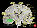
Transverse section through the midbrain showing the location of the red nuclei. The superior colliculi are at the top of image and the cerebral peduncles at the bottom of image – both in section.
Details
Part ofMidbrain
Identifiers
Latinnucleus ruber
MeSHD012012
NeuroNames505
NeuroLex IDbirnlex_1478
TA98A14.1.06.323
TA25898
FMA62407
Anatomical terms of neuroanatomy

The red nucleus or nucleus ruber is a structure in the rostral midbrain involved in motor coordination.[1] The red nucleus is pale pink, which is believed to be due to the presence of iron in at least two different forms: hemoglobin and ferritin.[2] The structure is located in the midbrain tegmentum next to the substantia nigra and comprises caudal magnocellular and rostral parvocellular components.[1] The red nucleus and substantia nigra are subcortical centers of the extrapyramidal motor system.

Function

[edit]

In a vertebrate without a significant corticospinal tract, gait is mainly controlled by the red nucleus.[3] However, in primates, where the corticospinal tract is dominant, the rubrospinal tract may be regarded as vestigial in motor function. Therefore, the red nucleus is less important in primates than in many other mammals.[1][4] Nevertheless, the crawling of babies is controlled by the red nucleus, as is arm swinging in typical walking.[5] The red nucleus may play an additional role in controlling muscles of the shoulder and upper arm via projections of its magnocellular part.[6][7] In humans, the red nucleus also has limited control over hands, as the rubrospinal tract is more involved in large muscle movement such as that for the arms (but not for the legs, as the tract terminates in the superior thoracic region of the spinal cord). Fine control of the fingers is not modified by the functioning of the red nucleus but relies on the corticospinal tract.[8] The majority of red nucleus axons do not project to the spinal cord but, via its parvocellular part, relay information from the motor cortex to the cerebellum through the inferior olivary complex, an important relay center in the medulla.[1]

Input and output

[edit]

The red nucleus receives many inputs from the cerebellum (interposed nucleus and the lateral cerebellar nucleus) of the opposite side and an input from the motor cortex of the same side.[9]

The red nucleus has two sets of efferents:[9]

  • In humans, the majority of the output goes to the bundle of fibers continues through the medial tegmental field toward the inferior olive of the same side, to form part of a pathway that ultimately influence the cerebellum.
  • The other output (the rubrospinal projection) goes to the rhombencephalic reticular formation and spinal cord of the opposite side, making up the rubrospinal tract, which runs ventral to the lateral corticospinal tract. As stated earlier, the rubrospinal tract is more important in non-primate species: in primates, because of the well-developed cerebral cortex, the corticospinal tract has taken over the role of the rubrospinal.

Additional images

[edit]

See also

[edit]

References

[edit]
[edit]
Revisions and contributorsEdit on WikipediaRead on Wikipedia
from Grokipedia
The red nucleus is a bilateral, ovoid-shaped nucleus located in the of the , specifically in its ventral portion at the level of the , and it plays a key role in and control. Structurally, the red nucleus consists of two main subdivisions: the caudal magnocellular red nucleus (RNm), which contains large neurons and projects primarily to the via the , and the rostral parvocellular red nucleus (RNp), featuring smaller neurons that connect to the through the . Its reddish appearance derives from the iron-rich in its neurons and their dense vascularization. The nucleus receives major afferent inputs from the cerebellar interposed and dentate nuclei, as well as from the motor and premotor cortices, enabling it to integrate cerebellar and cortical signals for refined motor output. Functionally, the red nucleus serves as a in the cerebellorubral and rubro-olivary pathways, facilitating limb movements, posture, and skilled actions such as reaching and grasping, particularly through the RNm's influence on proximal musculature in non-human . Recent studies (as of 2025) suggest the red nucleus also integrates reward and for goal-directed actions. In humans, where the predominates, the RNm is relatively vestigial, but the RNp contributes to and potentially cognitive-motor integration, including aspects of sensory discrimination and pain modulation via cerebellar loops. Evolutionarily, it originated as a primitive structure for locomotion in early vertebrates, with the RNp expanding in higher mammals to support complex behaviors, though its role diminishes with and enhanced direct cortical control. Clinically, lesions or dysfunctions in the red nucleus are associated with such as , syndromes like Benedikt's syndrome, and hypertrophic olivary degeneration, and it may play a compensatory role in motor recovery following injuries like . Additionally, iron accumulation in the red nucleus has been implicated in pathology, highlighting its relevance in neurodegenerative research.

Anatomy

Location and relations

The red nucleus is situated in the rostral , specifically within the ventral portion of this region. It lies ventral to the and dorsal to the . This structure exists as a bilateral paired formation, with one nucleus on each side of the midline, and is prominently visible in transverse cross-sections of the at the level of the . In fresh specimens, it appears as an oval-shaped mass approximately 5 mm in diameter, blending rostrally into the surrounding . The characteristic pale pink to red hue of the red nucleus arises from its high iron content, primarily stored as and within the neuronal , which contrasts with the surrounding gray matter. This pigmentation, along with dense vascularization, distinguishes it macroscopically. In terms of spatial relations, the red nucleus is positioned lateral to the and medial to the , while being anteriorly adjacent to the substantia nigra's medial pars reticulata and posteriorly bordered by elements of the oculomotor complex and . It is also integrated within the broader of the , facilitating its embedding in this diffuse network.

Internal structure

The red nucleus exhibits a distinct internal organization, subdivided into two primary parts: the caudal magnocellular division, characterized by large, sparsely distributed neurons that give rise to projections to the , and the rostral parvocellular division, featuring smaller neurons that project to the inferior olive. These subdivisions are cytoarchitectonically defined, with the magnocellular part displaying coarser Nissl substance and the parvocellular part showing finer granularity under Nissl staining. The neuronal population consists predominantly of projection neurons, which accumulate high levels of iron in their cell bodies, imparting the nucleus's reddish pigmentation visible in fresh tissue and accentuated by staining for ferric iron. are sparse throughout both divisions, primarily comprising small local Golgi type II cells. In humans, the red nucleus spans approximately 10 mm in rostrocaudal length, with neurons densely packed within the tegmental zone of the . The subdivisions demonstrate evolutionary conservation, with the magnocellular part linked to ancestral locomotor circuits across vertebrates.

Development and evolution

Embryonic development

The red nucleus originates at the diencephalon-mesencephalon boundary, specifically within prosomere 1, where its neurons emerge from the basal plate of the developing during early embryogenesis. These neurons form the foundational structure of the nucleus in the of the . This process is influenced by key signaling pathways, including Sonic hedgehog (Shh), which is expressed in ventral mesencephalic precursors that give rise to red nucleus neurons, and (FGF) signaling, particularly FGF8, which regulates patterning and the positioning of red nucleus precursors along mediolateral arcs. Differentiation of the red nucleus begins around gestational week 7, with initial and accumulation occurring alongside cerebellar outgrowth, leading to the formation of distinct neuronal populations by week 12. The magnocellular division develops earlier, with immature neurons appearing as dorsal islands to the parvocellular region at 12 weeks, progressing to a semilunar cluster by 16 weeks, and dispersing into larger clusters by 18-23 weeks. In contrast, the parvocellular division emerges slightly later, as an ovoid mass of immature neurons at 12 weeks, with large neurons differentiating by 16 weeks and small neurons by 21 weeks; both populations exhibit linear increases in perikaryonal size and exponential volume growth through 39 weeks. Iron accumulation, which contributes to the nucleus's characteristic pigmentation, initiates in the late fetal stage, with progressive deposition in magnocellular neurons. Postnatally, myelination of the , originating from the , continues, supporting the development of motor connectivity, though refinement persists. Full maturation of the nucleus, including stabilization of neuronal populations and tract myelination, occurs by approximately 2-3 years of age, aligning with broader development.

Evolutionary history

The red nucleus, a brainstem structure integral to motor coordination, traces its origins to early tetrapods, where it emerged to support basic limb-based locomotion. In fish and most amphibians, the red nucleus is absent or rudimentary, lacking a distinct rubrospinal tract, which aligns with their primarily aquatic, fin- or tail-driven movement. By contrast, in limbed amphibians such as anurans and urodeles, a crossed rubrospinal tract appears, facilitating the transition to terrestrial gait and serving as a primitive relay between cerebellar inputs and spinal outputs for appendicular control. This phylogenetic emergence correlates with the evolution of limbs or limb-like structures, as evidenced in certain chondrichthyans like rays, where enlarged pectoral fins may involve analogous tegmentospinal pathways. Comparative neuroanatomy reveals that the red nucleus's initial role was conserved for coordinating proximal limb movements essential for basic quadrupedal progression in these ancient lineages. In reptiles and birds, the red nucleus undergoes notable expansion, enhancing control and integrating more complex cerebellar connections. This development parallels advancements in , with the functioning as a primary descending pathway for limb modulation in non-mammalian amniotes, compensating for the limited corticospinal system. Reptilian red nuclei are predominantly magnocellular, projecting robustly to spinal to support rhythmic and adaptive movements, while avian counterparts show similar organization adapted to bipedal or flight-related motor demands. The increased is evident in experimental tract-tracing studies, which demonstrate denser rubro-olivary projections in these groups compared to amphibians, underscoring the red nucleus's role in refining locomotor precision amid diverse environmental challenges. Mammalian evolution features further adaptations in the red nucleus, with its prominence varying by locomotor style and the rise of the . In quadrupedal mammals like carnivores and , the magnocellular division remains well-developed and conserved, comprising a significant portion of the nucleus (e.g., about two-thirds in felines) to sustain skilled reaching and quadrupedal stability via the . However, in , the magnocellular red nucleus exhibits phylogenetic reduction, particularly in hominoids, where its relative volume shrinks dramatically (to ~0.0005% of volume in humans), reflecting the dominance of direct corticospinal projections for fine distal movements in and manual dexterity. Recent analyses confirm this reduction, alongside parvocellular expansion supporting goal-directed actions integrating behavioral valence and action plans beyond primary locomotion. This shift is accompanied by parvocellular expansion, linking the red nucleus more to higher-order sensory integration rather than primary locomotion. Key evidence for these evolutionary patterns derives from comparative neuroanatomy across over 20 primate species and broader clades, showing red nucleus size positively correlating with locomotor complexity—larger in species reliant on rubrospinal mediation and diminished where cortical pathways prevail. While endocasts provide indirect insights into brainstem scaling with overall evolution in early tetrapods, direct phylogenetic inferences rely on modern tract-tracing and cytoarchitectonic analyses, which confirm the red nucleus's adaptive trajectory from a simple locomotor hub to a multifaceted motor .

Neural connections

Afferent inputs

The red nucleus (RN) receives its primary afferent inputs from the contralateral and the ipsilateral . The cerebellar projections arise mainly from the interpositus nucleus (including globose and emboliform nuclei in ) and the dentate nucleus, traveling via the decussating fibers of the . These crossed cerebellar inputs are dominant and form excitatory synapses on RN neurons. The interpositus nucleus projects predominantly to the magnocellular division of the RN (RNm), providing topographic organization for proximal limb control, while the dentate nucleus targets the parvocellular division (RNp), influencing more distal and skilled movements. Cortical afferents originate from layer V pyramidal neurons in the ipsilateral primary motor, premotor, and supplementary motor areas via the corticorubral tract, with a mirroring the RN's internal organization. These uncrossed corticorubral fibers also utilize glutamate as the primary , facilitating excitatory drive. Secondary modulatory inputs to the RN include projections from the , particularly the , which contribute to extrapyramidal regulation of motor tone. The pontine and medullary provides additional diffuse afferents that influence RN activity during postural adjustments and locomotion. These secondary pathways integrate with primary inputs to fine-tune RN responsiveness, though they are less dense than the cerebellar and cortical projections. In terms of integration, the RNm receives a greater proportion of cerebellar input from the interpositus nucleus, emphasizing rubrospinal motor execution, whereas the RNp integrates more cortical afferents alongside dentate projections, supporting refined motor planning and coordination. This differential connectivity underlies the RN's role in processing parallel streams of sensorimotor information.

Efferent outputs

The red nucleus generates several key efferent pathways that integrate motor signals to downstream structures. The primary output from the magnocellular division is the , which originates in large neurons and decussates immediately to descend contralaterally through the lateral funiculus of the . This tract terminates primarily in the cervical and lumbosacral enlargements, synapsing with and alpha motoneurons to facilitate flexor muscle activation in the limbs, particularly the upper extremities. A second major pathway arises from the parvocellular division via the , which projects ipsilaterally to the , forming part of the dentato-rubro-olivary circuit essential for cerebellar modulation. Additional efferent projections from the red nucleus extend bilaterally—but with contralateral dominance—to the parvicellular and pontine nuclei, aiding in the coordination of posture and locomotion. These outputs are predominantly excitatory, mediated by glutamate-releasing neurons, though local interneurons within the nucleus provide inhibitory modulation that may influence branch-specific signaling. In , including humans, the is notably smaller and less prominent compared to other mammals, with projections more selectively targeting proximal muscles rather than distal ones, reflecting a diminished role relative to the dominant . This evolutionary shift underscores the red nucleus's retained but specialized contributions to gross in higher .

Function

Role in motor control

The red nucleus serves as a critical relay station, integrating signals from the and to facilitate smooth limb movements and maintenance of posture. It receives inputs from cerebellar nuclei and projects via the to influence spinal motor neurons, enabling coordinated motor output that refines voluntary actions. Within the extrapyramidal motor system, the red nucleus compensates for deficits in the by providing an alternative pathway for , particularly in scenarios involving recovery from impairments. It plays a pivotal role in the rubro-olivo-cerebellar loop, where parvocellular neurons project to the inferior olive, which relays error signals to the via climbing fibers, supporting adaptive error correction during ongoing movements. Mechanistically, rubrospinal projections provide excitatory drive to flexor muscles while exerting inhibitory effects on extensor muscles, promoting flexion-dominated responses essential for limb positioning. This modulation contributes to gait initiation, where red nucleus activity helps synchronize and steps, and to reaching behaviors, aiding precise endpoint control in . Evidence from animal models underscores these functions: unilateral lesions of the red nucleus in and induce contralateral , characterized by impaired coordination and during locomotion and skilled tasks, yet partial recovery occurs through neural plasticity, such as of remaining rubrospinal fibers or of alternative pathways within weeks. While reliance on the red nucleus for varies across , with stronger emphasis in quadrupeds for basic locomotion, its core mechanisms remain conserved in vertebrates. Recent studies as of 2025 have further elucidated the red nucleus's role in advanced motor functions, including integrating behavioral valence with action plans to support goal-directed in humans, providing error signals from the parvocellular division for in reaching movements, and initiating directional through excitatory neurons in mice.

Species-specific variations

In non-primate mammals such as cats and rats, the red nucleus plays a dominant role in locomotion through a prominent , which is essential for coordinating quadrupedal and postural adjustments during movement. In these species, lesions to the red nucleus significantly impair overground locomotion, highlighting its critical contribution to rhythmic motor patterns and flexor muscle control independent of higher cortical influences. This prominence underscores the red nucleus's primary function in facilitating basic limb propulsion and stability in quadrupedal animals. In contrast, and humans exhibit a diminished role for the red nucleus due to the evolutionary expansion of the direct , which assumes greater control over voluntary movements. The magnocellular division of the red nucleus, responsible for the , undergoes phylogenetic reduction in , shifting its influence primarily to proximal and movements rather than fine distal hand control. This adaptation allows for enhanced precision in manipulative tasks, with the rubrospinal pathway providing supplementary support for coarser motor actions. In birds and reptiles, the red nucleus aids in limb coordination and visuomotor integration without reliance on a developed , serving as a key premotor structure for basic appendicular movements. A crossed is present in these groups, facilitating spinal activation for locomotion and postural reflexes. The relative size of the red nucleus in such non-mammalian vertebrates inversely correlates with the extent of direct cortical-spinal projections, emphasizing its conserved role as a primary in species lacking advanced pyramidal systems. This species-specific variation reflects an evolutionary trade-off, where the reduced prominence of the red nucleus in higher enables greater dexterity through corticospinal dominance, optimizing fine motor skills at the expense of reliance on rubrospinal pathways for locomotion. Such adaptations trace back to the phylogenetic origins of the red nucleus as a cerebellar , evolving to accommodate diverse locomotor demands across vertebrates.

Clinical significance

Associated pathologies

Unilateral lesions of the red nucleus typically produce contralateral motor impairments, including , , and choreoathetotic movements, due to disruption of the . Lesions of the red nucleus can also lead to hypertrophic olivary degeneration due to disruption of the , resulting in inferior olivary and symptoms such as palatal . In cases involving adjacent structures, such as the fibers, the resulting syndrome—known as Claude's syndrome—manifests as ipsilateral oculomotor palsy combined with contralateral and . More extensive damage interrupting red nucleus outputs can contribute to decerebrate rigidity by inhibiting the and allowing unopposed vestibulospinal excitation of extensor muscles. Primary tumors originating in or involving the red nucleus are exceedingly rare, with gliomas representing the most common type among neoplasms; these often require careful surgical approaches to preserve the nucleus and avoid additional morbidity. Secondary involvement occurs more frequently through strokes, such as paramedian infarcts from occlusion, or demyelinating plaques in that lead to red nucleus and associated motor dysfunction. In pediatric populations, congenital of the red nucleus, often linked to broader posterior fossa malformations, is associated with developmental delays in , including and impaired fine motor skills. Experimental studies in animal models demonstrate that red nucleus lesions induce transient motor deficits, such as limb withdrawal impairments, which largely resolve over time; this recovery highlights the functional between the rubrospinal and corticospinal tracts in compensating for lost projections.

Diagnostic approaches

Magnetic resonance imaging (MRI) serves as a primary diagnostic tool for visualizing the red nucleus, particularly through T2-weighted sequences that highlight its hypointense appearance due to iron accumulation, providing contrast for detecting structural abnormalities or iron-related changes in neurodegenerative conditions. Diffusion tensor imaging (DTI), a specialized MRI technique, enables the reconstruction and assessment of fiber tracts connected to the red nucleus, such as the , by measuring and mean diffusivity to evaluate integrity in cases of injury or degeneration. (PET) offers insights into the metabolic activity of the red nucleus, with high-resolution systems capable of delineating its function in small structures and correlating it with cortical regions in . Electrophysiological assessments, including (TMS), evaluate the functional integrity of the originating from the red nucleus by measuring motor evoked potentials and connectivity changes, particularly in patients with damage where the red nucleus may compensate. Clinical neurological examinations focus on identifying signs of red nucleus involvement through targeted assessments of and rigidity; for instance, rubral or —characterized by low-frequency, irregular movements at rest, posture, and action—often correlates with lesions affecting the red nucleus, as seen in syndromes like Benedikt's, where contralateral hemitremor accompanies oculomotor . These exams integrate observation of motor deficits with correlation to pathology, aiding in the of syndromes. In research settings, provides a method to isolate and manipulate red nucleus activity in animal models, such as mice, by using light-sensitive proteins to activate or inhibit neurons in circuits involving the red nucleus, thereby elucidating its role in motor behaviors without invasive structural alterations.

References

Add your contribution
Related Hubs
User Avatar
No comments yet.