Hubbry Logo
Cone cellCone cellMain
Open search
Cone cell
Community hub
Cone cell
logo
8 pages, 0 posts
0 subscribers
Be the first to start a discussion here.
Cone cell
Cone cell
from Wikipedia

Cone cells
Normalized responsivity spectra of human cone cells, S, M, and L types
Details
LocationRetina of vertebrates
FunctionColor vision
Identifiers
MeSHD017949
NeuroLex IDsao1103104164
THH3.11.08.3.01046
FMA67748
Anatomical terms of neuroanatomy

Cone cells or cones are photoreceptor cells in the retina of the vertebrate eye. Cones are active in daylight conditions and enable photopic vision, as opposed to rod cells, which are active in dim light and enable scotopic vision. Most vertebrates (including humans) have several classes of cones, each sensitive to a different part of the visible spectrum of light. The comparison of the responses of different cone cell classes enables color vision. There are about six to seven million cones in a human eye (vs ~92 million rods), with the highest concentration occurring towards the macula and most densely packed in the fovea centralis, a 0.3 mm diameter rod-free area with very thin, densely packed cones. Conversely, like rods, they are absent from the optic disc, contributing to the blind spot.[1]

Cones are less sensitive to light than the rod cells in the retina (which support vision at low light levels), but allow the perception of color. They are also able to perceive finer detail and more rapid changes in images because their response times to stimuli are faster than those of rods.[2] In humans, cones are normally one of three types: S-cones, M-cones and L-cones, with each type bearing a different opsin: OPN1SW, OPN1MW, and OPN1LW respectively. These cones are sensitive to visible wavelengths of light that correspond to short-wavelength, medium-wavelength and longer-wavelength light respectively.[3] Because humans usually have three kinds of cones with different photopsins, which have different response curves and thus respond to variation in color in different ways, humans have trichromatic vision. Being color blind can change this, and there have been some verified reports of people with four types of cones, giving them tetrachromatic vision.[4][5][6] The three pigments responsible for detecting light have been shown to vary in their exact chemical composition due to genetic mutation; different individuals will have cones with different color sensitivity.

Structure

[edit]

Classes

[edit]

Most vertebrates have several different classes of cone cells, differentiated primarily by the specific photopsin expressed within. The number of cone classes determines the degree of color vision. Vertebrates with one, two, three or four classes of cones possess monochromacy, dichromacy, trichromacy and tetrachromacy, respectively.

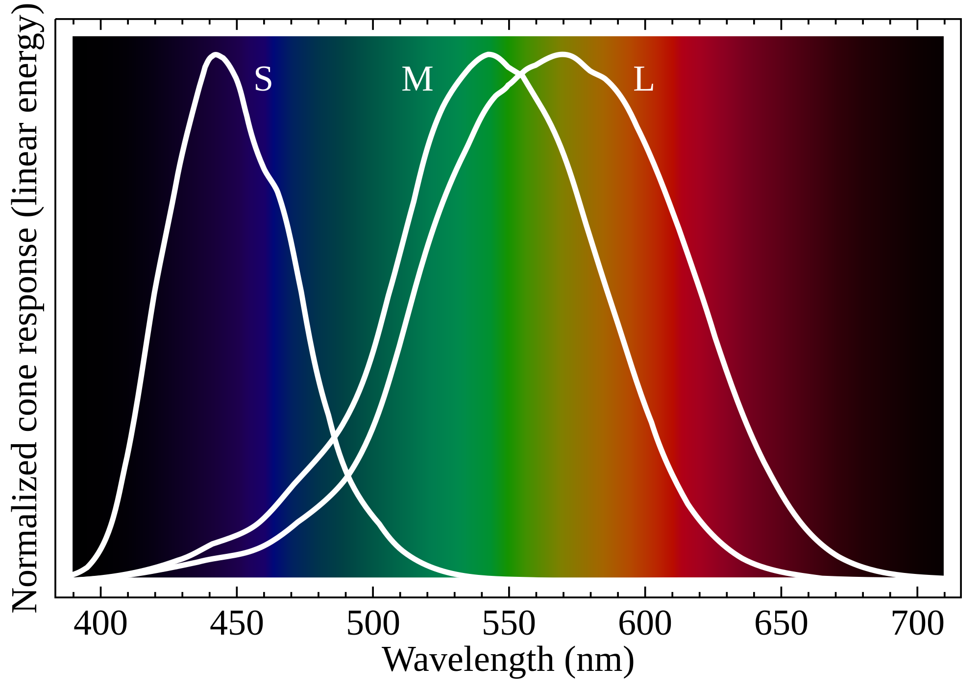
Humans normally have three classes of cones, designated L, M and S for the long, medium and short wavelengths of the visible spectrum to which they are most sensitive.[7] L cones respond most strongly to light of the longer red wavelengths, peaking at about 560 nm. M cones, respond most strongly to yellow to green medium-wavelength light, peaking at 530 nm. S cones respond most strongly to blue short-wavelength light, peaking at 420 nm, and make up only around 2% of the cones in the human retina. The peak wavelengths of L, M, and S cones occur in the ranges of 564–580 nm, 534–545 nm, and 420–440 nm nm, respectively, depending on the individual.[citation needed] The typical human photopsins are coded for by the genes OPN1LW, OPN1MW, and OPN1SW. The LMS color space is an often-used model of spectral sensitivities of the three cells of a typical human.[8][9]

Histology

[edit]
The structure of a cone cell

Cone cells are shorter but wider than rod cells. They are typically 40–50 μm long, and their diameter varies from 0.5–4.0 μm. They are narrowest at the fovea, where they are the most tightly packed. The S cone spacing is slightly larger than the others.[10]

Like rods, each cone cell has a synaptic terminal, inner and outer segments, as well as an interior nucleus and various mitochondria. The synaptic terminal forms a synapse with a neuron bipolar cell. The inner and outer segments are connected by a cilium.[2] The inner segment contains organelles and the cell's nucleus, while the outer segment contains the light-absorbing photopsins, and is shaped like a cone, giving the cell its name.[2]

The outer segments of cones have invaginations of their cell membranes that create stacks of membranous disks. Photopigments exist as transmembrane proteins within these disks, which provide more surface area for light to affect the pigments. In cones, these disks are attached to the outer membrane, whereas they are pinched off and exist separately in rods. Neither rods nor cones divide, but their membranous disks wear out and are worn off at the end of the outer segment, to be consumed and recycled by phagocytic cells.

Distribution

[edit]
Illustration of the distribution of cone cells in the fovea of an individual with normal color vision (left), and a color blind (protanopic) retina. Note that the center of the fovea holds very few blue-sensitive cones.
Distribution of rods and cones along a line passing through the fovea and the blind spot of a human eye[11]

While rods outnumber cones in most parts of the retina, the fovea, responsible for sharp central vision, consists almost entirely of cones. The distribution of photoreceptors in the retina is called the retinal mosaic, which can be determined using photobleaching. This is done by exposing dark-adapted retina to a certain wavelength of light that paralyzes the particular type of cone sensitive to that wavelength for up to thirty minutes from being able to dark-adapt, making it appear white in contrast to the grey dark-adapted cones when a picture of the retina is taken. The results illustrate that S cones are randomly placed and appear much less frequently than the M and L cones. The ratio of M and L cones varies greatly among different people with regular vision (e.g. values of 75.8% L with 20.0% M versus 50.6% L with 44.2% M in two male subjects).[12]

Function

[edit]
Bird, reptilian, and monotreme cone cells

The difference in the signals received from the three cone types allows the brain to perceive a continuous range of colors through the opponent process of color vision. Rod cells have a peak sensitivity at 498 nm, roughly halfway between the peak sensitivities of the S and M cones.

All of the receptors contain the protein photopsin. Variations in its conformation cause differences in the optimum wavelengths absorbed.

The color yellow, for example, is perceived when the L cones are stimulated slightly more than the M cones, and the color red is perceived when the L cones are stimulated significantly more than the M cones. Similarly, blue and violet hues are perceived when the S receptor is stimulated more. S Cones are most sensitive to light at wavelengths around 420 nm. At moderate to bright light levels where the cones function, the eye is more sensitive to yellowish-green light than other colors because this stimulates the two most common (M and L) of the three kinds of cones almost equally. At lower light levels, where only the rod cells function, the sensitivity is greatest at a blueish-green wavelength.

Cones also tend to possess a significantly elevated visual acuity because each cone cell has a lone connection to the optic nerve, therefore, the cones have an easier time telling that two stimuli are isolated. Separate connectivity is established in the inner plexiform layer so that each connection is parallel.[13]

The response of cone cells to light is also directionally nonuniform, peaking at a direction that receives light from the center of the pupil; this effect is known as the Stiles–Crawford effect.

S cones may play a role in the regulation of the circadian system and the secretion of melatonin, but this role is not clear yet. Any potential role of the S cones in the circadian system would be secondary to the better established role of melanopsin (see also Intrinsically photosensitive retinal ganglion cell).[14]

Color afterimage

[edit]

Sensitivity to a prolonged stimulation tends to decline over time, leading to neural adaptation. An interesting effect occurs when staring at a particular color for a minute or so. Such action leads to an exhaustion of the cone cells that respond to that color – resulting in the afterimage. This vivid color aftereffect can last for a minute or more.[15]

Associated diseases

[edit]

See also

[edit]

List of distinct cell types in the adult human body

References

[edit]
[edit]
Revisions and contributorsEdit on WikipediaRead on Wikipedia
from Grokipedia
Cone cells, also known as cones, are specialized photoreceptor cells in the of the vertebrate eye that mediate in bright light, enabling high spatial acuity and color discrimination. Unlike , which dominate in dim light and provide achromatic vision, cones are less sensitive to light—requiring at least 100 photons for activation—and are outnumbered by in the human by a ratio of approximately 20:1. In humans, there are about 6 to 7 million cone cells, concentrated densely in the central fovea of the macula lutea, where they form an exclusive layer to maximize resolution, while being sparser in the peripheral . Structurally, cone cells consist of an outer segment containing stacked, open membrane discs embedded in the plasma membrane and housing photopigments; an inner segment rich in mitochondria for energy; a cell body; and an that synapses in the outer plexiform layer. cones measure 41–50 μm in length and 1–1.2 μm in width, making them shorter and broader than , which contributes to their role in precise light detection. The photopigments in cones are opsins bound to : three types in enable trichromatic vision, with L-cones sensitive to long wavelengths (~555–565 nm, ), M-cones to medium wavelengths (~530–537 nm, ), and S-cones to short wavelengths (~415–430 nm, ). These types are distributed in a roughly 2:1 ratio of L- to M-cones in the fovea, with S-cones comprising only about 5–8% overall and absent from the foveal center. Functionally, cones initiate phototransduction by hyperpolarizing in response to , releasing glutamate onto bipolar and horizontal cells to convey wavelength-specific signals through parallel pathways to retinal ganglion cells. Their low convergence ratio—often 1:1 with midget bipolar cells—preserves fine spatial detail, supporting tasks like reading and , while their rapid adaptation (recovering in ~20 milliseconds) allows quick responses to changing illumination. Disruptions in cone function, such as in or cone dystrophies, lead to impaired and central vision loss, underscoring their essential role in daylight visual processing.

Overview

Definition and characteristics

Cone cells are specialized photoreceptor neurons situated in the outer nuclear layer of the , where their cell bodies reside, and they are primarily responsible for mediating high-acuity vision and color under photopic conditions of bright . Unlike rod photoreceptors, which facilitate in dim light, cone cells possess a higher activation threshold, requiring greater light intensity to function effectively, thus enabling detailed daylight vision while rendering them inactive in low-light environments. Morphologically, cone cells feature shorter, tapered outer segments that form a conical , in contrast to the longer, cylindrical outer segments of , which optimizes their role in high-resolution . In certain species, such as birds and reptiles, cone inner segments incorporate oil droplets that serve as filters, sharpening color discrimination by selectively transmitting specific wavelengths of light. The human retina harbors approximately 6 million cone cells, comprising about 5% of total photoreceptors and outnumbered by roughly 20 times the number of , with their highest concentration in the to support central, high-fidelity vision. Historically, cone cells were first identified and distinguished from as a separate class of retinal photoreceptors by in 1852 through microscopic examination of human retinal tissue. Their functional significance in was presaged by Thomas Young's 1802 trichromatic theory, which proposed the existence of three distinct types of light-sensitive receptors responsive to different spectral ranges, later understood to correspond to cone subtypes.

Evolutionary and comparative aspects

Cone cells, representing the ancestral photoreceptor type in s, emerged over 500 million years ago during the divergence of jawless and jawed fishes, with evolving later from cone-like precursors. Evidence from the , a jawless , reveals short photoreceptors that morphologically resemble cones but function as , supporting the view that the duplex retina—with both cone and types—arose prior to this split around the Cambrian-Ordovician boundary. gene duplications played a pivotal role in cone diversification, with early acquiring multiple visual pigment classes through successive genomic events, enabling the spectral tuning necessary for color discrimination in jawed fishes. These duplications, estimated to have occurred approximately 350–400 million years ago, expanded the ancestral single cone into lineages sensitive to different wavelengths, marking the transition from achromatic to chromatic vision. In comparison to , cones prioritize color discrimination and high-acuity vision in photopic conditions but exhibit lower sensitivity to dim light, reflecting their evolutionary primacy as the original photoreceptor adapted for diurnal environments. , derived via around 500 million years ago, dominate in nocturnal species for without color perception, whereas diurnal animals like birds possess four cone types for tetrachromatic vision, contrasting with the three in humans. Across species, cone variations highlight phylogenetic adaptations: evolved through LWS/MWS duplication for red-green-blue perception, while many mammals like dogs retain with only short- and medium-wavelength cones, limiting them to achromatic-like vision. Analogous photoreceptive structures evolved independently in non-s; feature rhabdomeric photoreceptors in compound eyes for motion detection and color, distinct from ciliary cones, and cephalopods achieve color sensitivity via in their convergently evolved camera eyes rather than specialized cone types. The adaptive significance of cones is evident in their density gradients, which correlate with ecological niches—particularly elevated in the fovea of predatory species to facilitate precise prey detection and tracking under varying light. Raptors and insectivorous birds, for instance, exhibit exceptionally high cone densities in deep foveal pits, enhancing for , a trait refined through evolutionary pressures favoring diurnal . evidence infers cone-like structures from Cambrian-era eyes, such as those in early arthropods and proto-s, where image-forming capabilities first appeared around 540 million years ago during the of visual systems. analyses further date diversification to the period (approximately 485–443 million years ago), aligning with the genomic expansions that underpinned cone-mediated vision in emerging lineages.

Anatomy

Types and classification

Cone cells are classified into subtypes primarily based on the peak absorption wavelengths of their photopigments, known as iodopsins or , which determine their sensitivity to different parts of the . In humans, there are three main types: S-cones (short-wavelength sensitive, also called cones), M-cones (medium-wavelength sensitive, or cones), and L-cones (long-wavelength sensitive, or cones). S-cones express the encoded by the and have a peak sensitivity at approximately 420 nm; M-cones express OPN1MW with a peak at 534 nm; and L-cones express OPN1LW with a peak at 564 nm. These photopigments are coupled with cone-specific alpha-transducin (encoded by GNAT2) to initiate signaling upon light absorption. The genetic basis of these subtypes influences their expression and susceptibility to variation. The OPN1LW and OPN1MW genes, which encode the L- and M-cone s, are located in a tandem array on the , making them X-linked and prone to mutations that cause red-green , such as deuteranomaly or protanomaly. In contrast, the OPN1SW gene for S-cone is autosomal, located on , and less commonly associated with congenital deficiencies. Allelic variations in the X-linked opsin genes can lead to subtle shifts in , contributing to individual differences in color perception among those with normal vision. In terms of abundance, S-cones constitute approximately 5-10% of the total cone population in the human retina, while M- and L-cones make up the majority, with an average M:L ratio of about 1:2 in the fovea. This ratio can vary individually, but it supports trichromatic color vision. The hypothesis of functional tetrachromacy in some females arises from X-chromosome mosaicism: heterozygous carriers of anomalous trichromacy alleles may express four distinct opsin variants (two L and two M types) across different cone populations due to random X-inactivation, potentially enabling expanded color discrimination, though behavioral evidence remains limited. Non-human mammals and other vertebrates exhibit variations in cone types. For example, many birds possess a fourth cone subtype that is (UV)-sensitive, with a peak absorption around 360 nm, in addition to S-, M-, and L-cones; this enables tetrachromatic vision, including UV detection for tasks like and mate selection.

Microscopic structure

Cone cells exhibit a distinctive tapered, conical morphology, with the outer segment measuring approximately 0.5-1 μm in diameter and 10-40 μm in length, distinguishing them from the more cylindrical rod photoreceptors. The inner segment features a mitochondria-rich region that supports high metabolic demands, while the synaptic terminal forms a broad pedicle specialized for synapses. This overall structure optimizes cones for daylight vision and acuity, with the cell body positioned just below the outer limiting . The outer segment consists of a stack of membranous discs, numbering around 1000 in human cones compared to approximately 2000 in , which are infoldings of the plasma membrane continuously connected to the ciliary stalk rather than free-floating. These discs are composed of bilayers embedding proteins, the photopigments responsible for light absorption, and undergo renewal at a rate of about 10% per day through basal addition and distal phagocytosis by the . Unlike rods, cone discs lack prominent rims in humans, contributing to their more open architecture. In the inner segment, the myoid region contains facilitating intracellular transport and is enriched with granules for , adjacent to the ellipsoid packed with elongated mitochondria. Avian cone inner segments include colored oil droplets that act as light filters to enhance , a feature absent in mammalian cones. The nucleus of cone cells is euchromatic, located in the outer nuclear layer, reflecting their active transcriptional state. At the synaptic pedicle, wide terminals (8-10 μm diameter) form ribbon synapses, containing dense ribbons associated with multivesicular bodies of synaptic vesicles and connecting to processes from bipolar and horizontal cells via postsynaptic invaginations. Histologically, cones can be distinguished from by their binding to agglutinin, which labels the surrounding the cone outer segments and pedicles. This staining highlights structural differences, such as the broader pedicle and connected disc morphology.

Distribution and organization

Cone cells are predominantly concentrated in the central region of the , particularly within the , a rod-free zone that enables high . In humans, cone density reaches up to 200,000 cones per square millimeter in the fovea, with an estimated total of approximately 120,000 cones in this area, while peripheral densities drop to less than 5,000 cones per square millimeter. This gradient ensures optimal resolution in the central . In terms of retinal layering, cone outer segments are located in the photoreceptor layer (layer of rods and cones), with their nuclei in the outer nuclear layer, and their axons extend to the outer plexiform layer, where they synapse with bipolar and horizontal cells. These connections often involve bipolar cells, which maintain a one-to-one relationship with individual cones, facilitating precise spatial sampling to ganglion cells. The topographic organization of cones features a hexagonal arrangement that maximizes sampling efficiency across the , with short-wavelength-sensitive (S-) cones more abundant in peripheral regions and long- (L-) and medium- (M-) wavelength-sensitive cones dominating the fovea. During development, cone cells originate near the and undergo tangential migration toward the fovea, completing relocation by birth in , which contributes to the formation of the foveal pit through the displacement of cells. Across species, diurnal animals exhibit higher cone-to-rod s compared to ; for instance, ground squirrels display a 90:10 , contrasting with the human ~1:20 cone-to-rod , reflecting adaptations to varying environments.

Physiology

Phototransduction process

In cone cells, phototransduction begins with the absorption of a by the visual , consisting of an protein bound to 11-cis-retinal in the outer segment discs. This absorption triggers the of 11-cis-retinal to all-trans-retinal within approximately 1 ms, forming the activated metarhodopsin state (R*) that initiates the signaling cascade. The activated then catalyzes the exchange of GDP for GTP on the G-protein , specifically the cone isoform GNAT2, leading to its activation. The activated (G*) subunit binds to and activates the cone-specific PDE6C, which (cGMP) into 5'-GMP. In the dark, high cGMP levels keep cyclic nucleotide-gated (CNG) channels, composed of CNGα3 subunits, open, allowing a depolarizing influx of Na⁺ and Ca²⁺ that maintains the resting at approximately -40 mV. Light-induced cGMP hydrolysis reduces these levels, causing the CNG channels to close and decreasing the inward current, which hyperpolarizes the cell by up to 20-30 mV. Recovery from the light response involves the restoration of cGMP levels and the regeneration of the visual pigment. (retinal outer segment guanylate cyclase, ROS-GC), regulated by guanylate cyclase-activating proteins (GCAPs) in a calcium-dependent manner, synthesizes cGMP to reopen CNG channels and repolarize the . Simultaneously, all-trans- is released from and transported to the , where isomerizes it back to 11-cis-retinal for reuse in the . Cone phototransduction exhibits distinct kinetics compared to , with faster response times of 50-100 ms versus 200 ms in , attributed to lower baseline cGMP concentrations and enhanced calcium feedback mechanisms that accelerate cascade deactivation. The amplification gain in cones is lower, typically requiring 10-100 photons to produce a detectable response, reflecting an overall sensitivity 10-100 times less than . Calcium ions play a key role in modulating the process, with buffers like recoverin binding Ca²⁺ to inhibit kinase and fine-tune by influencing GCAPs and PDE activity. The voltage change in response to light intensity follows a logarithmic relation: ΔV=log(IIsat)×10mV\Delta V = -\log\left(\frac{I}{I_{\text{sat}}}\right) \times 10 \, \text{mV} where II is the light intensity and IsatI_{\text{sat}} is the saturating intensity, yielding approximately 10 mV hyperpolarization per decade of intensity over 4-5 log units.

Role in color vision

Cone cells play a central role in trichromatic color vision through the Young-Helmholtz theory, which posits that the three types of cones—short-wavelength-sensitive (S), medium-wavelength-sensitive (M), and long-wavelength-sensitive (L)—respond to different spectral ranges, with their signals combining additively in the visual system to produce the perception of all colors. This model explains how overlapping cone sensitivities enable the discrimination of a vast array of hues from just three receptor types. The color matching functions derived from cone fundamentals, such as the Smith-Pokorny set, quantify this process, with peak sensitivities at approximately 420 nm for S-cones, 534 nm for M-cones, and 564 nm for L-cones. Beyond the receptors, color perception involves an at the post-receptoral stage, particularly in retinal ganglion cells of the parvocellular pathway, where signals form red-green (L-M) and blue-yellow (S-(L+M)) opponencies to encode chromatic differences. The broad overlap in cone spectral sensitivities allows for fine color discrimination, enabling humans to distinguish around 10 million hues through differential excitations of the cones. Metamerism, where different spectra appear identical, arises from this; the CIE XYZ tristimulus values, which standardize color representation, are computed from cone excitations via a linear transformation matrix, such as: (XYZ)=(0.40020.70750.08070.22631.16530.04570.00000.00000.9182)(LMS)\begin{pmatrix} X \\ Y \\ Z \end{pmatrix} = \begin{pmatrix} 0.4002 & 0.7075 & -0.0807 \\ -0.2263 & 1.1653 & 0.0457 \\ 0.0000 & 0.0000 & 0.9182 \end{pmatrix} \begin{pmatrix} L \\ M \\ S \end{pmatrix}
Add your contribution
Related Hubs
User Avatar
No comments yet.