

Community hub
Recent from talks
Contribute something
Nothing was collected or created yet.
This article needs additional citations for verification. (June 2008) |
| Rib | |
|---|---|
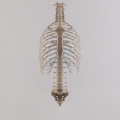
 Collection of single ribs in the Faculty of Education of Charles University | |
 Animation of all ribs, including the false ones in humans | |
| Details | |
| Identifiers | |
| Latin | costae |
| MeSH | D012272 |
| TA98 | A02.3.01.001 |
| TA2 | 1105, 1118 |
| FMA | 7574 |
| Anatomical terminology | |
In vertebrate anatomy, ribs (Latin: costae) are the long curved bones which form the rib cage, part of the axial skeleton.[1] In most tetrapods, ribs surround the thoracic cavity, enabling the lungs to expand and thus facilitate breathing by expanding the thoracic cavity. They serve to protect the lungs, heart, and other vital organs of the thorax. In some animals, especially snakes, ribs may provide support and protection for the entire body.
Human anatomy
[edit]Rib details
[edit]Human ribs are flat bones that form part of the rib cage to help protect internal organs. Humans usually have 24 ribs, in 12 pairs.[2] 1 in 500 people have an extra rib known as a cervical rib. People may have a cervical rib on the right, left or both sides.[3] All are attached at the back to the thoracic vertebrae and are numbered from 1 to 12 according to the vertebrae to which they attach. The first rib is attached to thoracic vertebra 1 (T1). At the front of the body, most of the ribs are joined by costal cartilage to the sternum. Ribs connect to vertebrae at the costovertebral joints.[4]
The parts of a rib includes the head, neck, body (or shaft), tubercle, and angle.
The head of the rib lies next to a vertebra. The ribs connect to the vertebrae with two costovertebral joints, one on the head and one on the neck. The head of the rib has a superior and an inferior articulating region, separated by a crest. These articulate with the superior and inferior costal facets on the connecting vertebrae.[5] The crest gives attachment to the intra-articulate ligament that joins the rib to the vertebra of the same number, at the intervertebral disc. Another ligament, the radiate ligament joins the head of the rib to both the body of the upper vertebra and to the body of the lower vertebra. The smaller middle part of the ligament connects to the intervertebral disc. This plane joint is known as the articulation of the head of the rib.
The other costovertebral joint is that between the tubercle on the neck and the transverse process of the corresponding thoracic vertebra, known as the costotransverse joint. The superior costotransverse ligament attaches from the non-articular facet of the tubercle to the transverse process of the vertebra.
The neck of the rib is a flattened part that extends laterally from the head. The neck is about 3 cm long. Its anterior surface is flat and smooth, whilst its posterior is perforated by numerous foramina and its surface rough, to give attachment to the ligament of the neck. Its upper border presents a rough crest (crista colli costae) for the attachment of the anterior costotransverse ligament; its lower border is rounded.
A tubercle of rib on the posterior surface of the neck of the rib, has two facets (surfaces) one articulating and one non-articulating. The articular facet, is small and oval and is the lower and more medial of the two, and connects to the transverse costal facet on the thoracic vertebra of the same rib number.[5] The transverse costal facet is on the end of the transverse process of the lower of the two vertebrae to which the head is connected. The non-articular portion is a rough elevation and affords attachment to the ligament of the tubercle. The tubercle is much more prominent in the upper ribs than in the lower ribs.
Rib cage
[edit]
The first seven sets of ribs, known as "true ribs", are attached to the sternum by the costal cartilages. The first rib is unique and easier to distinguish than other ribs. It is a short, flat, C-shaped bone, and attaches to the manubrium.[6] The vertebral attachment can be found just below the neck at the first thoracic vertebra, and the majority of this bone can be found above the level of the clavicle. Ribs 2 through 7 then become longer and less curved as they progress downwards.[7] The following five sets are known as "false ribs", three of these sharing a common cartilaginous connection to the sternum, while the last two (eleventh and twelfth ribs) are termed floating ribs.[2] They are attached to the vertebrae only, and not to the sternum or cartilage coming off of the sternum.
In general, human ribs increase in length from ribs 1 through 7 and decrease in length again through rib 12. Along with this change in size, the ribs become progressively oblique (slanted) from ribs 1 through 9, then less slanted through rib 12.[7]
The rib cage is separated from the lower abdomen by the thoracic diaphragm which controls breathing. When the diaphragm contracts, the thoracic cavity is expanded, reducing intra-thoracic pressure and drawing air into the lungs. This happens through one of two actions (or a mix of the two): when the lower ribs the diaphragm connects to are stabilized by muscles and the central tendon is mobile, when the muscle contracts the central tendon is drawn down, compressing the cavity underneath and expanding the thoracic cavity downward. When the central tendon is stabilized and the lower ribs are mobile, a contraction of the diaphragm elevates the ribs, which works in conjunction with other muscles to expand the thoracic indent upward.
Development
[edit]Early in the developing embryo, somites form and soon subdivide into three mesodermal components – the myotome, dermatome, and the sclerotome. The vertebrae and ribs develop from the sclerotomes.[8]
During the fourth week (fertilization age) costal processes have formed on the vertebral bodies. These processes are small, lateral protrusions of mesenchyme that develop in association with the vertebral arches. During the fifth week the costal processes on the thoracic vertebrae become longer to form the ribs. In the sixth week, the costovertebral joints begin to develop and separate the ribs from the vertebrae. The first seven pairs of ribs, the true ribs join at the front to the sternal bars. By the fetal stage the sternal bars have completely fused.[8]
The ribs begin as cartilage that later ossifies – a process called endochondral ossification. Primary ossification centers are located near the angle of each rib, and ossification continues in the direction away from the head and neck. During adolescence secondary ossification centers are formed in the tubercles and heads of the ribs.[8]
Other animals
[edit].jpg)

In jawed fish, there are often two sets of ribs attached to the vertebral column. One set, the dorsal ribs, are found in the dividing septum between the upper and lower parts of the main muscle segments, projecting roughly sideways from the vertebral column. The second set, the ventral ribs arise from the vertebral column just below the dorsal ribs, and enclose the lower body, often joining at the tips. Not all species possess both types of rib, with the dorsal ribs being most commonly absent. Sharks, for example, have no ventral ribs, and only very short dorsal ribs. In some teleosts, there may be additional rib-like bones within the muscle mass.[9]
Tetrapods, however, only ever have a single set of ribs which are probably homologous with the dorsal ribs of fishes. In the earlier choanates, every vertebra bore a pair of ribs, although those on the thoracic vertebrae are typically the longest. The sacral ribs were stout and short, since they formed part of the pelvis, connecting the backbone to the hip bones.[9]
In most true tetrapods, many of these early ribs have been lost, and in living amphibians and reptiles, there is great variation in rib structure and number. For example, turtles have only eight pairs of ribs, which are developed into a bony or cartilaginous carapace and plastron, while snakes have numerous ribs running along the full length of their trunk. Frogs typically have no ribs, aside from a sacral pair, which form part of the pelvis.[9]
In birds, ribs are present as distinct bones only on the thoracic region, although small fused ribs are present on the cervical vertebrae. The thoracic ribs of birds possess a wide projection to the rear; this uncinate process is an attachment for the shoulder muscles.[9] Usually dogs have 26 ribs. Mammals usually also only have distinct ribs on the thoracic vertebra, although fixed cervical ribs are also present in monotremes. In therian mammals, the cervical and lumbar ribs are found only as tiny remnants fused to the vertebrae, where they are referred to as transverse processes. In general, the structure and number of the true ribs in humans is similar to that in other mammals. Unlike reptiles, caudal ribs are never found in mammals.[9]
Ribs as food
[edit]Ribs as food are widely used from many animals. The ribs are the less meaty part of the meat chop and they are often cooked as part of a slab; five or more is known as a rack, as in a rack of lamb. Short ribs are ribs of beef either served singly or several as a plate. A rib steak from beef is a popular choice used in many cuisines. Pork ribs, including spare ribs are popular in European and Asian cuisine.
Animated images
[edit]-
 Thoracic cage with spine

See also
[edit]References
[edit]- ^ Gillen, Glen (2016-01-01), Gillen, Glen (ed.), "Chapter 18 - Trunk Control: Supporting Functional Independence", Stroke Rehabilitation (Fourth Edition), Mosby, pp. 360–393, doi:10.1016/b978-0-323-17281-3.00018-6, ISBN 978-0-323-17281-3, retrieved 2020-11-03
- ^ a b Sly, Peter D.; Collins, Rachel A. (2008-01-01), Taussig, Lynn M.; Landau, Louis I. (eds.), "Chapter 7 - Applied Clinical Respiratory Physiology", Pediatric Respiratory Medicine (Second Edition), Philadelphia: Mosby, pp. 73–88, doi:10.1016/b978-032304048-8.50011-6, ISBN 978-0-323-04048-8, retrieved 2020-11-03
- ^ Oner, Zulal; Oner, Serkan; Sahin, Necati Emre; Cay, Mahmut (26 January 2023). "Evaluation of congenital rib anomalies with multi-detector computed tomography in the Turkish population". Folia Morphologica. 83 (1): 182–191. doi:10.5603/FM.a2023.0006. PMID 36794687. S2CID 256899032.
- ^ Moore, Keith L.; Dalley, Arthur F.; Agur, Anne M. R. (2018). Clinically Oriented Anatomy (8th ed.). Philadelphia: Wolters Kluwer. pp. 293–297. ISBN 9781496347213.
- ^ a b Netter, Frank (2014). Atlas of human anatomy (Sixth ed.). Saunders. pp. 183–184. ISBN 9781455704187.
- ^ Sly, Peter D.; Collins, Rachel A. (2008-01-01), Taussig, Lynn M.; Landau, Louis I. (eds.), "Chapter 7 - Applied Clinical Respiratory Physiology", Pediatric Respiratory Medicine (Second Edition), Philadelphia: Mosby, pp. 73–88, doi:10.1016/b978-032304048-8.50011-6, ISBN 978-0-323-04048-8, retrieved 2020-11-03
- ^ a b Saladin, K. S. (2010). Anatomy and Physiology: The Unity of Form and Function (5th ed.). New York, NY: McGraw-Hill.
- ^ a b c Larsen, William (2001). Human embryology (3rd ed.). Churchill Livingstone. pp. 80–85. ISBN 0443065837.
- ^ a b c d e Romer, Alfred Sherwood; Parsons, Thomas S. (1977). The Vertebrate Body. Philadelphia, PA: Holt-Saunders International. pp. 170–173. ISBN 0-03-910284-X.
| International | |
|---|---|
| National | |
| Other | |
Human Anatomy
Structure of Individual Ribs
The human skeleton includes twelve pairs of ribs, forming the lateral and posterior boundaries of the thoracic cavity. These ribs are elongated, flat bones that vary in size and shape, with each pair numbered sequentially from superior (rib 1) to inferior (rib 12). A typical rib consists of several key components: the head, a wedge-shaped posterior end with one or two articular facets for connection to the thoracic vertebrae; the neck, a short constricted region adjacent to the head; the tubercle, a prominent elevation featuring an articular facet for the transverse process of the vertebra and a non-articular rough area for ligament attachment; the shaft, the long, curved main body that is flattened and twisted; and the costal groove, a shallow indentation along the inferior internal border of the shaft that houses the intercostal vein, artery, and nerve.[1][7][8] Ribs are classified into three categories based on their anterior attachments to the sternum via costal cartilages: true ribs (pairs 1 through 7), which connect directly and independently to the sternum; false ribs (pairs 8 through 10), which lack direct sternal attachment and instead connect indirectly to the costal cartilage of the seventh rib; and floating ribs (pairs 11 and 12), which end freely in the posterior abdominal musculature without any anterior cartilaginous or bony connection.[1][7][9] Rib 1 is notably short and broad with a single articular facet on its head and no costal groove, while ribs 2 and 10 are transitional with unique features like a rough tuberosity on rib 2 and a shorter tubercle on rib 10; ribs 11 and 12 lack tubercles and distinct necks. The lengths of the ribs progressively increase from the superior to the middle pairs, for example, with rib 1 measuring approximately 10–12 cm and rib 7 reaching about 25–30 cm as the longest, before decreasing inferiorly; curvature also intensifies from the flatter superior ribs to the more sharply angled inferior ones.[7][8]/07%3A_Skeletal_System_-_Parts_of_the_Skeleton/7.04%3A_The_Thorax/7.4B%3A_Thoracic_Cage%3A_Ribs) Posteriorly, each rib articulates with the thoracic vertebrae through two synovial joints: the costovertebral joints, where the head of the rib meets the inferior and superior demi-facets on adjacent vertebral bodies (or a single facet for atypical ribs 1, 11, and 12), and the costotransverse joints, where the articular surface of the tubercle connects to the transverse process of the corresponding vertebra (absent in ribs 11 and 12). These articulations are stabilized by intra-articular ligaments, radiate ligaments, and the costotransverse ligament.[1][10][11]Rib Cage
The rib cage, also known as the thoracic cage, forms the bony and cartilaginous framework that encloses and protects the thoracic cavity, integrating the skeletal elements of the chest wall. It consists of 12 pairs of ribs, the sternum, and the 12 thoracic vertebrae (T1–T12), which collectively provide structural support and define the boundaries of the upper respiratory and circulatory systems.[12][13] The sternum, located anteriorly, comprises three main parts: the superior manubrium, the central body, and the inferior xiphoid process, serving as the primary attachment site for the ribs via their costal cartilages. Posteriorly, the ribs articulate with the thoracic vertebrae, creating a closed skeletal ring that maintains the cage's integrity. The costal cartilages, composed of hyaline cartilage, extend from the anterior ends of the ribs to the lateral surface of the sternum, allowing for elastic connections that enhance the overall framework's adaptability. Between adjacent ribs lie the intercostal spaces, which accommodate muscles, nerves, and blood vessels essential to thoracic function.[13][14][15] In shape, the rib cage adopts a conical form, narrower at the superior aspect and widening inferiorly to enclose the thoracic cavity, which houses vital organs such as the heart and lungs. The superior aperture, or thoracic inlet, is bounded by the first thoracic vertebra, the first pair of ribs, and the manubrium, providing passage for structures entering the thorax from the neck. Conversely, the inferior aperture, or thoracic outlet, is formed by the twelfth thoracic vertebra, the eleventh and twelfth ribs, and the costal margin, allowing continuity with the abdominal cavity. This architecture ensures efficient containment while permitting necessary expansions.[16][17][13] The flexibility of the rib cage arises primarily from the costal cartilages, which provide a tough yet elastic connection, enabling the structure to accommodate volume changes without fracturing. Among the ribs, the true ribs (pairs 1–7) attach directly to the sternum via individual cartilages, contributing to upper stability, while the false ribs (pairs 8–10) and floating ribs (pairs 11–12) connect indirectly or float, allowing greater inferior mobility.[18][19][20]Function
Protective Role
The rib cage functions primarily as a bony enclosure that safeguards the heart, lungs, and great vessels from external trauma, acting as a semi-rigid barrier to blunt forces encountered in daily activities or accidents. This protective mechanism relies on the interconnected structure of the 12 pairs of ribs, sternum, and thoracic vertebrae, which collectively form a resilient cage capable of withstanding moderate impacts without compromising organ integrity.[21][22] Biomechanically, the curved, elliptical shape of the ribs plays a crucial role in absorbing and distributing impact forces across the entire thoracic framework, thereby reducing localized stress on any single component and enhancing overall injury tolerance. This design allows the rib cage to deform slightly under load while dissipating energy, preventing direct transmission to underlying viscera.[23] For instance, during blunt trauma, the intact rib cage maintains anterior chest wall stability, averting paradoxical collapse or flail segments that could otherwise occur if multiple ribs fracture and detach.[24] Furthermore, the rib cage reinforces thoracic spine stability, particularly in axial rotation, by increasing rigidity across all motion planes and limiting excessive vertebral movement.[25][26] In human evolution, the rib cage has adapted to support upright bipedal posture through a more cylindrical thorax with declined, curved ribs, optimizing protection for thoracic organs under gravitational loads and dynamic stresses associated with orthograde locomotion. This configuration, distinct from the conical shape in earlier hominins, balances rigidity for defense with flexibility, reflecting selective pressures for endurance and survival in open environments.[27]Respiratory Role
The rib cage facilitates respiration through coordinated movements that alter thoracic volume, enabling efficient ventilation. During inspiration, the external intercostal muscles contract to elevate the ribs, producing distinct motions: the upper ribs (typically 1 through 5) exhibit a pump-handle movement, where the anterior rib ends rise like the handle of a pump, increasing the anteroposterior diameter of the thorax.[5] The lower ribs (typically 6 through 10) perform a bucket-handle movement, swinging laterally outward like the side of a bucket, thereby expanding the transverse diameter.[5] These actions, combined with diaphragmatic descent, increase thoracic volume by approximately 500–1000 mL in normal to moderate breathing cycles, creating negative intrapleural pressure to draw air into the lungs.[28] Expiration reverses these dynamics primarily through passive elastic recoil, but active contraction of the internal intercostal muscles during forced exhalation depresses the ribs, reducing thoracic volume and aiding air expulsion.[29] The internal intercostals attach inferiorly to the costal grooves and superiorly to the inner surfaces near the rib tubercles, pulling adjacent ribs closer together to diminish both anteroposterior and transverse dimensions.[30] Similarly, the external intercostals originate from the inferior border and costal groove of the upper rib and insert on the superior border of the rib below, optimizing elevation for inspiration.[30] Specific muscle attachments enhance these mechanics, particularly for accessory respiration. The scalene muscles insert on the superior surfaces of the first and second ribs, near their tubercles, elevating these ribs during forced inspiration to further expand the upper thorax while providing a stable base for overall rib cage motion.[31] The articulated structure of the rib cage supports these expansions by allowing flexible yet constrained rib gliding at costovertebral and costochondral joints.[5] In vigorous breathing, the relatively fixed first rib anchors the superior cage against scalene pull, while the twelfth rib's posterior-only attachment limits its excursion, stabilizing the inferior boundary.[1]Development and Variations
Embryonic Development
The ribs originate from the sclerotomes, which are derived from the paraxial mesoderm forming somites during the third to fourth weeks of human gestation.[32] Somites segment into sclerotomes ventromedially and dermomyotomes dorsolaterally, with sclerotomal cells migrating around the notochord and neural tube to contribute to the axial skeleton, including the vertebral bodies and ribs.[33] This sclerotomal contribution ensures the ribs articulate with thoracic vertebrae, forming the foundational structure of the rib cage.[32] Chondrification centers emerge within the mesenchymal condensations of the prospective ribs around the sixth week of gestation, transforming the tissue into hyaline cartilage models that outline the future bony elements.[34] These cartilage anlagen provide a template for subsequent ossification, with primary centers appearing in the rib shafts starting in the eighth to ninth weeks.[35] Ossification proceeds via endochondral mechanisms, where hypertrophic chondrocytes are replaced by osteoblasts, beginning at multiple sites including the rib head and shaft.[36] By the end of the third month (approximately 12 weeks), primary ossification has extended through the shafts of all ribs, establishing the diaphyseal bone while the proximal and distal ends remain cartilaginous.[35] Secondary ossification centers in the costal cartilage develop postnatally, typically during adolescence, allowing for continued growth and articulation with the sternum.[37] Hox genes, a family of homeobox transcription factors, play a critical role in regulating somite segmentation, vertebral identity, and rib number along the anterior-posterior axis during embryogenesis.[38] Specifically, Hox9 and Hox10 paralogs promote thoracic identity and rib formation in the mid-axial region, while Hox10 suppression prevents rib development in lumbar segments, ensuring the typical human complement of 12 pairs of ribs.[39] Their collinear expression from 3' to 5' corresponds to progressive rostral-to-caudal patterning, with disruptions leading to homeotic transformations in rib morphology.[38]Anatomical Variations and Anomalies
The typical human rib cage consists of 12 pairs of ribs, but variations in the number of ribs are uncommon, with 11 pairs present in about 1% and 13 pairs (due to supernumerary lumbar ribs) in approximately 1-2% of individuals.[40][41] These numerical differences often stem from transitional vertebrae at the thoracolumbar junction, where the first lumbar vertebra may develop rudimentary ribs or the last thoracic vertebra may lack them.[42] One common variation is the cervical rib, an extra supernumerary rib arising from the seventh cervical vertebra (C7), with a prevalence of approximately 0.5-1% in the general population and a higher incidence in females (up to 1.09%) compared to males (0.5%).[43][44] Cervical ribs are typically rudimentary and unilateral but can be bilateral in about 40% of cases; they may compress neurovascular structures, contributing to thoracic outlet syndrome in symptomatic individuals.[45][46] Lumbar ribs, representing a thirteenth pair originating from the first lumbar vertebra, are rarer, with a pooled prevalence of 2.1%, often bilateral (65.4% of cases), and more frequently observed in European populations.[41] Other anomalies include bifid (forked) ribs, where a rib splits into two branches, typically at the anterior end and affecting the third or fourth rib; their prevalence ranges from 0.15% to 3.4% (mean 2%), with a possible right-sided and male predilection, and they account for up to 20% of congenital rib defects.[47][48] Fused ribs, involving synostosis or partial/complete bony union between adjacent ribs (most often the second and third), occur in 0.2-0.45% of cases and are usually asymptomatic incidental findings.[49][50] Absence or hypoplasia of ribs is less common and often associated with syndromes; for example, in Poland syndrome (prevalence 1 in 30,000 to 50,000 live births, more common in males and right-sided), unilateral absence or hypoplasia of 1-3 ribs (particularly the second to fourth) occurs in approximately 20-40% of affected individuals, leading to chest wall deformities.[51][52] These variations and anomalies are typically congenital, arising from disruptions in embryonic somitogenesis and chondrogenesis during the fourth to eighth weeks of gestation. Detection is primarily through imaging, with chest X-rays identifying most cases incidentally, while computed tomography (CT) provides detailed visualization of shape, attachment, and associated vertebral changes for confirmation.[49][53] Brief clinical implications include potential misdiagnosis on imaging or rare associations with compressive neuropathies, but most remain asymptomatic without intervention.[46]Clinical Significance
Injuries and Fractures
Rib fractures represent a common form of thoracic trauma, often resulting from high-energy impacts that exceed the structural integrity of the bony cage. These injuries can range from single, isolated breaks to multiple fractures affecting several ribs, with the latter increasing the risk of severe complications. Simple fractures, also known as closed or non-displaced fractures, occur when the bone breaks but does not pierce the skin or shift out of alignment, making them the most frequent type in blunt trauma scenarios.[54] In contrast, compound or open fractures are rarer in ribs due to the overlying soft tissue but involve penetration of the skin, potentially leading to infection.[55] Stress fractures, typically seen in athletes or from repetitive strain, develop gradually from microtrauma without acute injury, often in the posterior ribs due to muscle attachments.[56] The primary causes of rib fractures include blunt trauma from motor vehicle collisions, falls from height, physical assaults, and contact sports such as football or rugby, where direct force is applied to the chest wall.[57] In older adults, even low-impact events like prolonged or forceful coughing can precipitate fractures, particularly in those with underlying osteoporosis weakening the bone.[58] The fifth through tenth ribs are most prone to fracturing because they receive minimal protection from surrounding muscles and scapular structures, unlike the upper ribs shielded by the clavicle and shoulder girdle or the lower floating ribs with greater mobility.[59] A particularly dangerous pattern is flail chest, defined as fractures in three or more consecutive ribs, each broken in at least two places, creating a free-floating segment of the chest wall that moves paradoxically during respiration and impairs ventilation.[60] Healing of uncomplicated rib fractures generally involves an inflammatory phase followed by callus formation, with initial bony bridging occurring in 4 to 6 weeks under conservative management including pain control and respiratory exercises.[55] However, immediate effects can include sharp localized pain, shallow breathing, and complications such as pneumothorax, where a fractured rib punctures the lung, allowing air to enter the pleural space and cause lung collapse.[61]Associated Diseases
Costochondritis is an inflammatory condition affecting the cartilage connecting the ribs to the sternum, typically causing sharp chest pain that worsens with movement, deep breathing, or pressure on the affected area.[62] This pain often localizes to the upper ribs, particularly on the left side, and may mimic more serious cardiac or pulmonary issues.[63] Diagnosis relies on clinical history and physical examination, including palpation for tenderness at the costochondral junctions, with imaging such as X-rays or MRI used to exclude other pathologies like fractures or tumors.[64] Tietze syndrome represents a variant of costochondritis distinguished by localized swelling alongside pain and tenderness at the costochondral junction, usually affecting a single rib in young adults.[65] The swelling is nonsuppurative and self-limiting, though it can persist for months, with symptoms including visible or palpable enlargement near the second or third rib.[66] Diagnostic confirmation involves physical exam and imaging to rule out infection or malignancy, as the presentation may resemble abscesses or neoplasms.[65] Osteomyelitis of the ribs is a rare bacterial infection of the rib bone, often resulting from hematogenous spread or contiguous extension from nearby infections, presenting with localized pain, swelling, warmth, and systemic fever.[67] It predominantly affects children but can occur in adults, particularly following trauma or in immunocompromised states, with common pathogens including Staphylococcus aureus.[68] Diagnosis requires imaging such as CT or MRI to identify bone destruction and periosteal reaction, supported by blood cultures, elevated inflammatory markers, and sometimes biopsy for definitive microbiology.[67] Metastatic cancer commonly involves the ribs, with primary sources including breast and lung carcinomas, leading to bone pain, pathologic fractures, and hypercalcemia due to tumor-induced osteolysis.[69] Rib metastases from breast cancer often manifest as lytic lesions causing localized tenderness, while those from lung cancer may present with pleural effusions or respiratory symptoms secondary to chest wall involvement.[70] Diagnostic evaluation includes bone scintigraphy, CT, or MRI to detect lesions, with biopsy confirming the metastatic origin.[71] Hyperostosis refers to abnormal benign bone overgrowth affecting the ribs, often associated with conditions like diffuse idiopathic skeletal hyperostosis (DISH) or sternocostoclavicular hyperostosis, resulting in thickened cortical bone that may cause pain or restricted chest movement.[72] This reactive process alters rib mechanics and can mimic malignancy on imaging due to its expansive appearance.[73] Diagnosis is achieved through CT or MRI demonstrating ossification without aggressive features, with management focusing on symptom relief as the condition can be progressive.[72] Pathologic fractures of the ribs, where minimal or no trauma leads to breakage due to underlying disease, occur at higher prevalence in patients with osteoporosis, as weakened bone density predisposes to such events even during routine activities like coughing.[74] In these cases, osteoporosis exacerbates rib vulnerability, with imaging revealing fractures alongside reduced bone mineral density.[75] Slipping rib syndrome is a condition involving excessive mobility of the lower ribs (typically the 8th, 9th, or 10th), often due to rupture of the interchondral ligament, leading to intermittent sharp pain in the lower chest or upper abdomen that may radiate and worsen with movement or coughing. It is frequently underdiagnosed and can mimic gastrointestinal or cardiac issues. Diagnosis is clinical, aided by the "hooking maneuver" test, with treatment ranging from conservative measures to surgical stabilization in refractory cases.[76]Comparative Anatomy
In Mammals
In mammals, the number of rib pairs varies significantly across species, typically ranging from 9 to 24 pairs, with all ribs attaching to the thoracic vertebrae to form the protective rib cage. Humans possess 12 pairs, including floating ribs (pairs 11 and 12) that do not connect to the sternum. In contrast, dogs have 13 pairs, with the first 9 being true ribs that articulate directly with the sternum and the remaining 4 being asternal. This variation in rib count and attachment supports diverse body plans while maintaining the core function of enclosing vital organs.[77][78] Adaptations in rib structure reflect ecological and locomotor demands. In whales, ribs are broad, flattened, and loosely articulated with the vertebrae, forming a flexible cage that collapses under deep-sea pressure to facilitate diving and streamline the body for aquatic locomotion, rather than rigid fusion into plates. Giraffes exhibit elongated ribs and thoracic vertebrae that extend the functional neck length, providing structural support for their exceptionally long cervical region and aiding in maintaining posture during foraging at height. Bats, adapted for powered flight, have reduced and flattened ribs that minimize weight while preserving thoracic protection, contributing to their lightweight skeletal design essential for aerial maneuverability.[79][80][81] The number of rib pairs shows considerable variation across mammals. For instance, large herbivores like African elephants have 21 pairs, supporting their massive torsos, whereas some smaller mammals like humans have 12 pairs. Three-toed sloths exhibit the maximum of 24 pairs among mammals. This variability highlights how rib morphology evolves to balance protection, respiration, and locomotion across mammalian diversity.[82][83]In Non-Mammalian Vertebrates
In non-mammalian vertebrates, ribs exhibit diverse morphologies adapted to specific locomotor, respiratory, and protective needs, often differing from the more uniform thoracic cage seen in mammals. These structures, homologous across gnathostome lineages, originate from the axial skeleton and typically articulate with vertebrae to enclose and safeguard visceral organs, though their extent, composition, and additional features vary widely by class.[84] In birds, the rib cage is highly specialized for flight, featuring elongated thoracic ribs that articulate with a fused sternum. Most ribs bear uncinate processes—backward-projecting bony flaps that overlap adjacent ribs caudally, stiffening the thoracic basket against collapse during powerful downstrokes of the wings.[85] These processes also provide attachment sites for muscles supporting the scapula and aiding respiration, enhancing the efficiency of the avian bellows-like breathing mechanism.[86] The sternum, or keel bone (carina), is a prominent ventral fusion of sternal ribs that anchors the massive pectoral flight muscles, a key adaptation for powered flight absent in flightless species. Birds typically possess 6 to 8 pairs of ribs, with the anterior ones forming a rigid structure to protect the heart and lungs during aerial maneuvers.[87] Reptiles display varied rib configurations reflecting their ecological diversity. In crocodilians, such as alligators and crocodiles, the thoracic ribs form a robust cage similar to that in mammals, but they are supplemented by gastralia—ventral dermal bones or "belly ribs" embedded in the abdominal musculature that provide additional support to the ventral body wall and assist in costal rotation during lung ventilation.[88] These gastralia, numbering up to 7–8 rows, interrupt the ventral abdominal integument and facilitate the "cuirassal" breathing mode by expanding and contracting the thoracic and abdominal cavities.[89] In contrast, snakes possess an extensive series of ribs extending along nearly the entire vertebral column—often 200 or more pairs—lacking sternal connections and instead functioning in locomotion by deforming the body into lateral undulations or concertina movements.[90] These ribs, with movable capitula and tubercula, interdigitate with intercostal muscles to propel the elongate body without limbs.[91] Amphibians generally have fewer and shorter ribs than amniotes, with most species featuring 3–4 pairs of cartilaginous or weakly ossified structures confined to the anterior trunk, insufficient to form a complete rib cage.[92] In salamanders like Necturus, these primitive ribs articulate via caput and tuberculum heads but remain rudimentary, primarily protecting the viscera while allowing flexibility for aquatic or terrestrial undulation.[93] Frogs exhibit even greater reduction, often with only 1–2 pairs of short, cartilaginous ribs integrated into the pectoral girdle for limited thoracic support during jumping.[94] In fish, ribs are typically pleural elements arising from vertebral parapophyses, often cartilaginous in chondrichthyans and basal osteichthyans, enclosing the coelom with fewer pairs (e.g., 10–20 in teleosts) adapted to hydrodynamic pressures rather than terrestrial support.[95] Branchiostegal rays, slender bony supports along the gill covers, serve as functional analogs by bracing the opercular apparatus for gill ventilation, though they derive from the hyoid arch rather than axial elements.[96] Evolutionarily, ribs trace back to early gnathostomes as homologous serial elements derived from somitic mesoderm, providing a foundational axial scaffold that has been modified or lost in derived lineages.[84] For instance, in some legless squamates like amphisbaenians and certain anguids, ribs are reduced in the trunk or absent in caudal regions to accommodate burrowing, prioritizing flexibility over protection.[97] This variability underscores ribs' role in balancing structural integrity with locomotor demands across vertebrate evolution.[98]Culinary Uses
Rib Cuts and Preparation
Rib cuts used in culinary applications are primarily sourced from the rib sections of pigs, cows, and lambs, where the curvature and positioning of the ribs influence the final shape and meat-to-bone ratio of each piece.[99] Spare ribs, derived from the belly area in both pork and beef, are characterized by their meaty, rectangular form with a higher proportion of fat for flavor and moisture during cooking.[100] Back ribs, located adjacent to the loin in pork (often called baby back ribs due to their smaller size) and beef, feature a curved shape with leaner meat clinging closely to the bones.[100] Short ribs, specific to beef and cut from the chuck or plate primal near the belly, are typically portioned into bone-in segments (English style) or thin cross-cuts (flanken style) for versatile preparation.[101] In lamb, ribs are generally taken from the breast section, yielding smaller, fattier cuts like riblets or Denver-style ribs that are prized for their rich taste.[102] Butchery for these cuts begins with separating the rib rack from the carcass along natural seams, followed by trimming excess fat and, crucially, removing the translucent membrane (silverskin) from the bone side—accomplished by loosening a corner with a knife and pulling it away with a paper towel for better seasoning penetration and tenderness.[103] Preparation methods emphasize low-and-slow cooking to break down connective tissues, with grilling, smoking, and braising as the most common techniques; ribs are often seasoned with dry rubs containing salt, pepper, paprika, and garlic before cooking.[104] Grilling involves indirect heat at 250-300°F for 2-3 hours, spritzing with apple juice to maintain moisture, while smoking uses wood like hickory at 225-275°F for 3-5 hours until an internal temperature of 195°F is reached for pull-apart texture.[105] Braising starts with searing the ribs, then simmering in broth or wine for 2-4 hours at low heat to yield succulent results, particularly suited to short ribs.[106] Marinades enhance flavor, such as tangy barbecue sauce applied midway through cooking for American-style baby back ribs, which are smoked low and slow then glazed to create a caramelized exterior.[107] In Chinese cuisine, char siu ribs—typically made from pork spare or back ribs—are marinated overnight in a blend of hoisin, soy sauce, honey, and five-spice powder, then roasted or grilled at 350°F for about 2 hours, basted frequently for a glossy, lacquered finish reflective of Cantonese barbecue traditions.[108]Nutritional Aspects
Rib meat, particularly from beef and pork, is characterized by a high protein content, typically around 20-23 grams per 100 grams in cooked cuts, making it a valuable source for muscle repair and growth due to its complete amino acid profile.[109][110] Beef ribs also provide essential micronutrients such as vitamin B12 (approximately 2.3 micrograms per 100 grams in roasted large-end cuts, meeting nearly the daily value) and iron (about 2.4 milligrams per 100 grams), supporting red blood cell formation and energy metabolism.[111][112] However, the fat content varies significantly, with beef ribs often containing 30 grams of total fat per 100 grams, including 12 grams of saturated fat, which contributes to their caloric density of 250-400 kilocalories per 100 grams depending on the cut and trimming.[109] Health considerations for consuming rib meat include potential risks from elevated cholesterol levels, around 85 milligrams per 100 grams in beef ribs, which may contribute to cardiovascular issues if intake is excessive, particularly in diets high in saturated fats.[109] On the positive side, the heme iron and B vitamins in rib meat enhance bioavailability compared to plant sources, aiding in anemia prevention and neurological function. Cooking methods influence nutrient retention; for instance, grilling or broiling can lead to losses of up to 40% of water-soluble B vitamins and minerals due to drippings, while frying may increase overall fat content through added oils, though dry heat methods like roasting generally preserve more protein integrity. In comparisons between species, pork ribs tend to be leaner in certain cuts, such as country-style loin ribs, which average 18-25 grams of protein and 190-280 kilocalories per 100 grams with lower saturated fat (around 7 grams) than beef short ribs, offering a potentially heart-friendlier option while still providing comparable iron (about 1.2 milligrams per 100 grams) and B12 levels.[114][115][109]References
- https://www.[healthline](/page/Healthline).com/nutrition/cooking-nutrient-content