Community hub
Recent from talks
Knowledge base stats:
Talk channels stats:
Members stats:
Kyphosis
Kyphosis (from Greek κυφός (kyphos) 'hump') is an abnormally excessive convex curvature of the spine as it occurs in the thoracic and sacral regions. Abnormal inward concave lordotic curving of the cervical and lumbar regions of the spine is called lordosis.
It can result from degenerative disc disease; developmental abnormalities, most commonly Scheuermann's disease; Copenhagen disease, osteoporosis with compression fractures of the vertebra; multiple myeloma; or trauma.
A normal thoracic spine extends from the 1st thoracic to the 12th thoracic vertebra and should have a slight kyphotic angle, ranging from 20° to 45°. When the "roundness" of the upper spine increases past 45° it is called kyphosis or "hyperkyphosis". Scheuermann's kyphosis is the most classic form of hyperkyphosis and is the result of wedged vertebrae that develop during adolescence. The cause is not currently known and the condition appears to be multifactorial and is seen more frequently in males than females.
In the sense of a deformity, it is the pathological curving of the spine, where parts of the spinal column lose some or all of their lordotic profile. This causes a bowing of the back, seen as a slouching posture. Kyphosis is distinguished from scoliosis, a condition in which the spine has a sideways curve.
While most cases of kyphosis are mild and only require routine monitoring, serious cases can be debilitating. High degrees of kyphosis can cause severe pain and discomfort, breathing and digestion difficulties, cardiovascular irregularities, neurological compromise and, in the more severe cases, significantly shortened life spans. These types of high-end curves typically do not respond well to conservative treatment and almost always warrant spinal fusion surgery, which can restore the body's natural degree of curvature.
The risk of serious complications from spinal fusion surgery for kyphosis is estimated to be 5%, similar to the risks of surgery for scoliosis. Possible complications include inflammation of the soft tissue or deep inflammatory processes, breathing impairments, bleeding, and nerve injuries. According to the latest evidence, the actual rate of complications may be substantially higher. Even among those who do not develop serious complications, 5% of patients require reoperation within five years of the procedure, and in general it is not yet clear what one would expect from spine surgery during the long-term. Given that the signs and symptoms of spinal deformity cannot be changed by surgical intervention, surgery remains essentially a cosmetic choice. However, the cosmetic effects of surgery are not necessarily stable.
There are several kinds of kyphosis (ICD-10 codes are provided):
Kyphosis can be graded in severity by the Cobb angle. Also, sagittal balance can be measured. The sagittal balance is the horizontal distance between the center of C7 and the superior-posterior border of the endplate of S1 on a lateral radiograph.
Hub AI
Kyphosis AI simulator
(@Kyphosis_simulator)
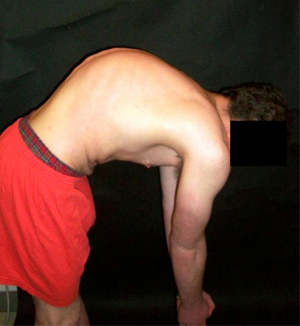
Kyphosis
Kyphosis (from Greek κυφός (kyphos) 'hump') is an abnormally excessive convex curvature of the spine as it occurs in the thoracic and sacral regions. Abnormal inward concave lordotic curving of the cervical and lumbar regions of the spine is called lordosis.
It can result from degenerative disc disease; developmental abnormalities, most commonly Scheuermann's disease; Copenhagen disease, osteoporosis with compression fractures of the vertebra; multiple myeloma; or trauma.
A normal thoracic spine extends from the 1st thoracic to the 12th thoracic vertebra and should have a slight kyphotic angle, ranging from 20° to 45°. When the "roundness" of the upper spine increases past 45° it is called kyphosis or "hyperkyphosis". Scheuermann's kyphosis is the most classic form of hyperkyphosis and is the result of wedged vertebrae that develop during adolescence. The cause is not currently known and the condition appears to be multifactorial and is seen more frequently in males than females.
In the sense of a deformity, it is the pathological curving of the spine, where parts of the spinal column lose some or all of their lordotic profile. This causes a bowing of the back, seen as a slouching posture. Kyphosis is distinguished from scoliosis, a condition in which the spine has a sideways curve.
While most cases of kyphosis are mild and only require routine monitoring, serious cases can be debilitating. High degrees of kyphosis can cause severe pain and discomfort, breathing and digestion difficulties, cardiovascular irregularities, neurological compromise and, in the more severe cases, significantly shortened life spans. These types of high-end curves typically do not respond well to conservative treatment and almost always warrant spinal fusion surgery, which can restore the body's natural degree of curvature.
The risk of serious complications from spinal fusion surgery for kyphosis is estimated to be 5%, similar to the risks of surgery for scoliosis. Possible complications include inflammation of the soft tissue or deep inflammatory processes, breathing impairments, bleeding, and nerve injuries. According to the latest evidence, the actual rate of complications may be substantially higher. Even among those who do not develop serious complications, 5% of patients require reoperation within five years of the procedure, and in general it is not yet clear what one would expect from spine surgery during the long-term. Given that the signs and symptoms of spinal deformity cannot be changed by surgical intervention, surgery remains essentially a cosmetic choice. However, the cosmetic effects of surgery are not necessarily stable.
There are several kinds of kyphosis (ICD-10 codes are provided):
Kyphosis can be graded in severity by the Cobb angle. Also, sagittal balance can be measured. The sagittal balance is the horizontal distance between the center of C7 and the superior-posterior border of the endplate of S1 on a lateral radiograph.