Hubbry Logo
Nasal boneNasal boneMain
Open search
Nasal bone
Community hub
Nasal bone
logo
8 pages, 0 posts
0 subscribers
Be the first to start a discussion here.
Nasal bone
Nasal bone
from Wikipedia

Nasal bone
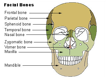
Nasal bone visible at center, in dark green.
Cartilages of the nose. Side view. (Nasal bone visible at upper left.)
Details
Identifiers
Latinos nasale
MeSHD009295
TA98A02.1.10.001
TA2748
FMA52745
Anatomical terms of bone

The nasal bones are two small oblong bones, varying in size and form in different individuals; they are placed side by side at the middle and upper part of the face and by their junction, form the bridge of the upper one third of the nose.

Each has two surfaces and four borders.

Structure

[edit]

There is heavy variation in the structure of the nasal bones, accounting for the differences in sizes and shapes of the nose seen across different people. Angles, shapes, and configurations of both the bone and cartilage are heavily varied between individuals. Broadly, most nasal bones can be categorized as "V-shaped" or "S-shaped" but these are not scientific or medical categorizations. When viewing anatomical drawings of these bones, consider that they are unlikely to be accurate for a majority of people.[1]

The two nasal bones are joined at the midline internasal suture and make up the bridge of the nose.

Surfaces

[edit]

The outer surface is concavo-convex from above downward, convex from side to side; it is covered by the procerus and nasalis muscles, and perforated about its center by the nasal foramen, a small passageway for the transmission of a small vein from the overlying soft tissues.

The inner surface is concave from side to side, and is traversed from above downward, by a groove for the passage of a branch of the nasociliary nerve.

Articulations

[edit]

The nasal articulates with four bones: two of the cranium, the frontal and ethmoid, and two of the face, the opposite nasal and the maxilla.

Other animals

[edit]

In primitive bony fish and tetrapods, the nasal bones are the most anterior of a set of four paired bones forming the roof of the skull, being followed in sequence by the frontals, the parietals, and the postparietals. Their form in living species is highly variable, depending on the shape of the head, but they generally form the roof of the snout or beak, running from the nostrils to a position short of the orbits. In most animals, they are generally therefore proportionally larger than in humans or great apes, because of the shortened faces of the latter. Turtles, unusually, lack nasal bones, with the prefrontal bones of the orbit reaching all the way to the nostrils.[2]

Additional images

[edit]

See also

[edit]

References

[edit]
[edit]
Revisions and contributorsEdit on WikipediaRead on Wikipedia
from Grokipedia
The nasal bones are a pair of small, oblong, rectangular bones situated in the midface that form the superior bony portion of the nose, specifically the bridge or root, contributing to the upper third of the nasal pyramid. These bones articulate medially with each other along the internasal suture, superiorly with the at the nasofrontal suture (defining the ), and inferiorly with the nasal processes of the , while their posterior surfaces help form part of the anterior nasal aperture and support the overlying nasal cartilages. On average, each nasal bone measures approximately 2.5 cm in length, with increasing thickness from the inferior free margin toward the thickest portion at the nasofrontal articulation, and they exhibit , being generally larger and more robust in males. Functionally, the nasal bones provide structural support to the external nose, maintaining its aesthetic contour and protecting the underlying during respiration and olfaction, while also serving as attachment sites for muscles and ligaments involved in nasal mobility. Embryologically, they develop from the fusion of the maxillary and frontonasal prominences during the fourth week of , with ossification centers appearing around the eighth to tenth weeks, progressing through four stages that transform nasal pits into primitive nasal cavities by the end of the embryonic period. Blood supply to the nasal bones derives primarily from branches of the (including the angular and superior labial arteries), the (anterior and posterior ethmoidal branches), and the , forming a rich vascular network that also nourishes the adjacent . Clinically, the nasal bones are the most commonly fractured facial bones in adults, often resulting from such as assaults or , with fractures classified into types I through V based on displacement and involvement of adjacent structures like the or orbital floor. typically involves conservative for nondisplaced fractures or closed/open reduction within 14 days for displaced ones to restore alignment and prevent complications like septal hematomas, airway obstruction, or cosmetic deformities. Variations in nasal bone morphology, including ethnic differences in and , influence surgical outcomes in and reconstructive procedures.

Anatomy

Gross anatomy

The nasal bones are a pair of small, rectangular, flat bones that form the superior aspect of the within the , or viscerocranium. They are positioned symmetrically on either side of the midline, medial to the frontal processes of the maxillae, and contribute to the bony framework of the external . These bones are oblong in overall shape, with a thicker superior portion that tapers inferiorly, exhibiting variability in form among individuals. The external surface of each nasal bone is transversely convex and serves as an attachment site for the procerus and nasalis muscles. In contrast, the internal surface is transversely concave and features a longitudinal groove, known as the sulcus ethmoidalis, which accommodates the anterior ethmoidal nerve. The superior border of the nasal bone is thick and serrated, articulating with the nasal part of the frontal bone to form the frontonasal suture. The inferior border is irregular and slopes upward, attaching to the lateral nasal cartilage and contributing to the margin of the nasal aperture. Laterally, it connects to the frontal process of the maxilla via the nasomaxillary suture, while the medial border articulates with the contralateral nasal bone through the internasal suture; the medial edges also project as a vertical crest that forms part of the nasal septum. In adults, the nasal bones measure approximately 2.5 cm in height (ranging from 1.8 to 3.1 cm) and 1.3 cm in breadth (ranging from 0.7 to 1.8 cm), with individual variations influenced by factors such as and . They are generally thicker at the superior end and become progressively thinner toward the inferior border.

Articulations and relations

The nasal bones articulate superiorly with the along the frontonasal suture, forming the superior aspect of the . Laterally, each nasal bone joins the frontal process of the via the nasomaxillary suture, contributing to the lateral walls of the . Medially, the two nasal bones meet at the internasal suture in the midline, creating a stable union that supports the nasal dorsum. Inferiorly, the nasal bones attach to the upper lateral nasal cartilages, marking the transition from the bony to the cartilaginous portion of the nasal vault. In terms of broader spatial relations, the nasal bones lie anterior to the anterior cranial fossa, contributing to the superior portion of the anterior wall of the nasal cavity alongside contributions from the ethmoid bone. They are positioned adjacent to the ethmoidal air cells posteriorly and medially, with the perpendicular plate of the ethmoid contributing to the nasal septum posterior to their medial edges. On their internal surface, the nasal bones feature a groove that accommodates the anterior ethmoidal nerve, facilitating its passage toward the external nose. The nasal bones are enveloped externally by soft tissues, including the skin and subcutaneous layer, as well as the overlying nasal musculature such as the procerus and nasalis muscles, which insert onto their anterior surfaces.

Development

Embryonic origins

The nasal bones originate from the frontonasal prominence, one of the five primary facial primordia that emerge during early embryonic development. This prominence develops ventral to the around the fourth week of , driven by the proliferation of mesenchymal cells derived from cranial . By weeks 4 to 7, the frontonasal prominence undergoes significant growth and differentiation, contributing to the formation of the midline nasal structures, including the foundational elements of the nasal bones. The initial formation of the nasal bones involves mesenchymal condensation surrounding the developing nasal placodes. These placodes appear as bilateral ectodermal thickenings on the inferolateral aspects of the frontonasal prominence by the end of the fourth week, marking the sites of future . Around week 5, the placodes invaginate to form nasal pits, inducing surrounding mesenchymal cells—primarily of origin—to condense and form the primitive nasal capsule. This cartilaginous framework provides the scaffold for the paired nasal bones, which arise from bilateral mesenchymal condensations within the capsule. By the eighth week, the precursors of the nasal bones integrate into the broader through the fusion and merger of the frontonasal prominence with the adjacent maxillary and frontal prominences. This process establishes the continuity of the with the and , completing the embryonic patterning of the nasal vault. The paired nature of the nasal bones reflects their bilateral origins from symmetric centers in the cartilaginous nasal capsule, ensuring symmetrical development of the nasal dorsum. Genetic regulation plays a crucial role in this embryonic patterning, particularly through transcription factors that govern cranial cell migration and differentiation. Genes such as DLX5 and DLX6 are essential for the proper development of the craniofacial , including nasal structures, by controlling mesenchymal cell fate in the frontonasal region; their inactivation in models leads to severe defects in nasal and calvarial bone formation. Similarly, MSX1 interacts with DLX5 to regulate osteogenic induction in -derived , influencing the migration and processes critical for nasal bone precursors. These genes ensure coordinated epithelial-mesenchymal interactions during weeks 4 to 8.

Ossification process

The ossification of the nasal bones proceeds via , in which mesenchymal cells within the frontonasal prominence directly differentiate into osteoblasts, forming bone tissue without an intervening cartilage model. This process is characteristic of most and facial bones, including the paired nasal bones. Each nasal bone arises from a single primary located near the midpoint of the future bone, within the mesenchymal condensation of the nasal capsule derived from the frontonasal process. These centers first become histologically visible at 9–10 weeks of , with radiographic and sonographic evidence appearing around 11-14 weeks . From this central point, ossification radiates outward, expanding superiorly toward the and inferiorly toward the nasal process of the , progressively mineralizing the thin, oblong plate characteristic of the adult nasal bone. By the end of , the nasal bones achieve substantial , reaching a mean length of approximately 12.3 mm at 40 weeks, though fine-tuning continues postnatally. The midline interosseous suture between the two nasal bones remains patent throughout life for articulation, without fusion. The superior border, initially smooth, develops its serrated morphology for interlocking with the nasal part of the around age 3 years, coinciding with the completion of sutural patterning. Adult dimensions and proportions, with length approximately doubling relative to slight increases in width, are attained by amid overall facial skeletal maturation. This ossification is modulated by local signaling pathways, notably bone morphogenetic protein (BMP) signaling, which drives mesenchymal cell commitment to osteogenesis in the craniofacial region through regulation of key transcription factors like Runx2. Mechanical stimuli from the expanding and surrounding soft tissues further guide bone deposition, aligning the nasal bones with the growing nasal dorsum. Variations in the process include delayed onset or progression, as seen in hypoplastic cases where centers may not mineralize until later in ; such delays are more common in premature infants due to truncated intrauterine development, though catch-up typically occurs with postnatal growth. Notably, the presence and length of the nasal bone at 11-14 weeks are evaluated in first-trimester screening for aneuploidies like , as or absence increases risk. Complete maturation aligns with pubertal facial changes in all cases.

Function

Structural support

The nasal bones form the rigid superior framework of the nose, constituting the bony vault of the upper third of the nasal pyramid in conjunction with the frontal processes of the maxillae. This structure provides critical to the , maintaining its elevation and preventing collapse of the nasal dorsum under gravitational or external forces. These bones also delineate the superior margin of the bony nasal (piriform aperture), establishing a stable transition from the rigid skeletal elements to the flexible lower lateral and alar cartilages that shape the nasal tip and nostrils. By defining this boundary, the nasal bones ensure the architectural integrity of the nasal entrance, supporting airflow and overall nasal projection. The nasal bones offer attachment sites for key nasal muscles, facilitating dynamic movements of the nasal soft tissues. For example, the transverse head of the inserts into the overlying the nasal dorsum, contributing to compression of the nasal aperture, while its alar head aids in alar flare by dilating the nostrils; the procerus muscle originates from the covering the superior nasal bones, enabling frowning and skin wrinkling over the bridge. Articulating superiorly with the at the nasofrontal suture and inferiorly with the maxillae at the nasomaxillary sutures, the nasal bones integrate into the broader nasomaxillary complex to distribute mechanical loads effectively. Finite element modeling reveals that peak principal stresses are concentrated at the nasomaxillary junction under perpendicular external forces. Variations in nasal bone shape and dimensions contribute to ethnic differences in nasal profiles; for instance, Caucasians often have narrower, more projecting nasal bones associated with leptorrhine (narrow) nose forms, in contrast to broader configurations in other groups.

Protective function

The nasal bones serve as a primary bony barrier, shielding the underlying , ethmoidal air cells, and portions of the from external trauma due to their anterior position in the midface. This rigid framework absorbs minor impacts, distributing forces away from more delicate internal structures and thereby reducing the risk of septal deviation or damage to adjacent sinuses. By maintaining the structural integrity of the nasal vault, the bones prevent inward collapse that could compromise the patency of nasal passages, ensuring unobstructed airflow essential for respiration. In respiratory function, the stabilizing role of the nasal bones supports efficient air warming and humidification as inhaled air passes through the , with their contribution to the overall framework facilitating and minimizing turbulence. This patency indirectly aids by allowing ciliated to effectively transport and trapped particles toward the nasopharynx, thereby protecting the lower from pathogens. Additionally, the protective positioning of the nasal bones safeguards the located in the superior nasal cavity roof, preserving the integrity of endings crucial for olfaction against minor frontal impacts.

Clinical significance

Trauma and fractures

Nasal bone fractures represent the most common type of fracture, accounting for up to 50% of all injuries due to the prominent and central position of the nasal pyramid, which makes it vulnerable to from assaults, , accidents, and falls. These fractures typically result from direct frontal or lateral impacts that disrupt the normal articulations of the nasal bones with adjacent structures like the and . Fractures are classified based on their pattern and degree of displacement, with simple linear fractures involving a clean break without fragmentation, comminuted fractures featuring multiple fragments, and depressed fractures where segments are pushed inward. They are further categorized as nondisplaced (bones remain aligned) or displaced (with lateral, medial, or posterior deviation), which influences management decisions; unilateral or bilateral involvement is also common, often extending to the in 42-96% of cases. Patients commonly present with immediate symptoms including , soft tissue swelling, periorbital ecchymosis (bruising), and nasal airway obstruction due to or deviation. Additional signs may include pain, (grating sensation), instability on , and visible deformity such as a crooked or broadened . relies primarily on clinical examination, with plain radiographs offering limited utility due to frequent superimposition of structures; computed tomography (CT) scans are preferred for confirming the extent of injury, assessing displacement, and evaluating associated fractures in complex cases. Management prioritizes restoring alignment and function while minimizing complications, beginning with conservative measures like ice application, analgesics, and elevation to reduce swelling in nondisplaced or minimally symptomatic fractures. For displaced fractures, closed reduction—using instruments like nasal elevators or under local or general —is indicated within 3-7 days post-injury for optimal outcomes, ideally before significant formation, and is successful in 60-90% of non-comminuted cases. Open reduction with is reserved for comminuted, severely displaced, or delayed fractures (>14 days), often requiring septorhinoplasty for cosmetic and functional correction after 3-6 months of healing. Post-reduction stabilization involves intranasal packing for 4-7 days and external splinting for 7-14 days, with prophylactic antibiotics to prevent during packing; healing typically occurs in 3-6 weeks, though full remodeling may take longer. Potential complications include septal , a blood collection between the and that requires urgent to prevent necrosis and saddle-nose . Other risks encompass persistent cosmetic , chronic nasal obstruction from septal deviation, infection or abscess formation, and, in rare extensions to the ethmoid or , cerebrospinal fluid or . Up to 20% of patients may experience dissatisfaction with outcomes, underscoring the importance of timely intervention and follow-up.

Congenital variations

Congenital variations of the nasal bones encompass developmental anomalies that arise during embryogenesis, potentially leading to hypoplasia, aplasia, bifidity, or hyperplasia, with significant implications for facial structure and associated syndromes. These anomalies can be detected prenatally through ultrasound imaging and are often linked to chromosomal or genetic disorders, influencing decisions on screening and intervention. Hypoplasia or aplasia of the nasal bones, characterized by underdeveloped or absent , is strongly associated with (trisomy 21). This variation results from delayed or incomplete of the nasal bone precursors and serves as a key marker, particularly when observed as an isolated finding or in combination with other soft markers. Prenatal detection is feasible via transabdominal or transvaginal between 11 and 14 weeks of gestation, where the nasal bone should normally be visible as a bright linear structure; its absence or marked prompts further evaluation. The incidence of nasal bone absence is approximately 0.5-3% in chromosomally normal fetuses but exceeds 60-70% in those with trisomy 21, providing a positive likelihood ratio of 20-60 for risk assessment. Bifid nasal bones represent a rare congenital anomaly involving a midline cleft or duplication of the nasal bone, often manifesting as a bifid nasal tip or widened . This condition is typically linked to , a spectrum of midline facial malformations arising from disruptions in the frontonasal prominence during early embryogenesis, and may occur in isolation or as part of syndromic with features such as and limb anomalies. The bifid appearance stems from incomplete fusion of the bilateral nasal bone anlagen, leading to aesthetic and occasionally functional concerns like nasal obstruction. Incidence is low, estimated at less than 1 in 100,000 births, underscoring its rarity. Hyperplasia of the nasal bones, involving excessive growth or thickening, can occur postnatally in conditions like , where elevated levels promote disproportionate bone enlargement, including prominence of the and alae. Similarly, may lead to hyperplastic changes in the nasal region through disorganized , resulting in and facial overgrowth. Although these are not strictly congenital, they represent postnatal exaggerations of bone development pathways that can mimic or exacerbate underlying congenital predispositions. Such overgrowth affects facial aesthetics and may contribute to respiratory issues if severe. Diagnosis of these congenital variations relies on prenatal for initial screening, followed by such as (NIPT), , or to confirm associations like trisomy 21. Postnatal confirmation involves clinical examination, three-dimensional computed tomography for bony detail, and . Management is tailored to severity and includes multidisciplinary approaches: for at-risk pregnancies, for mild that may resolve, and surgical correction—such as or —for bifid or hyperplastic cases to address cosmetic deformities or functional impairments like breathing difficulties, often performed after skeletal maturity. Early intervention improves outcomes, with surgical success rates exceeding 80% in correcting bifid nasal structures.

Comparative anatomy

In other mammals

The nasal bones are present in most terrestrial mammals as a pair of elongated, thin bones that form the dorsal bridge of the nose and contribute to the roof of the . In domestic dogs, these bones vary in length by breed but typically extend along the dorsal surface of the face, articulating with the frontal and maxillary bones to support the external nares and nasal planum. Similarly, in , the nasal bones are elongated and curved, forming the majority of the roof and integrating with the nasal conchae for airflow regulation. In aquatic mammals such as cetaceans, the nasal bones are highly modified and reduced compared to terrestrial forms, appearing as nodular, elevated, and anteroposteriorly compressed structures rather than flat plates, which facilitates the posterior migration of the nares to form the blowhole. For instance, in odontocetes like dolphins and pilot whales, computed tomography reveals asymmetric nasal bones with species-specific fossa morphology, adapted for underwater occlusion of the nasal passage during dives. In contrast, the nasal bones in are notably short and stout, supporting the base of the elongated while lacking any bony extension into the trunk itself, which is composed entirely of muscle and . Variations in nasal bone structure across mammals reflect adaptations to specific ecological niches. In rodents like rats and mice, the nasal bones are relatively small and contribute to a compact optimized for rapid olfaction, with shape under complex genetic control influencing overall morphology. Ruminants such as exhibit elongated nasal bones that form the snout's framework, potentially broadened to integrate with for respiratory efficiency during . In lagomorphs like rabbits, the rostrum—particularly the —is fenestrated with multiple small openings, reducing weight while maintaining structural integrity for burrowing and high-speed movement. Functional adaptations of the nasal bones often enhance olfaction and structural resilience. In carnivores such as canids, the nasal bones integrate closely with expanded turbinates—scroll-like bony projections—that increase surface area for scent detection, as seen in the intricate nasal skeleton of dogs where turbinates attach to the nasal and ethmoid bones. Herbivores like have thicker nasal bones that provide impact resistance during foraging or social interactions, with CT imaging highlighting robust fossa morphology for load distribution. In , nasal bone size varies phylogenetically, being smaller and less projecting in great apes like compared to more elongated and projecting forms in prosimians, paralleling human anatomy in supporting the external but with greater relative length in some species for enhanced facial projection.

Evolutionary history

The nasal bones, paired dermal elements forming the dorsal roof of the , trace their origins to the synapsid lineage, which diverged from sauropsid reptiles during the Late Carboniferous approximately 318 million years ago, inheriting a basic nasal architecture from ancestral amniotes but undergoing modifications in the therapsid radiation. In early synapsids like , the nasal bones were present as flat, elongate structures bounding the external nares, similar to those in contemporary reptiles, with associated septomaxillae facilitating nasal passages. As synapsids evolved toward mammaliamorphs during the Permian and , the nasal region adapted for enhanced olfaction and respiratory efficiency, with the frontonasal prominence—responsible for the rigid reptilian snout tip—repatterned in therians to form a protruding, flexible muzzle supported by elongated nasal and premaxillary bones. Fossil evidence indicates the first of key nasal-associated structures, such as respiratory turbinates within the nasal capsule, appeared in non-mammaliaform cynodonts by the around 225 million years ago, as seen in Brasilitherium riograndensis, where the nasal bones frame oval external nares and enclose a cavity with partially ossified ethmoturbinals, marking an early step toward mammalian endothermy by conserving and during . These adaptations specialized the nasal bones for supporting expanded olfactory epithelia, contrasting the rigid, less protrusive snouts of reptilian ancestors and enabling therian mammals to exploit diverse sensory niches. Convergent evolution produced dome-like enlargements of the nasal bones in unrelated lineages, such as the hollow nasal crests in hadrosaurid dinosaurs (e.g., ) and inflated nasal regions in some mammals like the extinct bovid Rusingoryx atopocranion, likely for vocal resonance and social communication rather than olfaction. In basal mammals, fossil records show variation: early monotremes exhibited relatively reduced nasal bones compared to therians, with shorter, less projecting snouts reflecting their semi-aquatic or specialized lifestyles, while placental mammals diversified these structures across ecological niches, with macrosmatic species like canids (e.g., dogs) featuring elongated nasal bones to accommodate voluminous turbinates for acute smell detection.

References

Add your contribution
Related Hubs
User Avatar
No comments yet.