Referred pain
Referred pain
Main page
2094740

Referred pain

logo
Community Hub0 subscribers
Read side by side
from Wikipedia
Referred pain
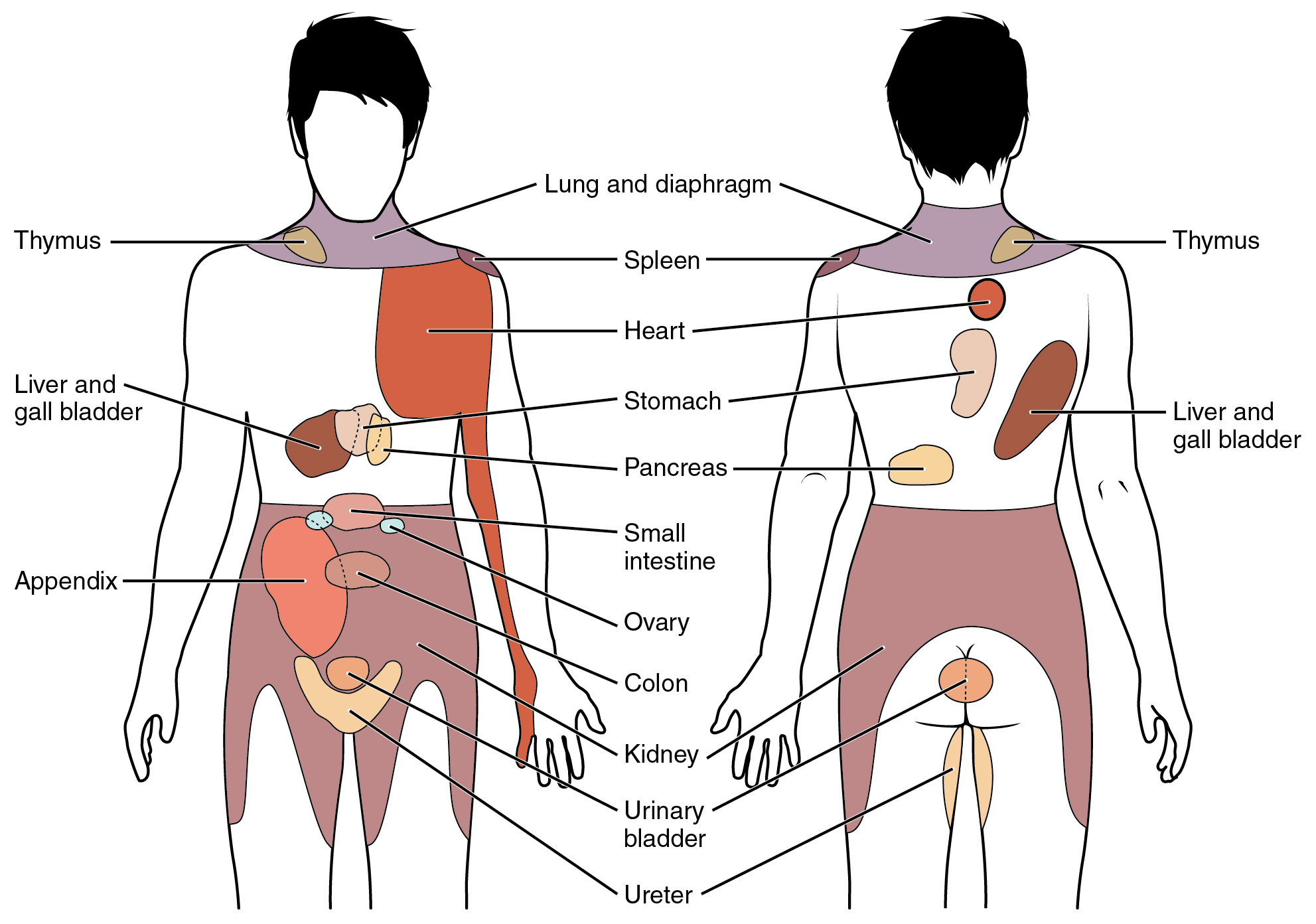
Conscious perception of visceral sensations map to specific regions of the body, as shown in this chart. Some sensations are felt locally, whereas others are perceived as affecting areas that are quite distant from the involved organ.
Identifiers
MeSHD053591
Anatomical terminology

Referred pain, also called reflective pain,[1] is pain perceived at a location other than the site of the painful stimulus. An example is the case of angina pectoris brought on by a myocardial infarction (heart attack), where pain is often felt in the left side of the neck, left shoulder, and back rather than in the thorax (chest), the site of the injury. The International Association for the Study of Pain has not officially defined the term; hence, several authors have defined it differently. Referred pain has been described since the late 1880s. Despite an increasing amount of literature on the subject, the biological mechanism of referred pain is unknown, although there are several hypotheses.

Radiating pain is slightly different from referred pain; for example, the pain related to a myocardial infarction could either be referred or radiating pain from the chest. Referred pain is when the pain is located away from or adjacent to the organ involved; for instance, when a person has pain only in their jaw or left arm, but not in the chest. Radiating pain would have an origin, where the patient can perceive pain, but the pain also spreads ("radiates") out from this origin point to cause the pain to be perceived in a wider area in addition.

Characteristics

[edit]
  • The size of referred pain is related to the intensity and duration of ongoing/evoked pain.[1]
  • Temporal summation is a potent mechanism for generation of referred muscle pain.[1]
  • Central hyperexcitability is important for the extent of referred pain.[1]
  • Patients with chronic musculoskeletal pains have enlarged referred pain areas to experimental stimuli.[vague] The proximal spread of referred muscle pain is seen in patients with chronic musculoskeletal pain and very seldom is it seen in healthy individuals.[1]
  • Modality-specific somatosensory changes occur in referred areas, which emphasize the importance of using a multimodal sensory test regime for assessment.[1]
  • Referred pain is often experienced on the same side of the body as the source, but not always.[2]

Mechanism

[edit]

There are several proposed mechanisms for referred pain.[3] Currently there is no definitive consensus regarding which is correct. The cardiac general visceral sensory pain fibers follow the sympathetics back to the spinal cord and have their cell bodies located in thoracic dorsal root ganglia 1-4(5). As a general rule, in the thorax and abdomen, general visceral afferent (GVA) pain fibers follow sympathetic fibers back to the same spinal cord segments that gave rise to the preganglionic sympathetic fibers. The central nervous system (CNS) perceives pain from the heart as coming from the somatic portion of the body supplied by the thoracic spinal cord segments 1-4(5). Classically the pain associated with a myocardial infarction is located in the mid or left side of the chest where the heart is actually located. The pain can radiate to the left side of the jaw and into the left arm. Myocardial infarction can rarely present as referred pain and this usually occurs in people with[4] diabetes or older age. Also, the dermatomes of this region of the body wall and upper limb have their neuronal cell bodies in the same dorsal root ganglia (T1-5) and synapse in the same second order neurons in the spinal cord segments (T1-5) as the general visceral sensory fibers from the heart. The CNS does not clearly discern whether the pain is coming from the body wall or from the viscera, but it perceives the pain as coming from somewhere on the body wall, i.e. substernal pain, left arm/hand pain, jaw pain.[citation needed]

Convergent-projection

[edit]

This represents one of the earliest theories on the subject of referred pain. It is based on the work of W.A. Sturge and J. Ross from 1888 and later TC Ruch in 1961. Convergent projection proposes that afferent nerve fibers from tissues converge onto the same spinal neuron, and explains why referred pain is believed to be segmented in much the same way as the spinal cord. Additionally, experimental evidence shows that when local pain (pain at the site of stimulation) is intensified the referred pain is intensified as well.[citation needed]

Criticism of this model arises from its inability to explain why there is a delay between the onset of referred pain after local pain stimulation. Experimental evidence also shows that referred pain is often unidirectional. For example, stimulated local pain in the anterior tibial muscle causes referred pain in the ventral portion of the ankle; however referred pain moving in the opposite direction has not been shown experimentally. Lastly, the threshold for the local pain stimulation and the referred pain stimulation are different, but according to this model they should both be the same.[1]

Convergence-facilitation

[edit]

Convergence facilitation was conceived in 1893 by J MacKenzie based on the ideas of Sturge and Ross. He believed that the internal organs were insensitive to stimuli. Furthermore, he believed that non-nociceptive afferent inputs to the spinal cord created what he termed "an irritable focus". This focus caused some stimuli to be perceived as referred pain. However, his ideas did not gain widespread acceptance from critics due to its dismissal of visceral pain.[citation needed]

Recently this idea has regained some credibility under a new term, central sensitization. Central sensitization occurs when neurons in the spinal cord's dorsal horn or brainstem become more responsive after repeated stimulation by peripheral neurons, so that weaker signals can trigger them. The delay in appearance of referred pain shown in laboratory experiments can be explained due to the time required to create the central sensitization.[1]

Axon-reflex

[edit]

Axon reflex suggests that the afferent fiber is bifurcated before connecting to the dorsal horn. Bifurcated fibers do exist in muscle, skin, and intervertebral discs. Yet these particular neurons are rare and are not representative of the whole body. Axon-Reflex also does not explain the time delay before the appearance of referred pain, threshold differences for stimulating local and referred pain, and somatosensory sensibility changes in the area of referred pain.[1]

Hyperexcitability

[edit]

Hyperexcitability hypothesizes that referred pain has no central mechanism. However, it does say that there is one central characteristic that predominates. Experiments involving noxious stimuli and recordings from the dorsal horn of animals revealed that referred pain sensations began minutes after muscle stimulation. Pain was felt in a receptive field that was some distance away from the original receptive field. According to hyperexcitability, new receptive fields are created as a result of the opening of latent convergent afferent fibers in the dorsal horn. This signal could then be perceived as referred pain.[citation needed]

Several characteristics are in line with this mechanism of referred pain, such as dependency on stimulus and the time delay in the appearance of referred pain as compared to local pain. However, the appearance of new receptive fields, which is interpreted to be referred pain, conflicts with the majority of experimental evidence from studies including studies of healthy individuals. Furthermore, referred pain generally appears within seconds in humans as opposed to minutes in animal models. Some scientists attribute this to a mechanism or influence downstream in the supraspinal pathways. Neuroimaging techniques such as PET scans or fMRI may visualize the underlying neural processing pathways responsible in future testing.[1]

Thalamic-convergence

[edit]

Thalamic convergence suggests that referred pain is perceived as such due to the summation of neural inputs in the brain, as opposed to the spinal cord, from the injured area and the referred area. Experimental evidence on thalamic convergence is lacking. However, pain studies performed on monkeys revealed convergence of several pathways upon separate cortical and subcortical neurons.[citation needed]

Examples

[edit]
Location Description
Upper chest/left limb Myocardial ischaemia (the loss of blood flow to a part of the heart muscle tissue) is possibly the best known example of referred pain; the sensation can occur in the upper chest as a restricted feeling, or as an ache in the left shoulder, arm or even hand.
Head "Ice-cream headache" or "brain freeze" is another example of referred pain, in which the vagus nerve or the trigeminal nerve in the throat and the palate, respectively, transmit pain signals following rapid cooling and rewarming of the capillaries in the sinuses.[5]
General Phantom limb pain, a type of referred pain, is the sensation of pain from a limb that has been lost or from which a person no longer receives physical signals. It is an experience almost universally reported by amputees and quadriplegics.
Right tip of scapula Liver, gallbladder[citation needed]
Umbilicus Pain in ovary
Left shoulder Thoracic diaphragm, spleen (Kehr's sign), lung[citation needed]
Back Low back pain[6]
Palm of hand Palmaris longus,[7] a problem originating in the forearm might be felt in the palm, and not in the forearm.

Laboratory testing methods

[edit]

Pain is studied in a laboratory setting due to the greater amount of control that can be exerted. For example, the modality, intensity, and timing of painful stimuli can be controlled with much more precision. Within this setting there are two main ways that referred pain is studied.[citation needed]

Algogenic substances

[edit]

In recent years several different chemicals have been used to induce referred pain including bradykinin, substance P, capsaicin,[8] and serotonin. However, before any of these substances became widespread in their use a solution of hypertonic saline was used instead. Through various experiments it was determined that there were multiple factors that correlated with saline administration such as infusion rate, saline concentration, pressure, and amount of saline used. The mechanism by which the saline induces a local and referred pain pair is unknown. Some researchers have commented that it could be due to osmotic differences, however that is not verified.[1]

Using electrical stimulation

[edit]

Intramuscular electrical stimulation (IMES) of muscle tissue has been used in various experimental and clinical settings. The advantage to using an IMES system over a standard such as hypertonic saline is that IMES can be turned on and off. This allows the researcher to exert a much higher degree of control and precision in terms of the stimulus and the measurement of the response. The method is easier to carry out than the injection method as it does not require special training in how it should be used. The frequency of the electrical pulse can also be controlled. For most studies a frequency of about 10 Hz is needed to stimulate both local and referred pain.[clarification needed][9]

Using this method it has been observed that significantly higher stimulus strength is needed to obtain referred pain relative to the local pain. There is also a strong correlation between the stimulus intensity and the intensity of referred and local pain. It is also believed that this method causes a larger recruitment of nociceptor units resulting in a spatial summation. This spatial summation results in a much larger barrage of signals to the dorsal horn and brainstem neurons.[1]

Use in clinical diagnosis and treatments

[edit]

Referred pain can be indicative of nerve damage. A case study done on a 63-year-old man with an injury sustained during his childhood developed referred pain symptoms after his face or back was touched. After even a light touch, there was a shooting pain in his arm. The study concluded that his pain was possibly due to a neural reorganization which sensitized regions of his face and back after the nerve damage occurred. It is mentioned that this case is very similar to what phantom limb syndrome patients experience. This conclusion was based on experimental evidence gathered by V. S. Ramachandran in 1993, with the difference being that the arm that is in pain is still attached to the body.[citation needed]

Orthopedic diagnosis

[edit]

From the above examples one can see why understanding of referred pain can lead to better diagnoses of various conditions and diseases. In 1981 physiotherapist Robin McKenzie described what he termed centralization. He concluded that centralization occurs when referred pain moves from a distal to a more proximal location. Observations in support of this idea were seen when patients would bend backward and forward during an examination.[citation needed]

Studies have reported that the majority of patients that experienced centralization were able to avoid spinal surgery through isolating the area of local pain. However, the patients who did not experience centralization had to undergo surgery to diagnose and correct the problems. As a result of this study there has been a further research into the elimination of referred pain through certain body movements.[citation needed]

One example of this is referred pain in the calf. McKenzie showed that the referred pain would move closer to the spine when the patient bent backwards in full extension a few times. More importantly, the referred pain would dissipate even after the movements were stopped.[10]

General diagnosis

[edit]

As with myocardial ischaemia, referred pain in a certain portion of the body can lead to a diagnosis of the correct local center. Somatic mapping of referred pain and the corresponding local centers has led to various topographic maps being produced to aid in pinpointing the location of pain based on the referred areas. For example, local pain stimulated in the esophagus is capable of producing referred pain in the upper abdomen, the oblique muscles, and the throat. Local pain in the prostate can radiate referred pain to the abdomen, lower back, and calf muscles. Kidney stones can cause visceral pain in the ureter as the stone is slowly passed into the excretory system. This can cause immense referred pain in the lower abdominal wall.[11]

Further, recent research has found that ketamine, a sedative, is capable of blocking referred pain. The study was conducted on patients with fibromyalgia, a disease characterized by joint and muscle pain and fatigue. These patients were looked at specifically due to their increased sensitivity to nociceptive stimuli. Furthermore, referred pain appears in a different pattern in fibromyalgic patients than non-fibromyalgic patients. Often this difference manifests as a difference in terms of the area that the referred pain is found (distal vs. proximal) as compared to the local pain. The area is also much more exaggerated owing to the increased sensitivity.[12]

See also

[edit]

References

[edit]
Revisions and contributorsEdit on WikipediaRead on Wikipedia
from Grokipedia
Referred pain is a clinical phenomenon characterized by the perception of pain at a site distant from the actual source of noxious stimulation, often without involvement of nerve root compression or direct injury to the referred area.[1] This type of pain typically arises from somatic structures such as muscles, joints, or viscera and is mediated by shared neural pathways in the central nervous system, leading to mislocalization by the brain.[2] The primary mechanism underlying referred pain is the convergence-projection theory, first proposed by Ruch in 1961, which posits that visceral or deep somatic nociceptive afferents converge with somatic afferents from superficial structures onto the same second-order neurons in the spinal cord or brainstem.[1] This convergence causes the central nervous system to attribute the pain signal to the more familiar somatic dermatome or myotome rather than the true origin, such as interpreting cardiac ischemia as arm or jaw discomfort.[3] Additional mechanisms include dichotomizing afferent fibers, where single nociceptors branch to both the primary painful site and a distant referral zone, potentially activating reflex arcs that amplify the sensation.[2] These processes often result in central sensitization, enhancing pain transmission and contributing to secondary hyperalgesia in the referred area.[2] Clinically, referred pain presents as a dull, aching, or pressing sensation that spreads across broad regions, frequently overlapping with dermatomal patterns but not strictly confined to them, and it is reported in 17% to 84% of cases involving low back pain or other musculoskeletal disorders.[2] Common examples include myocardial infarction pain radiating to the left shoulder and neck, or gallbladder disease perceived in the right scapular region.[1] Diagnosis can be challenging due to these overlapping patterns, often leading to misattribution and unnecessary interventions, such as dental extractions for jaw pain actually stemming from cardiac or temporomandibular sources.[3] Management typically targets the primary lesion through local anesthetic blocks, radiofrequency ablation, or surgical intervention, though outcomes remain variable and require further research for optimization.[2]

Fundamentals

Definition

Referred pain is the perception of pain in a somatic region distant from the site of noxious stimulation or tissue damage, arising from the shared innervation of visceral or deep somatic structures with superficial somatic areas via common spinal cord segments.[4] This occurs because afferent nerves from internal organs or deep tissues, such as those from the viscera or musculoskeletal structures, converge on the same dorsal horn neurons in the spinal cord as nerves from the skin or superficial muscles.[2] Nociception forms the foundational process in referred pain, involving the detection of potentially harmful stimuli—such as mechanical injury, extreme temperatures, or chemical irritants—by specialized peripheral sensory receptors known as nociceptors, which transmit signals through primary afferent fibers to the central nervous system.[4] In this context, the primary zone refers to the localized area of actual noxious stimulation or injury, where pain is initially generated, whereas the secondary zone denotes the remote somatic area where the pain is subsequently perceived due to neural overlap.[4] The scope of referred pain excludes conditions like radicular pain, which stems from direct irritation or compression of spinal nerve roots and manifests along a dermatomal distribution often with neurological deficits, and phantom pain, which involves sensations in an absent body part following amputation.[2]

Historical Development

The concept of referred pain emerged in the 19th century through early clinical and experimental observations of visceral-somatic pain patterns. French physiologist Claude Bernard, in his experiments during the 1850s, demonstrated that stimulation of anterior spinal roots could produce pain resembling visceral sensations, and that sectioning posterior roots failed to alleviate certain internal pains, suggesting alternative pathways for visceral afferent signals beyond traditional sensory routes.[5] This laid foundational insights into the non-localized nature of some pain experiences, shifting attention from purely somatic explanations. Building on such physiological groundwork, Louis Antoine Ranvier proposed an early theory of referred pain in 1875, attributing it to the branching of axons discovered via Camillo Golgi's silver staining method (the "black reaction" of 1873), which allowed irritated visceral fibers to converge on somatic nerve branches, displacing pain perception to the skin surface.[6] The late 19th century saw more systematic clinical mapping, with British neurologist Sir Henry Head's 1893 thesis on "Disturbances of Sensation with Especial Reference to the Pain of Visceral Disease" identifying zonal patterns of hyperalgesia (now known as Head's zones) through studies of herpes zoster and visceral disorders, establishing dermatomal overlaps as key to referral mechanisms.[7] Concurrently, physician James MacKenzie documented similar sensory referrals from viscera to somatic areas in 1893, linking them to deep nerve irritations.[8] In the 1920s, French surgeon René Leriche advanced surgical perspectives on pain management, observing through sympathectomies and nerve blocks that interrupting autonomic pathways could alter referred visceral pain, as detailed in his clinical reports on ischemia and neuralgia.[9] The 1930s marked a milestone with experimental validations, as John H. Kellgren's hypertonic saline injections in humans (1938–1939) confirmed segmental referral patterns from deep somatic and visceral sites, providing empirical maps that refined Head's zones. This era's pattern theory of pain, emphasizing temporal and spatial summation, influenced the 1930s–1950s research landscape, culminating in Ronald Melzack and Patrick Wall's gate control theory (1965), which integrated referral via spinal gating of convergent inputs from visceral and somatic afferents.[10] Post-1960s, understanding evolved toward neurophysiological models, with functional neuroimaging like fMRI revealing central convergence in areas such as the insula and thalamus during referred pain tasks, bridging anatomical observations with brain-level processing.[2] This progression highlighted a shift from static anatomical views to dynamic, integrative explanations, though gaps persisted in pre-20th-century mechanistic details.

Characteristics

Key Features

Referred pain exhibits distinct temporal characteristics compared to primary pain at the injury site. It often demonstrates a delayed onset, typically emerging seconds to minutes after the initial stimulus, once local pain has persisted for a period.[11] Furthermore, referred pain may continue for minutes to hours even after removal of the stimulus, sometimes becoming fixed in a specific region.[12][2] Spatially, referred pain manifests in areas remote from the primary zone, frequently aligning with dermatomal or myotomal distributions but not strictly following dermatomal boundaries like radicular pain.[13][2] These patterns can occur ipsilaterally or, less commonly, contralaterally to the origin.[14] In terms of intensity and quality, referred pain is generally milder and less localized than primary pain, presenting as dull, aching, gnawing, or burning sensations rather than sharp or shooting.[2] Its perception can be modulated by emotional factors, with higher negative emotionality—encompassing distress, fear, and catastrophic thinking—linked to increased odds of experiencing referred pain and greater local intensity.[15] Physiologically, episodes of referred pain are often accompanied by autonomic manifestations in the referral zones, including sudomotor changes like altered sweating and vasomotor responses such as skin temperature or color variations.[16] Recent studies highlight gender differences in referral patterns, with women tending to exhibit larger areas of referred pain and prolonged persistence compared to men in experimental models of visceral hypersensitivity.[17][18]

Distinctions from Other Pain Types

Referred pain differs from radicular pain primarily in its etiology and distribution pattern. Radicular pain arises from compression or irritation of a spinal nerve root, often due to conditions like herniated discs or spinal stenosis, resulting in sharp, shooting pain that radiates along a specific dermatome with associated neurological deficits such as numbness or weakness.[19] In contrast, referred pain lacks direct nerve root involvement and does not follow dermatomal boundaries; it typically manifests as a dull, aching sensation in a broader, non-segmental area away from the injury site, without motor or sensory loss.[2] Unlike phantom limb pain, which occurs in the absence of a limb following amputation and stems from maladaptive cortical reorganization in the somatosensory cortex, referred pain involves intact body parts and arises from actual nociceptive stimuli in visceral or somatic structures.[20] Phantom limb pain is characterized by perceptions of movement or sensation in the missing limb, driven by remapping of adjacent cortical areas, whereas referred pain reflects a misattribution of signals from an existing source to a distant site due to neural convergence.[21] Referred pain is also distinct from allodynia and hyperalgesia, which are hallmarks of central sensitization in neuropathic conditions. Allodynia involves pain elicited by non-noxious stimuli, such as light touch, due to lowered pain thresholds, while hyperalgesia denotes an exaggerated response to normally painful stimuli.[22] Referred pain, however, represents a location-specific mislocalization of nociceptive input without inherent amplification of stimulus intensity; it can coexist with these phenomena but is defined by the perceptual shift rather than altered sensory processing at the site of referral.[23] In comparison to sympathetically maintained pain (SMP), which is a form of neuropathic pain sustained by aberrant sympathetic efferent activity and relieved by sympathetic nerve blockade, referred pain does not depend on sympathetic outflow for its persistence.[24] SMP, often seen in complex regional pain syndrome, features autonomic changes like vasomotor instability and responds to interventions targeting the sympathetic chain, whereas referred pain typically resolves with treatment of the primary nociceptive source and shows no such blockade dependency.[22] Modern neuroimaging, particularly functional magnetic resonance imaging (fMRI) studies since 2000, has provided evidence for these phenomenological distinctions through differential neural activation patterns. For instance, visceral stimuli inducing referred pain activate distinct brainstem and cortical regions, such as the periaqueductal gray and anterior cingulate cortex, compared to the more localized somatosensory activations in radicular or somatic neuropathic pain.[25] Similarly, fMRI reveals that referred pain from visceral sources shares overlaps with somatic pain in the "pain matrix" but exhibits unique connectivity in the insula and thalamus, differentiating it from the cortical remapping seen in phantom limb pain or the sensitized pathways in allodynia/hyperalgesia.[26] These findings underscore how referred pain involves convergent projections without the structural or sensitization elements characteristic of other pain types.[27]

Mechanisms

Convergent Projection Theory

The convergent projection theory, first formalized by Ruch in 1961, explains referred pain as resulting from the anatomical convergence of nociceptive afferent fibers from visceral and somatic structures onto common second-order neurons in the dorsal horn of the spinal cord, particularly within the spinothalamic tract.[1] This shared projection modifies the classical labeled line theory, in which specific sensory pathways transmit distinct signals to the brain; instead, the brain attributes the pain to the somatic territory due to its greater density of sensory innervation and prior associative learning, leading to mislocalization of visceral pain signals.[28] Anatomically, this convergence occurs at specific spinal segments where visceral and somatic inputs overlap; for instance, cardiac visceral afferents entering at segments T1-T5 project to the same dorsal horn neurons as somatic afferents from the left arm and chest wall, resulting in referred pain during myocardial ischemia.[29] Early evidence supporting this theory came from animal experiments in the 1940s, such as those by Sinclair et al., who injected hypertonic saline into deep somatic tissues in humans and observed pain referral patterns consistent with multi-receptive fields in spinal neurons receiving inputs from both primary and secondary sites. Despite its foundational role, the theory has limitations in accounting for certain clinical observations, such as the delayed onset of referred pain after intense or prolonged visceral stimulation, which suggests additional central processing beyond simple projection overlap.[30] It also fails to fully explain modulatory influences from emotional states or higher cortical areas on referral patterns, indicating the involvement of supraspinal mechanisms not captured by spinal convergence alone.[30]

Convergence Facilitation Theory

The convergence facilitation theory posits that referred pain arises from the enhanced excitability of convergent neurons in the spinal cord, where subthreshold somatic inputs are amplified by visceral nociceptive signals, leading to the perception of pain in somatic regions. This mechanism builds upon the anatomical overlap of visceral and somatic afferents in the dorsal horn but emphasizes dynamic facilitation rather than mere convergence. Originally proposed by James Mackenzie in 1893, the theory suggests that irritation from internal organs lowers the threshold for somatic sensory activation in shared projection pathways, resulting in cutaneous or muscular pain referral without direct somatic injury.[31] At the core of this process is central sensitization in the dorsal horn, where somatic inputs facilitate visceral nociceptor signals through heterosynaptic potentiation. Repeated or sustained low-level visceral stimulation induces the wind-up phenomenon, a form of temporal summation that progressively lowers neuronal activation thresholds and amplifies responses to subsequent inputs. This involves N-methyl-D-aspartate (NMDA) receptor activation, triggered by glutamate release from primary afferents, which leads to intracellular calcium influx and phosphorylation of synaptic proteins, enhancing excitatory transmission in wide-dynamic-range neurons. Human studies from the 1990s using microneurography and psychophysical assessments demonstrated this temporal summation in referred pain zones, showing increased pain ratings and lowered thresholds in somatic areas during repeated muscle stimulation, indicative of facilitated central processing.[32][33][34] Recent research highlights the role of glial cells in this facilitation, particularly astrocytes and microglia in the dorsal horn, which contribute to sustained sensitization beyond acute neuronal changes. Activated glia release proinflammatory cytokines such as tumor necrosis factor-alpha (TNF-α) and interleukin-1β (IL-1β), which further potentiate NMDA receptor function and promote synaptic plasticity in response to visceral inputs. For instance, studies since 2015 have shown that TNF-α from spinal glia enhances visceral nociception and associated somatic referral in models of chronic inflammation, amplifying the disproportionate pain response to minor visceral irritation observed clinically, such as in irritable bowel syndrome where gut signals elicit widespread abdominal wall pain. This glial-neuronal interaction explains the persistence of referred pain and its resistance to peripheral interventions.[35]

Axon Reflex Theory

The axon reflex theory posits that referred pain arises from the peripheral branching of nociceptor axons, known as dichotomizing fibers, which innervate both the primary site of injury and remote tissues. When nociceptors are stimulated at the injury site, the impulse travels orthodromically along the main axon but also propagates antidromically through collateral branches to distant areas, activating sensory endings there without central nervous system involvement. This peripheral mechanism explains how local stimulation can elicit pain or sensations in anatomically separate but axonally connected regions, such as from a lumbar disc to the groin.[36] Physiologically, antidromic activation triggers the release of neuropeptides, including substance P and calcitonin gene-related peptide (CGRP), from the peripheral terminals of these branched axons. These neuropeptides induce neurogenic inflammation in the referral zone, manifesting as vasodilation, plasma extravasation, and a characteristic flare response that contributes to perceived pain. For instance, substance P sensitizes local nociceptors and promotes the release of inflammatory mediators, amplifying pain signals in the distant area. This process is mediated by unmyelinated C-fibers, which are prevalent in cutaneous and some deep tissues. Early evidence for this theory stems from observations of the triple response in human skin, where mechanical or chemical stimulation produces a localized red line, a surrounding flare, and a wheal, attributed to axon reflex-mediated neuropeptide release. This was first detailed in studies of cutaneous vascular responses, demonstrating antidromic vasodilation independent of central pathways. More recent double-labeling immunohistochemistry in animal models has confirmed dichotomizing axons innervating visceral structures like the lumbar disc and somatic sites such as the groin, supporting the theory's applicability to certain somatic referrals. Despite these findings, the axon reflex theory primarily accounts for superficial, cutaneous referred pain and has limited explanatory power for deep or visceral referrals, where dichotomizing fibers constitute less than 0.5% of afferents and central mechanisms predominate.[37]

Hyperexcitability Theory

The hyperexcitability theory posits that peripheral or central injury induces a state of heightened neuronal excitability in the dorsal horn of the spinal cord, resulting in the expansion of receptive fields for wide dynamic range (WDR) neurons. This expansion allows these neurons to respond to inputs from remote or non-injured areas, thereby generating referred pain. Unlike normal sensory processing, where receptive fields are localized, injury-triggered hyperexcitability causes dorsal horn neurons to integrate signals from broader somatic territories, mimicking pain referral patterns observed clinically.[38] The underlying process involves molecular changes such as the upregulation of voltage-gated sodium channels, particularly Nav1.3, in second-order dorsal horn neurons following nerve injury. This upregulation facilitates ectopic firing and sustained depolarization, amplifying synaptic inputs and leading to central sensitization. Consequently, this manifests as secondary hyperalgesia, where innocuous stimuli from surrounding areas evoke exaggerated pain responses, contributing to the spatial spread characteristic of referred pain.[39] Evidence for this theory derives primarily from animal models in the 1980s, where C.J. Woolf and colleagues demonstrated that conditioning stimulation of C-fibers in rats caused prolonged expansion of mechanical receptive fields in dorsal horn neurons, persisting beyond the stimulus duration. Post-nerve damage experiments further showed that dorsal horn WDR neurons developed novel receptive fields in remote dermatomes, correlating with behavioral signs of referred hyperalgesia. These findings established hyperexcitability as a key driver of receptive field plasticity in pain referral.[38][40] In clinical contexts, this theory explains chronic referred pain in conditions like fibromyalgia, where widespread musculoskeletal pain arises from augmented central excitability without identifiable peripheral injury. Patients exhibit expanded pain referral zones and secondary hyperalgesia, linked to sustained dorsal horn hyperexcitability that amplifies visceral and somatic inputs across multiple body regions.[41] Recent research from the 2010s has incorporated epigenetic mechanisms to account for the persistence of this hyperexcitability. For instance, injury-induced DNA methylation changes in dorsal horn neurons regulate genes like BDNF and Cdk5, promoting long-term upregulation of excitability-related proteins and maintaining expanded receptive fields in chronic pain states. These epigenetic modifications, such as histone acetylation and TET enzyme-mediated demethylation, provide a molecular basis for why hyperexcitability transitions from acute to enduring, influencing referred pain in neuropathic disorders.[42][43]

Thalamic Convergence Theory

The Thalamic Convergence Theory proposes that referred pain results from the integration of divergent somatosensory inputs within specific thalamic nuclei, particularly the ventral posterolateral (VPL) and ventral posteromedial (VPM) nuclei, which relay and process nociceptive signals to cortical areas, often leading to erroneous localization onto somatic body maps. This mechanism involves the convergence of visceral and somatic afferents at the thalamic level, where neurons fail to distinguish the origin of the input due to shared receptive fields, causing pain to be projected to anatomically unrelated but representationally adjacent regions in the somatosensory cortex. Originally articulated by Theobald in 1941, the theory emphasizes a central summation process in the thalamus as a key contributor to pain referral, distinct from peripheral or spinal-level convergence.[44][28][45] A critical aspect of this theory lies in the overlap of thalamo-cortical projections, where fibers from multiple peripheral sources terminate in overlapping thalamic territories, facilitating the blending of signals before cortical interpretation. Additionally, attentional modulation influences thalamic processing, as cognitive factors can amplify or alter the perceived location of pain by enhancing specific neural pathways within these nuclei. This higher-level integration builds upon prerequisite spinal convergence but shifts focus to supraspinal mislocalization. Evidence from functional magnetic resonance imaging (fMRI) studies in the 2000s, such as those examining visceral pain referral, has demonstrated increased thalamic activation in non-stimulated referral zones during experimentally induced angina-like stimuli, supporting the role of thalamic summation in generating referred sensations.[46][47][48] Recent advances, including diffusion tensor imaging (DTI) studies from 2022 onward, have revealed thalamic tract anomalies—such as reduced fractional anisotropy in thalamocortical pathways—in patients with chronic pain conditions, indicating microstructural disruptions that may perpetuate referral patterns by impairing precise signal localization. These findings underscore the theory's relevance to long-term pain disorders, where thalamic alterations contribute to sustained misreferral. However, the theory is primarily pertinent to the cognitive and perceptual dimensions of pain localization rather than initial nociceptive transmission, and it is considered secondary to foundational spinal mechanisms in most models of referred pain.[49][50] These theories are not mutually exclusive and often interact in the generation of referred pain.

Examples

Visceral Referred Pain

Visceral referred pain arises when nociceptive signals from internal organs are misinterpreted by the central nervous system as originating from somatic structures on the body surface, often due to shared neural pathways such as convergent projection.[51] In the cardiac system, myocardial ischemia, as seen in angina pectoris, commonly produces referred pain to the left arm, jaw, or chest wall. This pattern occurs because cardiac visceral afferents converge with somatic afferents from dermatomes T1-T5 in the spinal cord. Women are more likely to experience atypical cardiac referred pain, such as to the back, jaw, or epigastrium, compared to men who more often report classic left-arm radiation.[51][52][53] Gastrointestinal disorders provide classic examples of visceral referral. Acute appendicitis initially causes diffuse periumbilical pain due to midgut innervation at the T10 spinal segment, which later localizes to the right lower quadrant as parietal peritoneum involvement occurs.[54] Similarly, biliary colic from gallstones can refer pain to the right shoulder via irritation of the phrenic nerve (originating from C3-C5), as the inflamed gallbladder shares diaphragmatic innervation with somatic shoulder regions.[55] These patterns often follow embryological origins, where foregut structures like the biliary system refer to the epigastrium, midgut to the umbilicus, and hindgut to the suprapubic area.[56] In the urogenital system, renal colic from kidney stones typically manifests as severe flank pain that radiates to the groin or lower abdomen, reflecting the stone's migration along the ureter and convergence of visceral afferents with somatic nerves from T12-L1 segments.[57] This referral can intensify as the stone approaches the ureterovesical junction, mimicking testicular or labial pain in some cases.[58]

Somatic Referred Pain

Somatic referred pain arises from musculoskeletal structures, such as muscles, joints, ligaments, and deep tissues, and is perceived in distant somatic regions due to shared neural pathways.[2] Unlike localized pain, it typically manifests as a dull ache that does not follow the exact anatomical path of the affected structure, often complicating diagnosis. In hip osteoarthritis, pain originating from the joint frequently refers to the knee, mimicking primary knee pathology and delaying identification of the hip source.[59] This referral occurs via shared innervation from the L3-L4 spinal segments, where hip joint afferents converge with those from the knee.[2] Similarly, shoulder impingement syndrome, involving compression of the rotator cuff tendons under the acromion, can produce pain radiating down the arm to the elbow region.[60] Myofascial trigger points in the upper trapezius muscle commonly refer pain to the temple and jaw, contributing to tension-type headaches or temporomandibular symptoms.[61] Palpation of these points elicits pain in the temple area in approximately 80% of cases, following predictable referral zones along the muscle's fascial attachments.[62] Deep somatic irritation, such as in pleurisy affecting the diaphragm, often refers pain to the neck or scapular area via the phrenic nerve (C3-C5 origins).[63] This can present as sharp, pleuritic pain exacerbated by breathing, spreading ipsilaterally to the shoulder blade.[64] Temporomandibular joint (TMJ) disorders frequently cause referred pain to the ear, known as otalgia, due to proximity and shared trigeminal nerve branches.[65] Post-2010 studies confirm this in up to 70% of TMD patients, with ear pain persisting even after TMJ treatment resolves jaw symptoms. Evidence from 2021 highlights auriculotemporal nerve sensitization as a key mechanism.[66] These patterns of somatic referred pain often align with fascial planes, where myofascial tension transmits signals, or shared myotomes, reflecting segmental spinal convergence.[2] For instance, trapezius referrals follow upper cervical myotomes, while hip-to-knee pain traces lumbosacral distributions.[67] Somatic referred pain patterns are generally limited to segmentally related or nearby areas and do not include distant cross-limb referrals from lower to upper extremities. For example, knee pain does not refer to the thumb and index finger of the hand; such a pattern is not recognized in medical literature. Referred somatic pain typically involves shared innervation within the same or adjacent spinal segments (e.g., hip to knee via L3-L4), whereas pain in both the knee and hand usually arises from separate local conditions (such as multiple joint osteoarthritis) or systemic diseases rather than direct referral.[2]

Dental and Orofacial Referred Pain

In dentistry, referred pain commonly occurs after procedures like root canal treatment, where inflammation or nerve irritation in the treated tooth can cause perceived pain in adjacent teeth, front teeth, or even the opposite jaw side. This arises from convergence of nociceptive signals in the trigeminal nerve system, leading patients to report "random" tooth pain unrelated to visible issues in those teeth. Other examples include myofascial pain from jaw muscles referring to teeth, or sinus issues mimicking upper tooth pain.

Experimental Methods

Algogenic Substance Techniques

Algogenic substance techniques involve the administration of chemical agents that excite nociceptors to induce localized and referred pain in controlled human experiments, providing insights into pain referral pathways. Common substances include hypertonic saline, capsaicin, and bradykinin, which are injected subcutaneously or intramuscularly to simulate inflammatory nociceptor activation without causing tissue damage. These methods, pioneered in the late 1930s, allow researchers to quantify pain characteristics and referral patterns under standardized conditions.[68] Protocols typically entail injecting small volumes of the agent—such as 0.1–0.5 mL of 5–6% hypertonic saline, 0.01–1% capsaicin solution, or 10 μmol bradykinin—into targeted muscles like the tibialis anterior or masseter using a syringe or infusion pump for precise dosing. Pain onset occurs rapidly (within 10–60 seconds), with intensity rated on a 0–10 visual analog scale (VAS) and referred areas delineated through participant pain drawings at intervals up to 30 minutes post-injection. Measurements include latency to referral (often 20–30 seconds for saline), referral area size, and duration (typically 3–10 minutes), enabling dose-response analyses by varying concentrations or volumes. Bradykinin may be combined with serotonin to enhance effects, while capsaicin injections are guided by ultrasound in some protocols to ensure intramuscular placement.[68][69] Key findings demonstrate that these techniques replicate clinical referred pain patterns; for instance, hypertonic saline injected into the calf induces thigh referral, mirroring somatic-visceral convergence, with VAS intensities reaching 4–6/10 and referral areas expanding with repeated stimuli. Capsaicin evokes burning, cramplike pain with frequent referral to adjacent dermatomes, activating TRPV1 receptors on C-fibers, while bradykinin produces a deeper ache that sensitizes surrounding tissues, supporting central facilitation mechanisms. Quantitative studies show reproducible referral latencies and areas, with hypertonic saline yielding the most consistent results across sessions. These patterns align with convergent projection theory observed in prior mechanistic research.[70][68][69] The advantages of algogenic substance techniques lie in their controlled, reproducible nature, allowing ethical exploration of pain modulation, hyperalgesia, and analgesic efficacy without invasive procedures. They facilitate dose-response curves and mechanistic validation, with hypertonic saline being the most utilized due to its safety profile—over 6,000 injections reported without serious adverse events in aggregated studies. Ethical guidelines mandate that stimuli remain below individual tolerance limits, with immediate termination options and debriefing to minimize distress. Recent advancements include alternatives like ATP analogs targeting P2X receptors for purinergic pain signaling, though their human application remains limited compared to traditional agents.[71][72][73]

Electrical Stimulation Methods

Electrical stimulation methods involve the application of controlled electrical currents to peripheral nerves or muscles to experimentally induce and characterize referred pain, allowing researchers to map pain referral patterns and receptive fields in healthy human subjects. These techniques primarily utilize transcutaneous electrical nerve stimulation (TENS), which applies surface electrodes non-invasively, or percutaneous intramuscular electrical stimulation (IMES), which employs needle electrodes for deeper tissue targeting. Frequencies typically range from 2 to 100 Hz, with lower frequencies (e.g., 2-10 Hz) often used to activate nociceptive afferents and elicit sustained pain responses, while higher frequencies (e.g., 90-130 Hz) can modulate pain perception but are less common for induction in experimental settings.[74] The protocol generally begins with threshold determination to distinguish local pain at the stimulation site from referred pain in distant areas. Electrodes are placed to target specific dermatomes or myotomes, such as the tibialis anterior muscle or groin region, with stimulus intensity gradually increased from subthreshold levels (e.g., 0.02 mA) until pain ratings reach a predefined level, like 6/10 on a numerical rating scale (NRS). For IMES, rectangular pulses of 0.2-0.4 ms duration are delivered at intensities 50-150% above the local pain threshold, often for 10 minutes to assess temporal summation and referral onset, which is typically delayed by 20-40 seconds compared to local pain. Referred pain thresholds are consistently higher than local ones (e.g., 72% greater on average), and stimulation is repeated across sessions to evaluate reproducibility, with pain areas mapped via subject drawings or verbal reports.[74][74] Key findings demonstrate that high-frequency stimulation (e.g., 10-50 Hz) can mimic aspects of chronic referred pain by facilitating spatial and temporal summation, expanding pain referral areas (correlations of r=0.74-0.98 between intensity and area size), and altering cutaneous hyperalgesia thresholds by 2-4°C in both local and referred zones. In healthy subjects, these methods reliably map receptive fields, revealing reproducible referral patterns such as from the abdominal rectus to the thigh or testicle (90-100% consistency across sessions). Recent advancements post-2015 integrate microneurography with intraneural electrical microstimulation (INMS), enabling selective activation of specific afferent fibers (e.g., C-mechanosensitive nociceptors) to study fiber-type contributions to referred sensations, which often manifest as dull or sharp pain depending on skin type.[74][75] Advantages of electrical stimulation include non-invasive options like TENS for initial screening, precise segmental targeting without tissue damage or pharmacological side effects, and high reproducibility for longitudinal studies. These methods provide a controlled, quantifiable model for investigating pain convergence, superior to chemical induction in terms of rapid onset and adjustable parameters.[74][75]

Clinical Applications

Diagnostic Strategies

Referred pain patterns play a pivotal role in diagnosing underlying conditions across medical disciplines by enabling clinicians to trace symptoms to their visceral or somatic origins, often preventing misattribution to superficial issues. In orthopedics, knee pain may signal hip joint pathology, such as osteoarthritis, where pain from the hip is referred to the knee via shared neural pathways, necessitating evaluation of the hip to confirm the source.[76] In contrast, pain originating from the knee does not refer to distant upper limb areas such as the thumb and index finger, as such patterns are not recognized in medical literature; referred pain is typically segmental and involves nearby areas or specific visceral-somatic patterns, not cross-referral from lower to upper limbs. Pain in both the knee and hand is usually indicative of separate issues, such as arthritis affecting multiple joints, or systemic conditions, rather than direct referral from the knee.[2] Palpation serves as a key differentiator, with superficial tenderness suggesting joint or muscular sources while deeper, diffuse discomfort points to visceral referrals, allowing practitioners to distinguish between somatic and internal etiologies through careful assessment of pain quality and location.[77] In general diagnostic contexts, particularly emergency triage, referred pain from cardiac ischemia manifests as discomfort in the jaw, arm, or back, often identified by Levine's sign—a patient's instinctive clenched fist over the sternum—which has low sensitivity but high specificity for ischemic events when present.[78] For gastrointestinal disorders, Kehr's sign indicates diaphragmatic irritation from intra-abdominal pathology, such as splenic rupture, producing left shoulder pain that worsens with inspiration and guides urgent evaluation.[79] Core techniques for diagnosis include pain mapping, where patients delineate symptom distribution on body diagrams to reveal non-dermatomal patterns typical of visceral referrals, aiding localization beyond obvious sites.[80] Dermatome charts further assist by correlating reported pain areas with spinal nerve segments, helping differentiate radicular from convergent visceral pain in cases like cervical referrals.[81] Provocation tests, such as controlled hyperventilation, can elicit transient referred pain in cardiac conditions by inducing coronary spasm, providing dynamic confirmation when baseline symptoms are ambiguous.[82] Diagnostic challenges stem from the overlap between referred and localized pain, which can mimic unrelated conditions and contribute to misdiagnosis; for example, upper abdominal discomfort may represent cardiac ischemia rather than primary gastrointestinal issues, underscoring the importance of holistic history-taking and risk stratification to avert life-threatening delays.[83] Recent advancements integrate imaging modalities like positron emission tomography (PET) for confirmation, with 2010s studies using FDG-PET/MRI to visualize heightened metabolic activity in peripheral nerves and central pain pathways, thereby validating referred mechanisms in chronic cases and refining differential diagnoses.[84]

Therapeutic Interventions

Therapeutic interventions for referred pain primarily aim to address the underlying source of the nociceptive input or modulate the peripheral and central mechanisms that propagate the pain signal, thereby providing relief in both the primary and referred areas. Treating the primary site is a cornerstone approach, as resolution of the originating pathology often eliminates the referred component. For instance, in cases of cardiac ischemia causing referred pain to the left arm or jaw, percutaneous coronary intervention (PCI), such as angioplasty, has been shown to provide significant symptom relief by restoring blood flow and reducing ischemic pain, which in turn alleviates the associated referred sensations.[85] Similarly, surgical interventions targeting visceral sources, like laparoscopic cholecystectomy for acute cholecystitis, can normalize somatosensory function in the referred pain area, such as the right shoulder, by eliminating the primary inflammatory stimulus.[86] Pharmacological management of the primary site, including nitrates or anti-inflammatory agents for conditions like angina or pancreatitis, further supports this strategy by directly mitigating the nociceptive drive without solely relying on invasive procedures.[2] Symptom management strategies focus on alleviating pain in the referral zones to improve patient comfort and function while the primary issue is addressed. Local anesthetics applied directly to the referred pain areas can interrupt peripheral sensitization and provide temporary relief; for example, ketocaine compresses in labor-related referred pain has demonstrated efficacy in reducing discomfort in a randomized, double-blind study.[87] Transcutaneous electrical nerve stimulation (TENS) modulates pain transmission by activating non-nociceptive afferents, with evidence from experimental models showing that both high- and low-frequency TENS significantly reduce the intensity of referred hyperalgesia induced by intramuscular hypertonic saline, without affecting primary guarding behaviors.[88] Additionally, cognitive-behavioral therapy (CBT) targets pain perception and coping mechanisms, reducing overall distress and improving daily functioning in chronic pain conditions that include referred components, as supported by meta-analyses of behavioral interventions.[89] Mechanism-based interventions leverage neurophysiological insights, such as convergence and sensitization, to disrupt pain referral pathways. NMDA receptor antagonists, like ketamine, counteract central sensitization that amplifies referred pain signals in neuropathic and inflammatory contexts, offering analgesic effects in chronic pain states where wind-up phenomena contribute to referral.[90] Nerve blocks targeting convergence sites in the spinal cord or peripheral nerves can selectively inhibit shared pathways; for example, local anesthetic blocks have successfully abolished referred pain elicited by hypertonic saline injections, confirming their role in interrupting convergent projections from visceral to somatic territories.[2] Neuromodulation devices represent an advanced option for refractory referred pain, particularly in chronic myofascial or neuropathic cases. Spinal cord stimulators (SCS) deliver electrical impulses to the dorsal columns, modulating ascending pain signals and providing substantial relief (70-90%) in axial and extremity pain, including patterns consistent with referral from cervical or lumbar sources.[91] Post-2020 advancements, such as closed-loop SCS systems, enhance efficacy by adapting stimulation in real-time to neural activity, yielding superior pain reduction and functional improvements compared to traditional open-loop devices in chronic pain cohorts.[92] Clinical outcomes from randomized controlled trials (RCTs) underscore these benefits; for instance, a 2016 RCT on trigger point injections with lidocaine for myofascial pain syndrome demonstrated significant reductions in referred pain intensity and improved range of motion when combined with physical therapy, compared to either alone.[93] Overall, multimodal approaches integrating these interventions yield the most durable results, with RCTs from the 2000s highlighting up to 50% pain reduction in myofascial referral patterns following targeted injections.[94]

References

User Avatar
No comments yet.