Recent from talks
Physics of magnetic resonance imaging
Knowledge base stats:
Talk channels stats:
Members stats:
Physics of magnetic resonance imaging
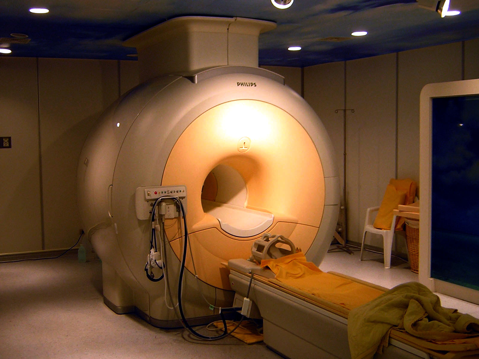
Magnetic resonance imaging (MRI) is a medical imaging technique mostly used in radiology and nuclear medicine in order to investigate the anatomy and physiology of the body, and to detect pathologies including tumors, inflammation, neurological conditions such as stroke, disorders of muscles and joints, and abnormalities in the heart and blood vessels among other things. Contrast agents may be injected intravenously or into a joint to enhance the image and facilitate diagnosis. Unlike CT and X-ray, MRI uses no ionizing radiation and is, therefore, a safe procedure suitable for diagnosis in children and repeated runs. Patients with specific non-ferromagnetic metal implants, cochlear implants, and cardiac pacemakers nowadays may also have an MRI in spite of effects of the strong magnetic fields. This does not apply on older devices, and details for medical professionals are provided by the device's manufacturer.
Certain atomic nuclei are able to absorb and emit radio frequency energy when placed in an external magnetic field. In clinical and research MRI, hydrogen atoms are most often used to generate a detectable radio-frequency signal that is received by antennas close to the anatomy being examined. Hydrogen atoms are naturally abundant in people and other biological organisms, particularly in water and fat. For this reason, most MRI scans essentially map the location of water and fat in the body. Pulses of radio waves excite the nuclear spin energy transition, and magnetic field gradients localize the signal in space. By varying the parameters of the pulse sequence, different contrasts may be generated between tissues based on the relaxation properties of the hydrogen atoms therein.
When inside the magnetic field (B0) of the scanner, the magnetic moments of the protons align to be either parallel or anti-parallel to the direction of the field. While each individual proton can only have one of two alignments, the collection of protons appear to behave as though they can have any alignment. Most protons align parallel to B0 as this is a lower energy state. A radio frequency pulse is then applied, which can excite protons from parallel to anti-parallel alignment; only the latter are relevant to the rest of the discussion. In response to the force bringing them back to their equilibrium orientation, the protons undergo a rotating motion (precession), much like a spun wheel under the effect of gravity. The protons will return to the low energy state by the process of spin-lattice relaxation. This appears as a changing magnetic flux, which yields a changing voltage in the receiver coils to give a signal. The frequency at which a proton or group of protons in a voxel resonates depends on the strength of the local magnetic field around the proton or group of protons, a stronger field corresponds to a larger energy difference and higher frequency photons. By applying additional magnetic fields (gradients) that vary linearly over space, specific slices to be imaged can be selected, and an image is obtained by taking the 2-D Fourier transform of the spatial frequencies of the signal (k-space). Due to the magnetic Lorentz force from B0 on the current flowing in the gradient coils, the gradient coils will try to move producing loud knocking sounds, for which patients require hearing protection.
The MRI scanner was developed from 1975 to 1977 at the University of Nottingham by Prof Raymond Andrew FRS FRSE following from his research into nuclear magnetic resonance. The full body scanner was created in 1978.
Subatomic particles have the quantum mechanical property of spin. Certain nuclei such as 1H (protons), 2H, 3He, 23Na or 31P, have a non–zero spin and therefore a magnetic moment. In the case of the so-called spin-1⁄2 nuclei, such as 1H, there are two spin states, sometimes referred to as up and down. Nuclei such as 12C have no unpaired neutrons or protons, and no net spin; however, the isotope 13C does.
When these spins are placed in a strong external magnetic field they precess around an axis along the direction of the field. Protons align in two energy eigenstates (the Zeeman effect): one low-energy and one high-energy, which are separated by a very small splitting energy.
Quantum mechanics is required to accurately model the behaviour of a single proton. However, classical mechanics can be used to describe the behaviour of an ensemble of protons adequately. As with other spin particles, whenever the spin of a single proton is measured it can only have one of two results commonly called parallel and anti-parallel. When we discuss the state of a proton or protons we are referring to the wave function of that proton which is a linear combination of the parallel and anti-parallel states.

In the presence of the magnetic field, B0, the protons will appear to precess at the Larmor frequency determined by the particle's gyro-magnetic ratio and the strength of the field. The static fields used most commonly in MRI cause precession which corresponds to a radiofrequency (RF) photon.[citation needed]
Hub AI
Physics of magnetic resonance imaging AI simulator
(@Physics of magnetic resonance imaging_simulator)
Physics of magnetic resonance imaging
Magnetic resonance imaging (MRI) is a medical imaging technique mostly used in radiology and nuclear medicine in order to investigate the anatomy and physiology of the body, and to detect pathologies including tumors, inflammation, neurological conditions such as stroke, disorders of muscles and joints, and abnormalities in the heart and blood vessels among other things. Contrast agents may be injected intravenously or into a joint to enhance the image and facilitate diagnosis. Unlike CT and X-ray, MRI uses no ionizing radiation and is, therefore, a safe procedure suitable for diagnosis in children and repeated runs. Patients with specific non-ferromagnetic metal implants, cochlear implants, and cardiac pacemakers nowadays may also have an MRI in spite of effects of the strong magnetic fields. This does not apply on older devices, and details for medical professionals are provided by the device's manufacturer.
Certain atomic nuclei are able to absorb and emit radio frequency energy when placed in an external magnetic field. In clinical and research MRI, hydrogen atoms are most often used to generate a detectable radio-frequency signal that is received by antennas close to the anatomy being examined. Hydrogen atoms are naturally abundant in people and other biological organisms, particularly in water and fat. For this reason, most MRI scans essentially map the location of water and fat in the body. Pulses of radio waves excite the nuclear spin energy transition, and magnetic field gradients localize the signal in space. By varying the parameters of the pulse sequence, different contrasts may be generated between tissues based on the relaxation properties of the hydrogen atoms therein.
When inside the magnetic field (B0) of the scanner, the magnetic moments of the protons align to be either parallel or anti-parallel to the direction of the field. While each individual proton can only have one of two alignments, the collection of protons appear to behave as though they can have any alignment. Most protons align parallel to B0 as this is a lower energy state. A radio frequency pulse is then applied, which can excite protons from parallel to anti-parallel alignment; only the latter are relevant to the rest of the discussion. In response to the force bringing them back to their equilibrium orientation, the protons undergo a rotating motion (precession), much like a spun wheel under the effect of gravity. The protons will return to the low energy state by the process of spin-lattice relaxation. This appears as a changing magnetic flux, which yields a changing voltage in the receiver coils to give a signal. The frequency at which a proton or group of protons in a voxel resonates depends on the strength of the local magnetic field around the proton or group of protons, a stronger field corresponds to a larger energy difference and higher frequency photons. By applying additional magnetic fields (gradients) that vary linearly over space, specific slices to be imaged can be selected, and an image is obtained by taking the 2-D Fourier transform of the spatial frequencies of the signal (k-space). Due to the magnetic Lorentz force from B0 on the current flowing in the gradient coils, the gradient coils will try to move producing loud knocking sounds, for which patients require hearing protection.
The MRI scanner was developed from 1975 to 1977 at the University of Nottingham by Prof Raymond Andrew FRS FRSE following from his research into nuclear magnetic resonance. The full body scanner was created in 1978.
Subatomic particles have the quantum mechanical property of spin. Certain nuclei such as 1H (protons), 2H, 3He, 23Na or 31P, have a non–zero spin and therefore a magnetic moment. In the case of the so-called spin-1⁄2 nuclei, such as 1H, there are two spin states, sometimes referred to as up and down. Nuclei such as 12C have no unpaired neutrons or protons, and no net spin; however, the isotope 13C does.
When these spins are placed in a strong external magnetic field they precess around an axis along the direction of the field. Protons align in two energy eigenstates (the Zeeman effect): one low-energy and one high-energy, which are separated by a very small splitting energy.
Quantum mechanics is required to accurately model the behaviour of a single proton. However, classical mechanics can be used to describe the behaviour of an ensemble of protons adequately. As with other spin particles, whenever the spin of a single proton is measured it can only have one of two results commonly called parallel and anti-parallel. When we discuss the state of a proton or protons we are referring to the wave function of that proton which is a linear combination of the parallel and anti-parallel states.
In the presence of the magnetic field, B0, the protons will appear to precess at the Larmor frequency determined by the particle's gyro-magnetic ratio and the strength of the field. The static fields used most commonly in MRI cause precession which corresponds to a radiofrequency (RF) photon.[citation needed]
Recent media