Hubbry Logo
VenuleVenuleMain
Open search
Venule
Community hub
Venule
logo
8 pages, 0 posts
0 subscribers
Be the first to start a discussion here.
Venule
Venule
from Wikipedia
Venule
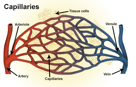
Types of blood vessels, including a venule, vein, and capillaries
Details
Identifiers
Latinvenula
MeSHD014699
TA98A12.0.00.037
TA23903
THH3.09.02.0.03002
FMA63130
Anatomical terminology

A venule is a very small vein in the microcirculation that allows blood to return from the capillary beds to drain into the venous system via increasingly larger veins. Post-capillary venules are the smallest of the veins with a diameter of between 10 and 30 micrometres (μm). When the post-capillary venules increase in diameter to 50 μm they can incorporate smooth muscle and are known as muscular venules.[1] Veins contain approximately 70% of total blood volume, while about 25% is contained in the venules.[2] Many venules unite to form a vein.

Structure

[edit]

Post-capillary venules have a single layer of endothelium surrounded by a basal lamina. Their size is between 10 and 30 micrometers and are too small to contain smooth muscle. They are instead supported by pericytes that wrap around them.[1] When the post-capillary venules increase in diameter to 50 μm they can incorporate smooth muscle and are known as muscular venules.[1] They have an inner endothelium composed of squamous endothelial cells that act as a membrane, a middle layer of muscle and elastic tissue and an outer layer of fibrous connective tissue. The middle layer is poorly developed so that venules have thinner walls than arterioles. They are porous so that fluid and blood cells can move easily from the bloodstream through their walls.

Short portal venules between the posterior pituitary and the anterior pituitary lobes provide an avenue for rapid hormonal exchange via the blood.[3] Specifically within and between the pituitary lobes is anatomical evidence for confluent interlobe venules providing blood from the anterior to the neural lobe that would facilitate moment-to-moment sharing of information between lobes of the pituitary gland.[3]

In contrast to regular venules, high endothelial venules are a special type of venule where the endothelium is made up of simple cuboidal cells. Lymphocytes exit the blood stream and enter the lymph nodes via these specialized venules when an infection is detected. Compared with arterioles, the venules are larger with much weaker muscular coat. They are the smallest united common branch in the human body.

See also

[edit]

References

[edit]
[edit]
Revisions and contributorsEdit on WikipediaRead on Wikipedia
from Grokipedia
A venule is a small in the that collects from beds and channels it into larger veins, facilitating the return of to the heart. In the systemic circulation, this is typically deoxygenated, whereas in the , it is oxygenated. These vessels typically range in diameter from 10 to 100 micrometers and are formed by the convergence of multiple capillaries. Unlike arteries, venules have thin, highly permeable walls that allow for fluid and cellular exchange, making them integral to both circulation and immune responses. Structurally, larger venules consist of three primary layers: the innermost lined with squamous endothelial cells, a thin tunica media with sparse and elastic fibers, and an outer tunica of . Smaller postcapillary venules, however, primarily feature an endothelial layer supported by , without a distinct tunica media. The endothelial layer is particularly porous in smaller venules, enabling the movement of fluids, nutrients, and leukocytes, while provide limited in postcapillary types. Their walls are thinner and more compliant than those of arterioles, rendering them susceptible to rupture under or volume. Venules are classified into three main types based on size and composition: postcapillary venules (10–30 μm, primarily endothelial with , key for leukocyte diapedesis), collecting venules (larger, with added ), and muscular venules (50–100 μm, featuring 2–3 layers of for enhanced contractility). A specialized subtype, high-endothelial venules (HEVs), found in lymph nodes, have cuboidal endothelial cells that promote lymphocyte trafficking during immune responses. These distinctions reflect their progressive transition from capillary-like permeability to vein-like durability. In function, venules play a critical role in the venous system's low-pressure reservoir, holding a significant portion of the body's while regulating flow through post-capillary sphincters. They serve as primary sites for transendothelial migration of during or , linking circulation with immunity. Additionally, their permeability supports the exchange of oxygen, nutrients, and waste, ensuring efficient microcirculatory throughout the body.

Anatomy

Definition and Characteristics

Venules are small vessels that connect to larger veins, serving as the initial segment of the venous system in the . They collect deoxygenated directly from capillary beds after nutrient and has occurred in tissues. These vessels typically range in diameter from 10 to 200 micrometers, with postcapillary venules being the smallest at the lower end of this spectrum. Compared to arteries, venules possess thin walls consisting of and minimal surrounding , and the smallest venules lack a layer entirely. In the circulatory system, venules are positioned downstream from capillaries and upstream from collecting veins, facilitating the unidirectional flow of blood from peripheral tissues back toward the heart under low pressure.

Histological Structure

Venules exhibit a three-layered histological structure similar to that of larger veins, consisting of the tunica intima, tunica media, and tunica adventitia, though these layers are notably thinner and less developed compared to those in arteries or major veins. The innermost comprises a continuous layer of endothelial cells resting on a thin , with minimal subendothelial . These endothelial cells are typically flattened and squamous in shape, forming a that lines the vessel lumen. However, in specialized high endothelial venules (HEVs) found in lymphoid tissues, the endothelial cells adopt a distinctive cuboidal or plump morphology, facilitating . In some venules, particularly those in salivary glands or under the influence of (VEGF), the features fenestrations—small pores approximately 50-100 nm in diameter—that enhance permeability for fluid and solute exchange. The middle in venules is sparse and consists primarily of a few layers of cells in larger () venules, along with that provide structural support and regulate vascular tone; in the smallest postcapillary venules, this layer may be absent or rudimentary, relying instead on embedded in the . The outermost tunica adventitia is composed of rich in and fibers, anchoring the venule to surrounding tissues; it is often indistinct in smaller venules due to its thinness and fusion with adjacent . Certain venules, especially small ones in the lower limbs, muscles, and other tissues, contain microscopic venous valves (MVVs) to prevent backflow. These valves are typically bicuspid, with delicate leaflets formed by covered in , containing fibrils and occasional elastic fibers; the cusps are oriented to direct flow toward larger vessels. Compared to capillaries, venules possess thicker walls due to the presence of tunica media and , transitioning from the single endothelial layer of capillaries to a more structured vessel. In contrast to larger veins, venules have significantly less in the tunica media, lacking the robust contractile apparatus that enables venoconstriction.

Classification and Types

Venules are classified primarily based on their , structural composition, and anatomical , with subtypes including post-capillary venules, collecting venules, venules, and specialized variants such as high endothelial venules (HEVs). Post-capillary venules represent the smallest subtype, typically ranging from 10 to 50 micrometers in , and serve as the immediate continuation of beds. These venules feature a thin wall consisting primarily of supported by a and occasional , contributing to their notably high permeability for fluid and solute exchange. They are the primary site for leukocyte diapedesis, where migrate across the into surrounding tissues. Collecting venules are larger structures, measuring approximately 50 to 100 micrometers in , and function as transitional vessels that aggregate from multiple post-capillary venules. These venules possess a slightly thicker wall with emerging cells around the , aiding in the gradual propulsion of toward larger veins while maintaining some permeability. They are distributed throughout most tissues, bridging the microcirculatory network to more robust venous drainage. Muscular venules, with diameters of 100 to 200 micrometers, exhibit a more developed layer in their tunica media, similar to small veins, which allows for active regulation of blood flow and vessel tone. These venules are particularly prominent in organs such as the skin and mucosa, where their contractile properties support localized hemodynamic control. The presence of this muscle layer distinguishes them from smaller venules, marking a structural progression toward veins. High endothelial venules (HEVs) constitute a specialized subtype of post-capillary venules, characterized by cuboidal rather than flattened endothelial cells and a thickened basement membrane. Found predominantly in secondary lymphoid organs like lymph nodes, HEVs facilitate the trafficking of lymphocytes from blood into lymphoid tissues through distinct adhesion molecule expression on their endothelium. This unique morphology and location adapt them for immune cell homing rather than general blood collection.

Physiology

Role in Blood Circulation

Venules function as low-resistance conduits in the venous system, collecting deoxygenated blood directly from the venous ends of capillaries and channeling it into progressively larger veins for return to the heart. This role facilitates the transition from the microcirculation to the macrocirculation, minimizing energy loss in blood flow while maintaining continuity in the systemic and pulmonary circuits. Due to their thin walls and sparse smooth muscle, venules offer negligible resistance to flow compared to arterioles, allowing efficient drainage of capillary beds across tissues. A key aspect of venules' circulatory role involves the that drives venous return. At the exit of capillaries, hydrostatic is approximately 20 mmHg, which drops to about 10 mmHg within venules as encounters the compliant venous network. This modest pressure decline—coupled with the overall low venous pressures—prevents excessive and supports steady propulsion toward the vena cava, aided by external factors like contractions. Venules thus handle a significant portion of the total pressure dissipation in the postcapillary segment, ensuring balanced systemic flow. In terms of volume management, venules and associated small veins serve as primary capacitance vessels, accommodating roughly 60% of the total circulating at rest. This high storage capacity buffers fluctuations in and venous return, distending under normal conditions to maintain preload without substantially elevating pressure. The compliance of venules allows them to expand or contract dynamically, optimizing distribution during physiological demands like exercise. Flow dynamics in venules are characterized by laminar patterns, with average velocities ranging from 0.5 to 1 mm/s, far slower than in arteries due to the larger cumulative cross-sectional area of the venous bed. This low-velocity flow, influenced by venular compliance and minimal , promotes efficient volume handling and reduces on vessel walls. Regulation of venular tone is subdued relative to arterioles, with limited intrinsic vasoregulation, but venules remain responsive to extrinsic neural and humoral signals. Sympathetic activation releases norepinephrine, which binds to α-adrenergic receptors on venular , inducing that reduces capacitance and mobilizes stored . This mechanism supports overall circulatory by fine-tuning venous return in response to systemic needs.

Permeability and Fluid Exchange

Venules exhibit high endothelial permeability, primarily due to loose intercellular junctions and, in some cases, fenestrations, which allow for greater transendothelial passage of , solutes, and small proteins compared to capillaries. This structural feature facilitates efficient exchange in the , where postcapillary venules serve as key sites for and . The process is governed by forces, which balance hydrostatic pressure—favoring from the vascular lumen to the —and —opposing it by drawing back via plasma proteins. The net rate of fluid movement across the venular wall is described by the Starling equation: Jv=Kf[(PcPi)σ(πcπi)]J_v = K_f \left[ (P_c - P_i) - \sigma (\pi_c - \pi_i) \right] where JvJ_v represents the fluid flux per unit surface area, KfK_f is the (reflecting venular wall permeability), PcP_c and PiP_i are (venular) and interstitial hydrostatic pressures, σ\sigma is the for proteins (typically ≥0.9 in venules, indicating partial protein restriction), and πc\pi_c and πi\pi_i are plasma and interstitial oncotic pressures. This equation derives from for porous media flow, where hydraulic pressure drives bulk fluid movement, combined with van't Hoff's osmotic law, adjusted by σ\sigma to account for semipermeable barriers; in venules, KfK_f is elevated (e.g., 10-100 times higher than in continuous capillaries) due to larger pore sizes, and typical pressures include venular PcP_c of 12-25 mmHg, with πc\pi_c around 25 mmHg and πi\pi_i lower at 10-15 mmHg, resulting in net under normal conditions. Under , slight net filtration occurs, balanced by lymphatic drainage. Venules play a central role in formation, acting as the primary site for interstitial fluid leakage or , particularly when increases permeability via the "stretched pore" mechanism, where elevated venular pressure enlarges endothelial gaps, reducing σ\sigma and allowing protein-rich fluid . In venous , postcapillary venules experience heightened hydrostatic pressure, shifting balance toward and promoting tissue swelling. Permeability varies regionally across venules to meet organ-specific demands; for instance, intestinal venules display higher permeability (transendothelial electrical resistance of 1-3 Ω cm², allowing passage of molecules up to 4 ) to support nutrient absorption, whereas cerebral venules maintain low permeability (resistance ~2000 Ω cm², restricting molecules >800 Da) as part of the blood-brain barrier.

Involvement in Immune Response

Venules, particularly post-capillary venules, serve as critical sites for leukocyte during , facilitating the transition of immune cells from the bloodstream into tissues. This process, known as , is essential for mounting effective and adaptive immunity. Leukocyte extravasation occurs in distinct steps orchestrated by endothelial cells in venules. Initially, leukocytes undergo rolling along the venular mediated by selectins, such as P-selectin and expressed on activated endothelial cells, which bind to carbohydrate ligands on leukocytes. This is followed by firm adhesion, where like LFA-1 () on leukocytes interact with intercellular adhesion molecule-1 () on the endothelium, arresting leukocyte movement. Finally, transmigration, or diapedesis, allows leukocytes to cross the endothelial barrier, often through paracellular routes between endothelial cells or transcellularly via specialized pores. High endothelial venules (HEVs), a specialized subset found in lymphoid tissues, play a pivotal role in naive homing. These venules express peripheral node addressin (PNAd), a scaffold that serves as a for on lymphocytes, enabling selective tethering and recruitment of naive T and B cells to lymph nodes for immune surveillance. This /PNAd interaction, combined with gradients like CCL19 and CCL21, ensures efficient lymphocyte entry into lymphoid organs. Inflammatory mediators further enhance venule involvement in acute immune responses by promoting leukocyte influx. and , released from mast cells and other sources during or , bind to receptors on venular endothelial cells, inducing rapid cytoskeletal rearrangements that increase permeability and expression of molecules, thereby facilitating into inflamed tissues. Tissue-specific adaptations highlight venules' versatility in immunity; for instance, dermal venules in the skin respond to allergens by upregulating molecules and permeability in allergic reactions, enabling rapid and T-cell recruitment to combat type 2 immune threats like .

Pathophysiology and Clinical Significance

Common Disorders

Venules, as the smallest postcapillary vessels, are susceptible to various pathologies that disrupt their structural integrity and functional role in . Common disorders include inflammatory conditions, dilations, and occlusive events, which can lead to localized tissue damage and systemic manifestations. These pathologies often stem from immune-mediated processes, hemodynamic factors, or genetic predispositions, highlighting the venule's vulnerability in the microvascular network. Recent studies (as of 2024-2025) have highlighted venular contributions to cerebral small vessel disease and in . Venulitis refers to specifically targeting venules, typically as part of small-vessel syndromes. In Henoch-Schönlein (HSP), also known as IgA , immune complex deposition triggers leukocytoclastic in dermal and mucosal venules, resulting in non-thrombocytopenic characterized by palpable, purpuric lesions on the lower extremities and buttocks. Associated symptoms include due to increased and extravascular fluid leakage, often accompanied by and from gastrointestinal involvement. This condition predominantly affects children and is the most common childhood , with venular involvement confirmed histologically by IgA deposits in vessel walls. Telangiectasia involves permanent dilation of venules, leading to visible, branching networks of small red vessels on the skin surface, commonly termed . These ectatic venules arise from chronic venous hypertension or and are frequently observed in , where facial venular dilation contributes to persistent and flushing. Hereditary factors, such as in (HHT), promote abnormal venular and arteriolar connections, resulting in recurrent epistaxis and mucocutaneous lesions due to fragile, dilated venules. Telangiectasias measure less than 1 mm in diameter and primarily cause cosmetic concerns, though they may indicate underlying disorders. Venule occlusion occurs when thrombi or compressive forces block venular flow, impairing drainage and causing upstream ischemia. In retinal venule thrombosis, often a manifestation of branch retinal vein occlusion (BRVO), fibrin-platelet aggregates obstruct venules at arteriovenous crossings, leading to edema, hemorrhages, and ischemic damage to the inner . This results in sectoral loss and macular ischemia if the central venule is involved, with prevalence increasing with age and cardiovascular risk factors. Venular occlusion disrupts the low-pressure return system, exacerbating fluid and tissue hypoxia. The study of venular pathologies traces back to early microvascular research by , who in 1919 described the and functional dynamics of venules and capillaries in mammalian tissues, laying foundational insights into their in pathological states like and occlusion.

Diagnostic Methods

Diagnostic methods for assessing venule health primarily involve non-invasive techniques and microscopic examinations that evaluate structural , flow dynamics, and endothelial function. These approaches are essential for detecting abnormalities such as dilation, , or in venules, which are small post-capillary vessels critical for . Nailfold capillaroscopy is a widely used non-invasive technique employing to visualize microvascular changes at the nailfold, where capillaries drain into post-capillary venules, revealing venule dilation and often associated with diseases. This method involves placing an oil-immersion lens or digital videocapillaroscope against the nailfold after gentle warming to enhance visibility, allowing observation of morphological patterns like enlarged venular loops or avascular areas indicative of microvascular damage. High-resolution images obtained through this approach provide qualitative and semi-quantitative assessments, with standardized scoring systems evaluating parameters such as capillary density and hemorrhage, aiding early detection in conditions affecting venular . Intravital enables real-time imaging of venule blood flow and cellular interactions, particularly leukocyte rolling along the , using labeling in animal models or transcutaneous setups in . This technique employs epifluorescence or multiphoton to track dynamic processes in post-capillary venules, quantifying parameters like rolling velocity (typically 10-50 μm/s under ) and efficiency, which are altered in inflammatory states. It is particularly valuable for studying venular in accessible tissues like the or dermal microvasculature, providing insights into leukocyte-endothelial interactions without invasive procedures. Biopsy followed by histological analysis remains a definitive invasive method for evaluating venule , involving punch or incisional sampling of affected or tissue, with to identify endothelial in suspected venulitis. Hematoxylin and eosin highlights leukocytoclastic changes, fibrinoid necrosis, and perivenular infiltrates in venular walls, while special stains like periodic acid-Schiff or for endothelial markers (e.g., ) confirm extent and cellular composition. This approach is crucial when non-invasive methods are inconclusive, offering histopathological confirmation of venulitis through observation of neutrophilic or lymphocytic involvement in post-capillary venules. Imaging modalities such as Doppler ultrasound and (OCT) provide non-invasive assessment of venular function in specific vascular beds. High-frequency pulsed-wave Doppler ultrasound detects low-velocity blood flow (less than 5 mm/s, down to approximately 0.5 mm/s) in small venules, measuring shear rates and volume flux in peripheral or cerebral microvasculature through spectral analysis of backscattered echoes. Complementarily, OCT, particularly , visualizes retinal venules by capturing motion contrast from blood flow, enabling quantification of vessel diameters (typically 50-150 μm for retinal venules) and perfusion density without dyes, useful for detecting venular occlusions or caliber changes.

Therapeutic Approaches

Therapeutic approaches for venule dysfunction primarily target inflammation, dilation, and thrombosis in these small postcapillary vessels, aiming to restore normal permeability, reduce leukocyte infiltration, and prevent clot formation. Pharmacological interventions form the cornerstone, with corticosteroids widely used to manage venulitis, a condition characterized by inflammation of venules often seen in small-vessel vasculitides. These agents suppress inflammatory cascades, decreasing vascular permeability and inhibiting leukocyte adhesion to endothelial surfaces, thereby alleviating edema and tissue damage. Systemic corticosteroids, such as prednisone at doses of 0.5–1 mg/kg/day, are administered for acute flares, with tapering over weeks to months to minimize side effects while achieving remission. Procedural options like address structural abnormalities in venules, particularly in cases of where superficial venules dilate and become visible. This minimally invasive technique involves injecting sclerosing agents, such as (0.25–1%) or sodium tetradecyl , directly into the affected venules to induce endothelial damage and , leading to vessel collapse and resolution of the dilation. Performed under magnification with fine needles (e.g., 30-gauge), the procedure is highly effective for lower extremity , with compression therapy post-injection enhancing outcomes and reducing recurrence. Patient satisfaction is high, though transient or matting may occur. Anticoagulants play a key role in preventing venule , especially in hypercoagulable states or microvascular occlusion risks, such as post-surgical settings. (LMWH), like enoxaparin, inhibits factor Xa and , reducing formation and propagation in small vessels including venules. Administered subcutaneously at prophylactic doses (e.g., 40 mg daily), LMWH has demonstrated efficacy in preventing microvascular during peripheral vascular procedures, with a favorable safety profile compared to unfractionated , including lower risk. Emerging therapies focus on modulating immune cell trafficking across venules, particularly in inflammatory diseases where excessive contributes to . Anti-selectin monoclonal antibodies target adhesion molecules like P-selectin and on venular , inhibiting leukocyte rolling and migration. For instance, anti-P-selectin antibodies have reduced vein wall and formation in experimental models by limiting accumulation. Similarly, anti-E-selectin monoclonal antibodies block infiltration in LPS-induced , decreasing tissue damage by 50–70% in murine studies and showing promise in conditions like ischemia-reperfusion injury. These biologics represent a targeted approach to venule-specific immune responses, potentially complementing traditional therapies in cases.

References

Add your contribution
Related Hubs
User Avatar
No comments yet.