Hubbry Logo
Stimulator of interferon genesStimulator of interferon genesMain
Open search
Stimulator of interferon genes
Community hub
Stimulator of interferon genes
logo
8 pages, 0 posts
0 subscribers
Be the first to start a discussion here.
Stimulator of interferon genes
from Wikipedia

STING1
Available structures
PDBOrtholog search: PDBe RCSB
Identifiers
AliasesSTING1, ERIS, MITA, MPYS, NET23, SAVI, STING, hMITA, hSTING, Stimulator of interferon genes, transmembrane protein 173, STING-beta, TMEM173, stimulator of interferon response cGAMP interactor 1
External IDsOMIM: 612374; MGI: 1919762; HomoloGene: 18868; GeneCards: STING1; OMA:STING1 - orthologs
Orthologs
SpeciesHumanMouse
Entrez
Ensembl
UniProt
RefSeq (mRNA)

NM_001301738
NM_198282
NM_001367258

NM_001289591
NM_001289592
NM_028261

RefSeq (protein)

NP_001288667
NP_938023
NP_001354187

NP_001276520
NP_001276521
NP_082537

Location (UCSC)Chr 5: 139.48 – 139.48 MbChr 18: 35.87 – 35.87 Mb
PubMed search[3][4]
Wikidata
View/Edit HumanView/Edit Mouse

Stimulator of interferon genes (STING), also known as transmembrane protein 173 (TMEM173) and MPYS/MITA/ERIS is a regulator protein that in humans is encoded by the STING1 gene.[5]

STING plays an important role in innate immunity. STING induces type I interferon production when cells are infected with intracellular pathogens, such as viruses, mycobacteria and intracellular parasites.[6] Type I interferon, mediated by STING, protects infected cells and nearby cells from local infection by binding to the same cell that secretes it (autocrine signaling) and nearby cells (paracrine signaling.) It thus plays an important role, for instance, in controlling norovirus infection.[7]

STING works as both a direct cytosolic DNA sensor (CDS) and an adaptor protein in Type I interferon signaling through different molecular mechanisms. It has been shown to activate downstream transcription factors STAT6 and IRF3 through TBK1, which are responsible for antiviral response and innate immune response against intracellular pathogen.[8]

Structure

[edit]
Human STING Protein Architecture

Amino acids 1–379 of human STING include the 4 transmembrane regions (TMs) and a C-terminal domain. The C-terminal domain (CTD: amino acids 138–379) contains the dimerization domain (DD) and the carboxy-terminal tail (CTT: amino acids 340–379).[8]

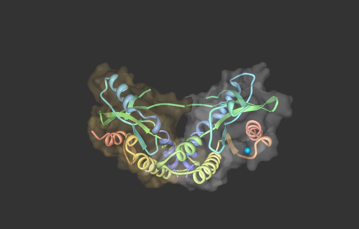
The STING forms a symmetrical dimer in the cell. STING dimer resembles a butterfly, with a deep cleft between the two protomers. The hydrophobic residues from each STING protomer form hydrophobic interactions between each other at the interface.[8][9]

Expression

[edit]

STING is expressed in hematopoietic cells in peripheral lymphoid tissues, including T lymphocytes, NK cells, myeloid cells and monocytes. It has also been shown that STING is highly expressed in lung, ovary, heart, smooth muscle, retina, bone marrow and vagina.[10][11]

Localization

[edit]

The subcellular localization of STING has been elucidated as an endoplasmic reticulum protein. Also, it is likely that STING associates in close proximity with mitochondria associated ER membrane (MAM)-the interface between the mitochondrion and the ER.[12] During intracellular infection, STING is able to relocalize from endoplasmic reticulum to perinuclear vesicles potentially involved in exocyst mediated transport.[12] STING has also been shown to colocalize with autophagy proteins, microtubule-associated protein 1 light chain 3 (LC3) and autophagy-related protein 9A, after double-stranded DNA stimulation, suggesting its presence in the autophagosome.[13]

Function

[edit]

STING mediates the type I interferon production in response to intracellular DNA and a variety of intracellular pathogens, including viruses, intracellular bacteria and intracellular parasites.[14] Upon infection, STING from infected cells can sense the presence of nucleic acids from intracellular pathogens, and then induce interferon β and more than 10 forms of interferon α production. Type I interferon produced by infected cells can find and bind to Interferon-alpha/beta receptor of nearby cells to protect cells from local infection.

Antiviral immunity

[edit]

STING elicits powerful type I interferon immunity against viral infection. After viral entry, viral nucleic acids are present in the cytosol of infected cells. Several DNA sensors, such as DAI, RNA polymerase III, IFI16, DDX41 and cGAS, can detect foreign nucleic acids. After recognizing viral DNA, DNA sensors initiate the downstream signaling pathways by activating STING-mediated interferon response.[15]

Adenovirus, herpes simplex virus, HSV-1 and HSV-2, as well as the negative-stranded RNA virus, vesicular stomatitis virus (VSV), have been shown to be able to activate a STING-dependent innate immune response.[14]

STING deficiency in mice led to lethal susceptibility to HSV-1 infection due to the lack of a successful type I interferon response.[16]

Point mutation of serine-358 dampens STING-IFN activation in bats and is suggested to give bats their ability to serve as reservoir hosts.[17]

Against intracellular bacteria

[edit]

Intracellular bacteria, Listeria monocytogenes, have been shown to stimulate host immune response through STING.[18] STING may play an important role in the production of MCP-1 and CCL7 chemokines. STING deficient monocytes are intrinsically defective in migration to the liver during Listeria monocytogenes infection. In this way, STING protects host from Listeria monocytogenes infection by regulating monocyte migration. The activation of STING is likely to be mediated by cyclic di-AMP secreted by intracellular bacteria.[18][19]

Other

[edit]

STING may be an important molecule for protective immunity against infectious organisms. For example, animals that cannot express STING are more susceptible to infection from VSV, HSV-1 and Listeria monocytogenes, suggesting its potential correlation to human infectious diseases.[20]

Role in host immunity

[edit]

Although type I IFN is absolutely critical for resistance to viruses, there is growing literature about the negative role of type I interferon in host immunity mediated by STING. AT-rich stem-loop DNA motif in the Plasmodium falciparum and Plasmodium berghei genome and extracellular DNA from Mycobacterium tuberculosis have been shown to activate type I interferon through STING.[21][22] Perforation of the phagosome membrane mediated by ESX1 secretion system allows extracellular mycobacterial DNA to access host cytosolic DNA sensors, thus inducing the production of type I interferon in macrophages. High type I interferon signature leads to the M. tuberculosis pathogenesis and prolonged infection.[22] STING-TBK1-IRF mediated type I interferon response is central to the pathogenesis of experimental cerebral malaria in laboratory animals infected with Plasmodium berghei. Laboratory mice deficient in type I interferon response are resistant to experimental cerebral malaria.[21]

STING signaling mechanisms

[edit]
STING signaling

STING mediates type I interferon immune response by functioning as both a direct DNA sensor and a signaling adaptor protein. Upon activation, STING stimulates TBK1 activity to phosphorylate IRF3 or STAT6. Phosphorylated IRF3s and STAT6s dimerize, and then enter nucleus to stimulate expression of genes involved in host immune response, such as IFNB, CCL2, CCL20, etc.[8][23]

Several reports suggested that STING is associated with the activation of selective autophagy.[13] Mycobacterium tuberculosis has been shown to produce cytosolic DNA ligands which activate STING, resulting in ubiquitination of bacteria and the subsequent recruitment of autophagy related proteins, all of which are required for 'selective' autophagic targeting and innate defense against M. tuberculosis.[24]

In summary, STING coordinates multiple immune responses to infection, including the induction of interferons and STAT6-dependent response and selective autophagy response.[8]

As a cytosolic DNA sensor

[edit]

Cyclic dinucleotides-second-messenger signaling molecules produced by diverse bacterial species were detected in the cytosol of mammalian cells during intracellular pathogen infection; this leads to activation of TBK1-IRF3 and the downstream production of type I interferon.[8][25] STING has been shown to bind directly to cyclic di-GMP, and this recognition leads to the production of cytokines, such as type I interferon, that are essential for successful pathogen elimination.[26]

As a signaling adaptor

[edit]

DDX41, a member of the DEXDc family of helicases, in myeloid dendritic cells recognizes intracellular DNA and mediates innate immune response through direct association with STING.[27] Other DNA sensors- DAI, RNA polymerase III, IFI16, have also been shown to activate STING through direct or indirect interactions.[15]

Cyclic GMP-AMP synthase (cGAS), which belongs to the nucleotidyltransferase family, is able to recognize cytosolic DNA contents and induce STING-dependent interferon response by producing secondary messenger cyclic guanosine monophosphate–adenosine monophosphate (cyclic GMP-AMP, or cGAMP). After cyclic GMP-AMP bound STING is activated, it enhances TBK1's activity to phosphorylate IRF3 and STAT6 for downstream type I interferon response.[28][29]

It has been proposed that intracellular calcium plays an important role in the response of the STING pathway.[30]

See also

[edit]
  • STING agonist – Component of the innate immune system

References

[edit]

Further reading

[edit]
Revisions and contributorsEdit on WikipediaRead on Wikipedia
from Grokipedia
The Stimulator of interferon genes (STING), also known as TMEM173, MITA, MPYS, or ERIS, is an endoplasmic reticulum-resident adaptor protein that serves as a central mediator in the innate immune system's cytosolic DNA-sensing pathway, triggering robust production of type I interferons and proinflammatory cytokines upon detection of microbial or self-derived DNA. Encoded by the TMEM173 gene on human chromosome 5, STING is a 379-amino-acid, approximately 42 kDa protein characterized by a transmembrane domain anchoring it to the ER membrane, a ligand-binding domain, and a C-terminal tail that recruits downstream signaling molecules; it functions primarily as a symmetrical dimer that oligomerizes upon activation. Discovered independently by four research groups in 2008, STING was identified as essential for type I interferon induction in response to DNA viruses or transfected double-stranded DNA, marking a pivotal advance in understanding non-nuclear DNA surveillance in mammalian cells. Its activation begins when cyclic GMP-AMP synthase (cGAS) detects cytosolic double-stranded DNA from pathogens or damaged cells, producing the second messenger 2'3'-cyclic GMP-AMP (2'3'-cGAMP), which binds to STING's ligand-binding domain, inducing a conformational change— including a 180° rotation of the domain—and translocation from the ER through the Golgi to punctate perinuclear structures. This process recruits and activates tank-binding kinase 1 (TBK1), which phosphorylates interferon regulatory factor 3 (IRF3) and nuclear factor κB (NF-κB), leading to their nuclear translocation and transcriptional upregulation of interferon-β (IFN-β) and other inflammatory genes. Beyond interferon signaling, STING activation promotes autophagy, metabolic reprogramming, and regulated cell death, thereby bridging innate immunity with adaptive responses and influencing processes like T-cell priming and antibody production. STING's roles extend to diverse physiological and pathological contexts, including antiviral defense against DNA and some RNA viruses (e.g., ), antitumor immunity by enhancing tumor immunogenicity and response to checkpoint inhibitors, and regulation of . Dysregulation of STING contributes to , such as STING-associated vasculopathy with onset in infancy (SAVI) caused by gain-of-function mutations, as well as chronic inflammation in conditions like systemic lupus erythematosus, neurodegenerative disorders, , and metabolic syndromes. In cancer, STING agonists—such as cyclic dinucleotides and non-nucleotide small molecules like MSA-2—are under clinical investigation to boost antitumor immunity, while inhibitors like H-151 target excessive inflammation; recent structural insights from cryo-electron microscopy have revealed additional regulatory pockets and mechanisms, informing next-generation therapeutics.

Discovery and background

Identification and initial characterization

The stimulator of interferon genes (STING), encoded by the TMEM173 gene, was independently identified in 2008 by four research groups as a critical adaptor in innate immune signaling. Ishikawa and employed an expression strategy using approximately 5,500 and 9,000 murine full-length cDNAs transfected into embryonic kidney 293T cells harboring an IFN-β promoter-driven reporter to screen for factors that potently induce type I production. This approach identified STING as a predominantly localized to the (ER), capable of activating the and transcription factors to drive IFN-β expression and establish an antiviral state. Concurrently, Zhong et al. described the same protein, initially termed mediator of IRF3 activation (), through a similar expression screen in HEK293 cells using an IFN-β promoter reporter. They demonstrated that MITA overexpression activates and induces type I s, while its knockdown via impairs IFN-β production in response to viral infections, such as vesicular stomatitis virus (VSV), and reduces antiviral activity. MITA was characterized as associating with the (VISA, also known as MAVS) and recruiting TBK1 kinase for IRF3 phosphorylation, thereby linking RIG-I-like receptors to downstream interferon induction. Independently, Sun et al. identified the protein as ERIS (endoplasmic reticulum IFN stimulator) and showed that it activates innate immune signaling through dimerization, leading to type I IFN production in response to viral infection. Jin et al. termed it (MHC class II plasma membrane protein with stimulatory function) and characterized it as a novel tetraspanin-like associated with , involved in transducing apoptotic and inflammatory signals in immune cells. Early functional studies further established STING/MITA as an ER-resident protein with multiple transmembrane domains that interacts with components of the translocon complex, such as SSR2 and SEC61β, facilitating its role in innate signaling. Subsequent work confirmed its essential function in cytosolic DNA sensing; STING-deficient mouse embryonic fibroblasts exhibited defective type I interferon responses to transfected B-form DNA or herpes simplex virus-1 (HSV-1) infection, highlighting its broader involvement in DNA-mediated immunity beyond RNA virus detection. The nomenclature evolved from MITA to the widely adopted STING following recognition of the protein's identity across studies and its specific stimulatory effect on interferon genes.

Evolutionary aspects

The stimulator of interferon genes (STING) protein is widely conserved across vertebrates, from teleost fish such as (Danio rerio) to mammals including humans and mice, underscoring its fundamental role in innate immunity. Homologs of STING are also present in certain , notably the cnidarian (Nematostella vectensis), where it functions in cyclic dinucleotide-mediated signaling, though without the response seen in vertebrates. In arthropods like , a STING homolog exists and contributes to antiviral defense through NF-κB-dependent pathways, but it lacks direct cytosolic DNA-sensing capabilities and instead responds to double-stranded via cGAS-like receptors (cGLRs). Core structural features of STING, including the four N-terminal transmembrane domains that anchor it to the and the C-terminal tail (CTT) involved in , exhibit high conservation across metazoan species, enabling the binding of cyclic dinucleotides such as 2'3'-cGAMP. This domain architecture supports the protein's role as an adaptor in innate immune responses, with the ligand-binding domain showing particular invariance from cnidarians to mammals. The CTT, while present in , has undergone refinements in vertebrates to recruit specific kinases like TBK1. Phylogenetic studies reveal that STING emerged over 500 million years ago in the last common ancestor of cnidarians and bilaterians, predating the evolution of adaptive immunity in jawed vertebrates. This ancient origin aligns with the of major metazoan lineages and highlights STING's co-evolution with cytosolic sensing mechanisms. In mammals, STING has adapted with heightened sensitivity to the vertebrate-specific second messenger 2'3'-cGAMP, produced by cGAS, compared to bacterial cyclic di-GMP sensors, allowing more efficient activation against intracellular pathogens.

Molecular properties

Protein structure

The human STING protein consists of 379 and is anchored to the (ER) membrane via an N-terminal comprising four transmembrane helices (TM1–TM4, approximately residues 1–137). The C-terminal domain (CTD, residues 138–379) extends into the and houses a ligand-binding pocket within its ligand-binding subdomain (residues 155–340). This architecture positions STING as an ER-resident adaptor for cytosolic sensing. STING functions as a homodimer, adopting a symmetrical "butterfly-like" fold where the two CTDs assemble in a head-to-head orientation, stabilized primarily by hydrophobic interactions at the dimer interface. The C-terminal tail (CTT, residues 340–379) protrudes flexibly from the CTD and serves as a recruitment platform for TBK1 through a conserved PLPLRT/SD motif. Crystal structures of the STING CTD reveal conformational dynamics central to its function; the apo form (PDB: 4F5W) displays an open ligand-binding pocket with the CTDs oriented parallel to the . Ligand binding, such as to 2'3'-cGAMP (PDB: 4KSY), closes the pocket and induces a ~180° rotation of the -binding domain relative to the transmembrane domains, repositioning the CTT for downstream interactions. Post-translational modifications further regulate STING structure and stability. Palmitoylation at Cys88 and Cys91 within the cytoplasmic loop between TM2 and TM3 enhances membrane association and is required for proper ER-to-Golgi trafficking. Ubiquitination at key residues, including Lys224, Lys289, and Lys370, influences dimer stability, oligomerization, and threshold by modulating protein interactions and degradation.

Expression and regulation

The STING1 gene, encoding the stimulator of interferon genes protein, is located on human chromosome 5q31.2. Basal expression of STING1 is prominent in various immune cells, including dendritic cells and macrophages, where it supports innate immune surveillance, while it remains low in non-immune tissues such as hepatocytes. STING expression exhibits distinct tissue distribution, with elevated levels observed in the and under steady-state conditions. In the , STING is constitutively present in respiratory epithelial and immune cells to facilitate rapid responses to pathogens. Placental expression is notable in and stromal cells, contributing to antiviral defense at the maternal-fetal interface, while in cardiac tissue, it is detected in cardiomyocytes and fibroblasts, potentially aiding in the regulation of during . These patterns are derived from proteomic and transcriptomic analyses across tissues. STING expression is dynamically regulated and inducible by type I interferons through binding to a specific site in the STING1 promoter, establishing a loop that amplifies antiviral signaling. This induction enhances STING levels in response to initial detection, ensuring sustained immune activation. Regulatory mechanisms further fine-tune STING expression, including epigenetic silencing via promoter , which is frequently observed in various cancers and suppresses innate immune responses to evade antitumor immunity. Additionally, microRNAs such as miR-24 post-transcriptionally suppress STING by targeting its 3' , thereby dampening excessive in viral infections. Type I interferons also mediate feedback inhibition of the STING pathway by inducing suppressors of cytokine signaling like SOCS1, which attenuates downstream signaling to prevent . Species-specific differences in STING regulation influence experimental models, with mice displaying higher constitutive expression in immune cells compared to humans, where expression is more predominantly inducible; this disparity can affect the translation of murine findings to human therapeutics.

Cellular localization

Steady-state distribution

In resting cells, the stimulator of interferon genes (STING) protein primarily localizes to the (ER) membrane, where its four transmembrane domains anchor it such that both the N-terminal tail and the C-terminal ligand-binding domain face the cytosol.00243-5) This topology enables STING to maintain a dimeric structure in the while poised for activation. Additionally, STING associates with mitochondria-associated ER membranes (MAMs) through interactions with lipids such as , which help regulate its positioning at ER-mitochondria contact sites. STING retention at the ER in the steady state is maintained by interactions that counteract anterograde trafficking, including binding to the ER calcium sensor STIM1, which sequesters it on the ER membrane.00243-5) Furthermore, STING binds to the cargo receptor Surf4 and the coat protein complex I (COPI) component α-COP, facilitating retrograde transport from the Golgi back to the ER to prevent ectopic localization. STING also colocalizes with Sec24C, a component of COPII vesicles involved in ER export, allowing quality control mechanisms to balance its trafficking and ensure proper folding and retention. Immunofluorescence studies in HeLa cells demonstrate STING's punctate distribution consistent with ER localization, often co-staining with ER markers such as calreticulin. These images further reveal close proximity between STING puncta and sarco/endoplasmic reticulum Ca²⁺-ATPase (SERCA) pumps, underscoring its integration into ER calcium homeostasis networks. In humans, STING exists in three main isoforms (STING1, STING2, and STING3), with STING1 (the canonical 379-amino-acid form) predominating and exhibiting the characteristic ER membrane localization due to its intact transmembrane domains. Isoforms 2 and 3, which lack portions of the C-terminal domain, show reduced or altered ER association but are less abundant in most cell types. A novel isoform, STING-ΔN, lacking the N-terminal transmembrane region (amino acids 1-120), is expressed in various human tissues and cell lines but does not associate with the ER; it functions as a negative regulator of STING signaling by disrupting interactions with TBK1 and 2'3'-cGAMP.

Activation-induced trafficking

Upon activation, STING undergoes ligand-induced conformational changes that promote its oligomerization, including tetramer formation, which is essential for initiating its exit from the (ER).00243-5) This oligomerization recruits the coat protein complex II (COPII) machinery, specifically the SAR1 and the cargo adaptor SEC24C, to form vesicles that facilitate STING's anterograde transport from the ER. In contrast to its steady-state retention in the ER, this dynamic process ensures rapid relocalization to downstream compartments for signaling competence. The primary trafficking route involves STING's incorporation into COPII-coated vesicles at ER exit sites, followed by transit through the ER-Golgi intermediate compartment (ERGIC) to the Golgi apparatus. From the Golgi, activated STING forms punctate structures that accumulate near autophagosomes, where it colocalizes with autophagy markers such as 1 light chain 3 (LC3) and autophagy-related protein 9A (ATG9A). This association supports non-canonical induction, linking STING trafficking to cellular during immune activation. Activated STING further accumulates in perinuclear regions, a process dependent on microtubule-based along the secretory pathway.00196-1) This perinuclear positioning facilitates efficient signaling before STING's termination via the autophagy-lysosome pathway, which degrades the protein approximately 2-4 hours post-activation to prevent prolonged responses. Live-cell imaging studies have visualized this trafficking, demonstrating that STING egress from the ER occurs within about 30 minutes following stimulation with cyclic GMP-AMP (cGAMP), highlighting the rapid kinetics of the process.

Activation and sensing

Ligand recognition

STING directly recognizes endogenous cyclic dinucleotides, primarily 2'3'-cyclic GMP-AMP (2'3'-cGAMP), through a binding pocket located in its C-terminal domain (CTD). This pocket forms at the interface of the STING homodimer and accommodates the V-shaped conformation of the , with key interactions involving bonds and stacking with residues such as Tyr167 and Ser243. The binding affinity is high, with a (Kd) of approximately 4 nM as measured by using the STING CTD (residues 139–379). In addition to endogenous ligands, STING binds bacterial cyclic dinucleotides, including cyclic di-GMP (c-di-GMP) and cyclic di-AMP (c-di-AMP), which are produced by intracellular pathogens such as . These prokaryotic ligands fit into the same V-shaped pocket in the CTD, though with lower affinity compared to 2'3'-cGAMP; for example, c-di-GMP binding induces partial pocket closure and cooperative activation. The recognition of these bacterial second messengers enables STING to detect microbial invasion directly. Ligand engagement triggers conformational rearrangements in STING, including closure of a β-sheet lid over the binding pocket to seal the dinucleotide and an approximately 180° rotation of the ligand-binding domain relative to the transmembrane domain. This rotation repositions the dimer and exposes the C-terminal tail for subsequent interactions. Specificity for activating ligands relies on precise phosphate positioning and the 2'-3' phosphodiester linkage; non-canonical variants like 3'3'-cGAMP bind with reduced affinity due to suboptimal interactions with pocket residues, resulting in lower activation potency.

Upstream sensors and regulators

The primary upstream sensor of the STING pathway is cyclic GMP-AMP synthase (cGAS), a nucleotidyltransferase that detects double-stranded DNA (dsDNA) in the , often derived from pathogens or damaged host cells. Upon binding dsDNA, cGAS undergoes conformational changes that enable it to catalyze the synthesis of the second messenger 2'3'-cyclic GMP-AMP (2'3'-cGAMP) through a two-step reaction: first forming the linear intermediates (amidate and ) from ATP and GTP, followed by cyclization to the canonical mixed-linkage dinucleotide. This cGAMP then binds directly to STING, inducing its oligomerization and activation. Other DNA sensors contribute to STING activation independently or in cooperation with cGAS. The DEAD-box DDX41 directly binds cytosolic dsDNA and recruits STING to initiate signaling, bypassing cGAS in certain contexts such as infections. Similarly, the PYHIN protein IFI16 functions as a nuclear sensor, particularly for herpesvirus genomes like those of HSV-1, where it detects viral DNA in the nucleus, promotes assembly, and cooperates with cGAS to enhance cytosolic 2'3'-cGAMP production and STING activation in and other cells. STING activity is modulated by various upstream regulators that fine-tune its activation. Positive regulators include TRAF3, which is recruited to ligand-bound STING to facilitate TBK1 and phosphorylation, thereby amplifying interferon induction. TRIM32, an E3 , promotes STING signaling by mediating K63-linked ubiquitination on STING, enhancing its stability and interaction with downstream effectors during antiviral responses. Negative regulators counteract excessive activation; for instance, RNF5 (an E3 ligase) induces K48-linked ubiquitination of STING, targeting it for proteasomal degradation and attenuating pathway activity post-stimulation. Pathogens have evolved mechanisms to evade these upstream sensors and regulators. Herpes simplex virus 1 (HSV-1) employs its virion host shutoff protein UL41, an RNase, to degrade cGAS at both the mRNA and protein levels, thereby suppressing 2'3'-cGAMP production and STING activation during infection. Certain bacteria, such as , secrete phosphodiesterases (e.g., PdeA1) that hydrolyze bacterial cyclic di-nucleotides like c-di-GMP, preventing their accumulation and subsequent STING activation in host cells, while others like modulate cyclic di-AMP levels via specific hydrolases to limit immune detection.

Signaling pathways

Canonical interferon induction

Upon activation, STING oligomerizes and employs its C-terminal tail (CTT) to recruit (TBK1), promoting TBK1 trans-autophosphorylation at Ser172 within the activation loop to initiate kinase activity. This recruitment occurs following STING's activation-induced trafficking to perinuclear compartments, where the oligomeric platform concentrates TBK1 for efficient signaling. The activated TBK1 then phosphorylates STING at Ser366 in the CTT, enhancing further assembly of the signaling complex, and subsequently phosphorylates () at key C-terminal residues Ser396 and Ser398. Phosphorylation of IRF3 induces a conformational change that enables its homodimerization, followed by nuclear translocation via interaction with importins. In the nucleus, the dimeric binds to interferon-stimulated response elements (ISREs) in the promoter regions of type I genes, including IFNA and IFNB, thereby driving their transcription and subsequent production of IFN-α and IFN-β proteins. This core TBK1- axis represents the primary mechanism by which STING elicits type I responses. The canonical STING pathway operates independently of the (MAVS), distinguishing it from RNA-sensing pathways like RIG-I-MDA5. In macrophages, STING activation induces upregulation of IFN-β mRNA, establishing rapid antiviral signaling.

Non-canonical outputs

Beyond the canonical induction of type I interferons via , STING engages non-canonical signaling pathways that drive proinflammatory production through activation. Upon activation, STING recruits TBK1, which, along with IKKε, redundantly phosphorylates components of the IKK complex, including indirect promotion of IKKβ phosphorylation to form a loop that releases from IκB inhibition. This enables translocation to the nucleus, where it transcribes proinflammatory genes such as those encoding TNF and IL-6, contributing to broader inflammatory responses independent of production. STING also activates the STAT6 pathway via TBK1-mediated at Ser407, facilitating STAT6 dimerization and nuclear translocation to induce genes involved in immune responses. This non-canonical arm supports Th2 cytokine production, such as IL-4 and IL-13, which is implicated in , contrasting the antiviral focus of the IRF3 pathway. In metabolic regulation, STING links innate sensing to cellular by inducing (ER) stress and processes, including mitophagy. Activated STING triggers the PERK-eIF2α axis, repressing global translation while promoting selective autophagy to alleviate ER stress; this also involves STX17 interaction to modulate autophagosome-lysosome fusion during energy stress. In the liver, STING activation exacerbates lipid accumulation and steatosis by coupling ER stress to impaired , as evidenced in models of where STING deficiency prevents buildup. STING contributes to inflammatory cell death through crosstalk with the AIM2 , promoting in contexts like cytosolic DNA sensing or . AIM2 activation by dsDNA limits excessive STING signaling, but in AIM2-deficient settings, heightened STING activity amplifies inflammasome-independent via gasdermin-mediated pore formation, enhancing IL-1β release and tissue .

Physiological functions

Antiviral immunity

The cGAS-STING pathway serves as a critical sensor in antiviral immunity, primarily detecting cytosolic double-stranded DNA (dsDNA) intermediates produced during viral replication, such as those from DNA viruses, as well as certain RNA virus-derived nucleic acids. Upon binding these ligands, cyclic GMP-AMP synthase (cGAS) catalyzes the production of the second messenger 2'3'-cyclic GMP-AMP (cGAMP), which activates stimulator of interferon genes (STING) by promoting its oligomerization and translocation from the endoplasmic reticulum to perinuclear compartments. This activation recruits TANK-binding kinase 1 (TBK1), which phosphorylates both STING and interferon regulatory factor 3 (IRF3), culminating in the transcriptional induction of type I interferons (IFN-α and IFN-β). These type I IFNs bind to IFNAR receptors on infected and neighboring cells, triggering the kinase-signal transducer and activator of transcription (JAK-STAT) signaling cascade. Phosphorylated /STAT2 dimers translocate to the nucleus, forming the interferon-stimulated gene factor 3 (ISGF3) complex with IRF9 to drive expression of hundreds of s (ISGs). Key antiviral ISGs include myxovirus resistance protein A (MxA), which traps viral nucleocapsids to prevent replication, and 2'-5'-oligoadenylate synthetase (OAS), which activates RNase L to degrade viral and host RNAs, thereby restricting viral spread and establishing a cellular antiviral state. This mechanism exemplifies the canonical interferon induction pathway, where STING acts as a central hub for innate antiviral defense. STING-mediated responses are essential for controlling specific viral infections, particularly DNA viruses. In STING-deficient mice, herpes simplex virus 1 (HSV-1) infection leads to markedly elevated viral loads in the and other tissues compared to wild-type controls, underscoring STING's role in limiting HSV-1 dissemination. For , STING contributes to restricting infection in neural tissues, where its deficiency exacerbates viral burdens and in susceptible models. Beyond innate responses, STING bridges to adaptive immunity by enhancing in dendritic cells, where activated STING upregulates presentation of viral antigens and costimulatory molecules, promoting the priming and activation of + T cells for cytotoxic clearance of infected cells. This process has been demonstrated in models, where STING agonists like cGAMP amplify + T cell responses against viral challenges. Recent insights into highlight viral evasion strategies, with the accessory protein ORF9b interacting directly with STING to inhibit its and trafficking to signaling sites, thereby suppressing IFN production and facilitating immune escape during infection.

Antibacterial and other defenses

STING plays a critical role in sensing intracellular bacteria through recognition of bacterial cyclic dinucleotides, particularly cyclic di-AMP (c-di-AMP), which is secreted by pathogens such as Listeria monocytogenes and Chlamydia trachomatis. Upon cytosolic release, c-di-AMP binds directly to STING, triggering its activation and subsequent downstream signaling that induces type I interferon production and chemokine secretion, including CCL5 (also known as RANTES) and MCP-1 (CCL2), to promote immune cell recruitment and chemokinesis. In L. monocytogenes infection, STING-dependent detection of c-di-AMP enhances host defense by facilitating early innate responses, though excessive type I interferon can limit adaptive immunity. Similarly, during C. trachomatis infection, STING senses c-di-AMP produced by the bacterial diadenylate cyclase DacA, leading to interferon-beta induction and control of bacterial replication in epithelial cells. Against intracellular bacteria like , STING contributes to host defense by promoting , a process that targets and degrades pathogens within phagosomes. Bacterial DNA released into the via the ESX-1 system activates cGAS to produce cGAMP, which binds STING and initiates TBK1-dependent signaling, resulting in ubiquitination of bacteria and recruitment to autophagosomes marked by LC3. This STING-mediated pathway restricts M. tuberculosis growth in macrophages and enhances bacterial clearance in vivo, as evidenced by increased susceptibility in STING-deficient models. Beyond microbial threats, STING detects self-DNA in micronuclei formed due to genomic instability, linking DNA damage to anti-tumor immunity. Micronuclei containing chromatinized self-DNA activate cGAS-STING signaling, which induces type I interferons and promotes an inflammatory microenvironment that recruits and activates cytotoxic T cells against tumors. This pathway enhances anti-tumor responses by alerting the to nascent cancer cells with chromosomal aberrations. In dendritic cells (DCs), STING activation drives maturation, upregulating co-stimulatory molecules like and , and facilitating of tumor antigens to prime + T cell responses. Selective STING stimulation in DCs potentiates this effect, licensing type I conventional DCs to orchestrate robust anti-tumor immunity without . STING also participates in defenses against other pathogens, such as fungi and parasites, though outcomes can vary. In Candida albicans infection, STING senses cytosolic DNA released during host-pathogen interactions, potentially including mitochondrial DNA (mtDNA) from damaged cells, to modulate innate responses via translocation to phagosomes and regulation of type I interferon signaling. This activation supports anti-fungal immunity but can limit excessive inflammation. For parasitic infections like those caused by Plasmodium species, STING detects parasite DNA in infected cells, inducing type I interferons that aid in controlling replication; however, hyperactivation risks immunopathology, including exacerbated cerebral malaria through endothelial inflammation and T cell dysregulation.

Pathological implications

Autoimmune disorders

Dysregulated activation of the stimulator of interferon genes (STING) pathway has been implicated in several autoinflammatory and autoimmune disorders characterized by excessive type I production and chronic inflammation. One prominent example is STING-associated vasculopathy with onset in infancy (SAVI), a monogenic autoinflammatory disease caused by heterozygous gain-of-function mutations in the STING1 gene (formerly TMEM173). These mutations, such as the common V155M variant in the dimerization domain, lead to constitutive STING activation independent of upstream ligands, resulting in persistent type I interferon signaling and downstream inflammatory responses. Clinically, SAVI manifests in infancy with severe , cutaneous vasculopathy, and , often progressing to and vasculitic lesions without infectious triggers. Aicardi-Goutières syndrome (AGS), a hereditary interferonopathy, also involves hyperactivity of the cGAS-STING axis due to genetic defects in . Mutations in genes such as TREX1, RNASEH2A/B/C, or SAMHD1 cause intracellular accumulation of self-nucleic acids, including , which aberrantly activates cGAS to produce cyclic GMP-AMP (cGAMP) and subsequently stimulates STING. This mimics a chronic viral infection state, driving robust type I interferon production and . Patients typically present in early childhood with , calcifications, and elevated expression, underscoring the pathway's role in distinguishing self from non-self nucleic acids. In systemic lupus erythematosus (SLE), STING contributes to disease pathogenesis by promoting type I production, particularly in plasmacytoid dendritic cells (pDCs). Circulating immune complexes containing self-nucleic acids activate cGAS-STING in pDCs, amplifying interferon-alpha secretion that sustains production and immune complex deposition in tissues. This pathway exacerbates and cutaneous manifestations, with STING inhibition shown to reduce levels and inflammation in preclinical models.30722-7) Recent investigations have highlighted STING's involvement in (RA), where pathway activation in synovial fibroblasts and macrophages drives release and joint destruction. Elevated STING signaling in RA synovium, triggered by damage-associated molecular patterns, correlates with increased disease activity and synovial , suggesting a contributory role in RA risk through enhanced local inflammation.

Cancer and chronic inflammation

The stimulator of interferon genes (STING) pathway exhibits a dual role in cancer, acting primarily as a tumor suppressor through the induction of immunogenic (ICD) in tumor cells, which releases damage-associated molecular patterns (DAMPs) such as and ATP to activate dendritic cells and promote priming and infiltration into the . This mechanism enhances antitumor immunity, particularly in immunogenic cancers like and non-small cell , where STING activation correlates with improved patient outcomes and increased T cell infiltration. Furthermore, synthetic cyclic GMP-AMP (cGAMP) analogs, as STING agonists, synergize with PD-1 checkpoint inhibitors to boost therapeutic efficacy by amplifying type I signaling and + T cell responses, leading to tumor regression in preclinical models. In contrast, chronic or dysregulated STING activation can promote tumorigenesis by shifting toward -dependent proinflammatory signaling, which fosters tumor cell survival, proliferation, and immune evasion rather than interferon-mediated immunity. This pro-tumor effect is evident in , where STING-driven activation upregulates anti-apoptotic genes and supports castration-resistant tumor growth, contributing to disease progression. Sustained STING signaling in the also elevates immunosuppressive cytokines like IL-6 and TGF-β, inhibiting T and functions while promoting and . Beyond cancer, chronic STING activation contributes to metabolic and vascular inflammation. In obesity, mitochondrial DNA leakage into the cytosol activates the cGAS-STING pathway in , driving proinflammatory production and , which exacerbates systemic metabolic dysfunction. Similarly, in , STING signaling in endothelial cells, triggered by oxidized lipids or free fatty acids, induces adhesion molecule expression (e.g., , ) and inflammatory responses that promote plaque formation and vascular dysfunction. Recent studies highlight STING hyperactivity in neurodegeneration, particularly in (ALS), where it drives microglial type I responses to cytosolic DNA accumulation, amplifying and motor neuron loss in both familial and sporadic cases. This pathway's overactivation in ALS underscores its broader role in chronic inflammatory pathologies beyond .

Therapeutic applications

Agonist development

Development of agonists for the stimulator of interferon genes (STING) has focused on enhancing innate immune responses, particularly for , by directly or indirectly activating the pathway to promote production and antitumor immunity. Small-molecule STING agonists represent an early class of compounds, with 5,6-dimethylxanthenone-4-acetic acid (DMXAA) being the first to enter clinical trials as a mouse STING-specific activator that induced tumor regression in preclinical models but failed in human studies due to lack of activity on human STING variants. Subsequent efforts led to human-compatible agonists like ADU-S100 (also known as MIW815), a cyclic dinucleotide (CDN) analog that entered phase I and II trials for intratumoral treatment of glioblastoma and other solid tumors, demonstrating safety but limited clinical efficacy, leading to discontinuation of the program in 2019. In 2025, proteolysis-targeting chimeras (PROTACs) emerged for targeted STING pathway activation, such as dual PROTAC nanocarriers that degrade DNA repair proteins like and , amplifying cytosolic DNA accumulation to indirectly trigger STING signaling and enhance antitumor T-cell infiltration in models. Cyclic GMP-AMP (cGAMP) analogs, the natural STING , have been chemically modified to overcome rapid degradation by ectonucleotide pyrophosphatase/ 1 (ENPP1). These include phosphorothioate-linked variants that exhibit increased resistance to and prolonged stability in serum while maintaining potent STING activation in immune cells. To improve delivery and bioavailability, such analogs are often encapsulated in nanoparticles, which enhance tumor retention, endosomal escape, and localized STING stimulation in the , reducing off-target effects. Clinical advancement of STING agonists has emphasized combinations with inhibitors (ICIs) to boost efficacy, with phase II trials of compounds like ulevostinag (MK-1454) plus showing antitumor activity in head and neck by increasing responses and T-cell activation. As of 2025, other agonists like IMSA101 are in Phase II trials combined with radiotherapy and nivolumab for advanced solid tumors, and E7766 is in Phase I evaluating safety and antitumor activity. However, challenges persist, including systemic toxicity from excessive cytokine release, which limits intravenous dosing and necessitates localized administration strategies. Intratumoral injection remains the primary delivery method to confine induction to the tumor site and minimize adverse events, as seen in trials of ADU-S100. Additionally, viral vectors have been explored for approaches, engineering oncolytic viruses to deliver STING-activating CDNs or express pathway components directly in tumors to sustain immune activation.

Antagonist strategies

Small-molecule inhibitors represent a primary strategy for antagonizing STING activity, particularly in conditions involving pathway hyperactivity such as . H-151, a selective covalent inhibitor, targets cysteine 91 (Cys91) in the STING transmembrane domain, preventing its and subsequent recruitment of TBK1, thereby blocking downstream production. Similarly, C-176 functions as a mouse-specific analog that covalently binds Cys91, inhibiting STING palmitoylation and activation in preclinical models of . These compounds have demonstrated efficacy in reducing type I responses in cellular assays and animal models of , with H-151 showing particular promise in suppressing STING-driven responses without broad off-target effects. Biologic approaches to STING antagonism include antibodies and designed to neutralize or competitively inhibit the protein. Anti-STING monoclonal antibodies are under investigation to block binding or oligomerization, potentially offering high specificity for extracellular or surface-exposed STING in diseased tissues. , such as modified analogs of 2'3'-cGAMP, act as competitive antagonists by occupying the STING cyclic dinucleotide-binding pocket in its inactive conformation, preventing endogenous -induced conformational changes and signaling. These biologics aim to provide reversible inhibition with reduced systemic compared to small molecules, though their development remains largely preclinical. As of 2025, STING antagonists are in early clinical development, with ASP5502 in Phase I trials for autoimmune conditions such as Sjögren's syndrome. Preclinical studies, including orthosteric inhibitors of the C-terminal domain (CTD) and gene editing technologies such as CRISPR/Cas9, show promise for correcting gain-of-function STING mutations like N154S in SAVI and AGS, restoring wild-type-like regulation in patient-derived cells and reducing interferon signatures in disease models. These approaches hold promise for monogenic disorders but require validation in ongoing preclinical and early-phase studies. Key challenges in STING antagonist development include the risk of immunosuppression, as pathway inhibition may impair antiviral defenses and increase susceptibility to infections, necessitating careful dosing to preserve baseline immunity. Tissue-specific delivery remains a hurdle, particularly for central nervous system disorders like (ALS), where STING hyperactivity drives ; nanoparticle-based or brain-penetrant formulations are being investigated to enhance targeting while minimizing peripheral effects.

References

Add your contribution
Related Hubs
User Avatar
No comments yet.