Hubbry Logo
OvulationOvulationMain
Open search
Ovulation
Community hub
Ovulation
logo
8 pages, 0 posts
0 subscribers
Be the first to start a discussion here.
Ovulation
Ovulation
from Wikipedia
Ovulation
Following a surge of luteinizing hormone (LH), an oocyte (immature egg cell) will be released into the uterine tube, where it will then be available to be fertilized by a male's sperm within 12 hours. Ovulation marks the end of the follicular phase of the ovarian cycle, and the start of the luteal phase.
Identifiers
MeSHD010060
TEE1.0.0.0.0.0.7
Anatomical terminology

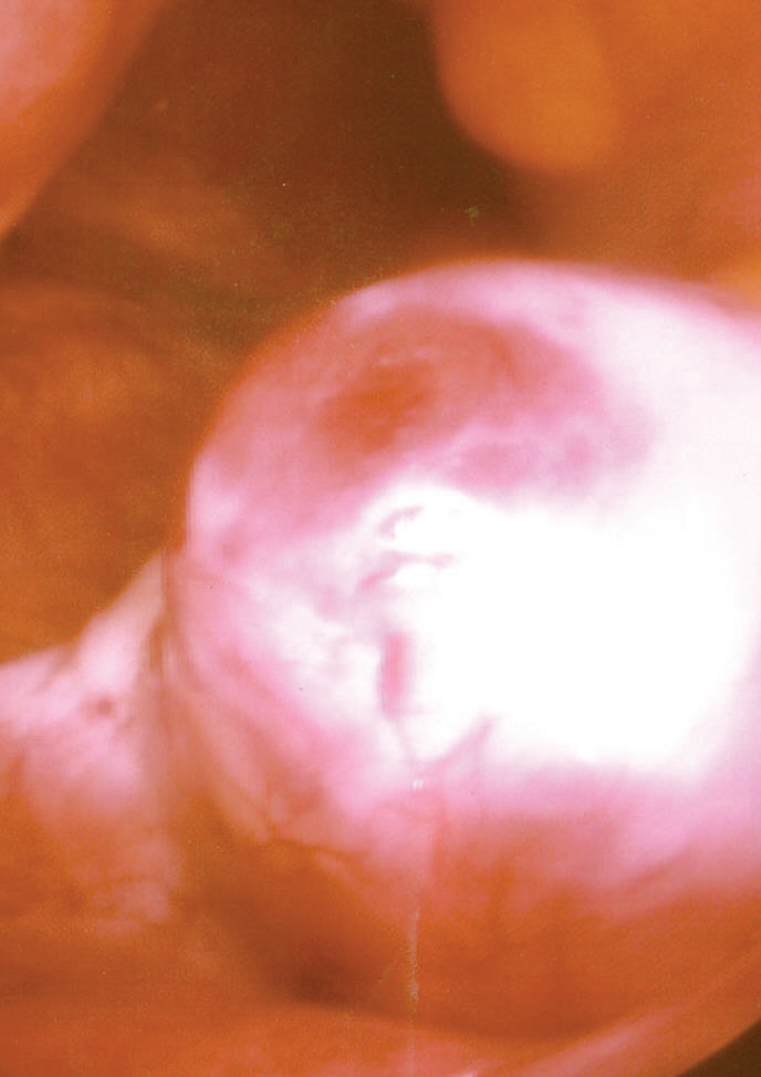
Ovulation is an important part of the menstrual cycle in female vertebrates where the egg cells are released from the ovaries as part of the ovarian cycle. In female humans ovulation typically occurs near the midpoint in the menstrual cycle and after the follicular phase. Ovulation is stimulated by an increase in luteinizing hormone (LH). The ovarian follicles rupture and release the secondary oocyte ovarian cells.[1]

After ovulation, during the luteal phase, the egg will be available to be fertilized by sperm. If it is not, it will break down in less than a day. Meanwhile, the uterine lining (endometrium) continues to thicken to be able to receive a fertilized egg. If no conception occurs, the uterine lining will eventually break down and be shed from the body via the vagina during menstruation.[2]

Some people choose to track ovulation in order to improve or aid becoming pregnant by timing intercourse with their ovulation.[3] The signs of ovulation may include cervical mucus changes, mild cramping in the abdominal area, and a small rise in basal body temperature. Medication is also sometimes required by those experiencing infertility to induce ovulation.[3]

Process

[edit]
Ovulation occurs about midway through the menstrual cycle, after the follicular phase, and is followed by the luteal phase. Note that ovulation is characterized by a sharp spike in levels of luteinizing hormone (LH) and follicle-stimulating hormone (FSH), resulting from the peak of estrogen levels during the follicular phase.

Ovulation occurs about midway through the menstrual cycle, after the follicular phase. The days in which a woman is most fertile can be calculated based on the date of the last menstrual period and the length of a typical menstrual cycle.[4] The few days surrounding ovulation (from approximately days 10 to 18 of a 28-day cycle), constitute the most fertile phase.[5][6][7][8] The time from the beginning of the last menstrual period (LMP) until ovulation is, on average, 14.6 days, but with substantial variation among females and between cycles in any single female, with an overall 95% prediction interval of 8.2 to 20.5 days.[9]

The process of ovulation is controlled by the hypothalamus of the brain and through the release of hormones secreted in the anterior lobe of the pituitary gland, luteinizing hormone (LH) and follicle-stimulating hormone (FSH).[10] In the preovulatory phase of the menstrual cycle, the ovarian follicle will undergo a series of transformations called cumulus expansion, which is stimulated by FSH. After this is done, a hole called the stigma will form in the follicle, and the secondary oocyte will leave the follicle through this hole. Ovulation is triggered by a spike in the amount of FSH and LH released from the pituitary gland. During the luteal (post-ovulatory) phase, the secondary oocyte will travel through the fallopian tubes toward the uterus. If fertilized by a sperm, the fertilized secondary oocyte or ovum may implant there 6–12 days later.[11]

Follicular phase

[edit]

The follicular phase (or proliferative phase) is the phase of the menstrual cycle during which the ovarian follicles mature. The follicular phase lasts from the beginning of menstruation to the start of ovulation.[12][13]

For ovulation to be successful, the ovum must be supported by the corona radiata and cumulus oophorous granulosa cells.[14] The latter undergo a period of proliferation and mucification known as cumulus expansion. Mucification is the secretion of a hyaluronic acid-rich cocktail that disperses and gathers the cumulus cell network in a sticky matrix around the ovum. This network stays with the ovum after ovulation and has been shown to be necessary for fertilization.[15][16]

Ovulation

[edit]

Estrogen levels peak towards the end of the follicular phase, around 12 and 24 hours. This, by positive feedback, causes a surge in levels of luteinizing hormone (LH) and follicle-stimulating hormone (FSH). This lasts from 24 to 36 hours, and results in the rupture of the ovarian follicles, causing the oocyte to be released from the ovary.[17]

Through a signal transduction cascade initiated by LH, which activates the pro-inflammatory genes through cAMP secondary messenger, proteolytic enzymes are secreted by the follicle that degrade the follicular tissue at the site of the blister, forming a hole called the stigma. The secondary oocyte leaves the ruptured follicle and moves out into the peritoneal cavity through the stigma, where it is caught by the fimbriae at the end of the fallopian tube. After entering the fallopian tube, the oocyte is pushed along by cilia, beginning its journey toward the uterus.[10]

By this time, the oocyte has completed meiosis I, yielding two cells: the larger secondary oocyte that contains all of the cytoplasmic material and a smaller, inactive first polar body. Meiosis II follows at once but will be arrested in the metaphase and will so remain until fertilization. The spindle apparatus of the second meiotic division appears at the time of ovulation. If no fertilization occurs, the oocyte will degenerate between 12 and 24 hours after ovulation.[18] Approximately 1–2% of ovulations release more than one oocyte. This tendency increases with maternal age. Fertilization of two different oocytes by two different spermatozoa results in fraternal twins.[10]

The precise moment of ovulation was captured on film for the first time in 2008, coincidentally, during a routine hysterectomy procedure. According to the attending gynecologist, the ovum's emergence and subsequent release from the ovarian follicle occurred within a 15-minute timeframe.[19]

Luteal phase

[edit]

The follicle proper has met the end of its lifespan. Without the oocyte, the follicle folds inward on itself, transforming into the corpus luteum (pl. corpora lutea), a steroidogenic cluster of cells that produces estrogen and progesterone. These hormones induce the endometrial glands to begin production of the proliferative endometrium and later into secretory endometrium, the site of embryonic growth if implantation occurs. The action of progesterone increases basal body temperature by one-quarter to one-half degree Celsius (one-half to one degree Fahrenheit). The corpus luteum continues this paracrine action for the remainder of the menstrual cycle, maintaining the endometrium, before disintegrating into scar tissue during menses.[20]

Clinical presentation

[edit]

The start of ovulation may be detected by signs that are not readily discernible other than to the ovulating female herself, thus humans are said to have a concealed ovulation.[21] In many animal species there are distinctive signals indicating the period when the female is fertile. Several explanations have been proposed to explain concealed ovulation in humans.

Females near ovulation experience changes in the cervical mucus, and in basal body temperature. Furthermore, many females experience secondary fertility signs including Mittelschmerz (pain associated with ovulation) and a heightened sense of smell, and can sense the precise moment of ovulation.[22][23] However, midcycle pain may also not be due to Mittelschmerz, but due to other factors such as cysts, endometriosis, sexually transmitted infections, or an ectopic pregnancy.[24] Other possible signs of ovulation include tender breasts, bloating, and cramps, although these symptoms are not a guarantee that ovulation is taking place.[25][26]

Many females experience heightened sexual desire in the several days immediately before ovulation.[27] One study concluded that females subtly improve their facial attractiveness during ovulation.[28]

Chance of fertilization by day relative to ovulation[29]

Symptoms related to the onset of ovulation, the moment of ovulation and the body's process of beginning and ending the menstrual cycle vary in intensity with each female but are fundamentally the same. The charting of such symptoms — primarily basal body temperature, mittelschmerz and cervical position — is referred to as the sympto-thermal method of fertility awareness, which allow auto-diagnosis by a female of her state of ovulation. Once training has been given by a suitable authority, fertility charts can be completed on a cycle-by-cycle basis to show ovulation. This gives the possibility of using the data to predict fertility for natural contraception and pregnancy planning.

Urine levels of the hormone pregnanediol 3-glucuronide of over 5 μg/mL has been used to confirm ovulation. This test has a 100% specificity over 107 women.[30]

Disorders

[edit]

Disorders of ovulation, also known as ovulatory disorders are classified as menstrual disorders and include oligoovulation (infrequent or irregular ovulation) and anovulation (absence of ovulation):[31]

  • Oligoovulation is infrequent or irregular ovulation (usually defined as cycles of greater than 36 days or fewer than 8 cycles a year)
  • Anovulation is absence of ovulation when it would be normally expected (in a post-menarchal, premenopausal female). Anovulation usually manifests itself as irregularity of menstrual periods, that is, unpredictable variability of intervals, duration, or bleeding. Anovulation can also cause cessation of periods (secondary amenorrhea) or excessive bleeding (dysfunctional uterine bleeding).

The World Health Organization (WHO) has developed the following classification of ovulatory disorders:[32]

Menstrual disorders can often indicate ovulatory disorder.[34]

Ovulation induction

[edit]

Ovulation induction is a promising assisted reproductive technology for patients with conditions such as polycystic ovary syndrome (PCOS) and oligomenorrhea. It is also used in in vitro fertilization to make the follicles mature before egg retrieval. Usually, ovarian stimulation is used in conjunction with ovulation induction to stimulate the formation of multiple oocytes.[35] Some sources[35] include ovulation induction in the definition of ovarian stimulation.

A low dose of human chorionic gonadotropin (HCG) may be injected after completed ovarian stimulation. Ovulation will occur between 24 and 36 hours after the HCG injection.[35]

By contrast, induced ovulation in some animal species occurs naturally, ovulation can be stimulated by coitus.[36]

Ovulation suppression

[edit]

Combined hormonal contraceptives inhibit follicular development and prevent ovulation as a primary mechanism of action.[37] The ovulation-inhibiting dose (OID) of an estrogen or progestogen refers to the dose required to consistently inhibit ovulation in women.[38] Ovulation inhibition is an antigonadotropic effect and is mediated by inhibition of the secretion of the gonadotropins, LH and FSH, from the pituitary gland.

In assisted reproductive technology including in vitro fertilization, cycles where a transvaginal oocyte retrieval is planned generally necessitates ovulation suppression, because it is not practically feasible to collect oocytes after ovulation. For this purpose, ovulation can be suppressed by either a GnRH agonist or a GnRH antagonist, with different protocols depending on which substance is used.

Fertility and timing of ovulation

[edit]

Most women who are able to conceive are fertile for an estimated five days before ovulation and one day after ovulation.[39] There is some evidence that for couples who have been trying to conceive a child for less than 12 months, and the female is under 40 years old, practicing timed intercourse (timing intercourse with ovulation using urine tests that predict ovulation) may help improve the rate of pregnancy and live births.[39] The role that stress plays in ovulation, fertility, and understanding the biological basis for stress-induced anovulation and the role of cortisol is not entirely clear.[40]

See also

[edit]

Notes

[edit]

Further reading

[edit]
[edit]
Revisions and contributorsEdit on WikipediaRead on Wikipedia
from Grokipedia
Ovulation is the physiologic process in which a mature , or ovum, is released from the dominant into the , marking a critical phase of the essential for . This event typically occurs once per cycle, approximately 14 days before the onset of in a standard 28-day cycle, though cycle lengths vary among individuals. The released ovum remains viable for fertilization by for about 12 to 24 hours, during which conception is possible if intercourse occurs. The , which encompasses ovulation, is divided into the , ovulation itself, and the , all regulated by a complex interplay of hormones from the , , and ovaries. During the , (FSH) from the stimulates the growth of several ovarian follicles, one of which becomes dominant and secretes increasing levels of . Rising initially exerts on the pituitary to suppress FSH, promoting of subordinate follicles, but eventually triggers that induces a surge in both (LH) and FSH around mid-cycle. The LH surge, peaking for 24 to 48 hours, initiates final maturation, weakens the follicular wall through enzymatic degradation, and causes follicular rupture approximately 36 to 44 hours later, expelling the ovum into the where it is captured by the fimbriae of the . Following ovulation, the ruptured follicle transforms into the under LH stimulation, which produces progesterone to prepare the uterine for potential implantation. If fertilization does not occur, the degenerates, progesterone levels decline, and ensues, restarting the cycle. Disruptions in ovulation, such as , can lead to or conditions like , highlighting its central role in reproductive health. Understanding ovulation is vital for fertility tracking, contraception, and assisted reproductive technologies, with methods like charting or ovulation predictor kits relying on its predictable hormonal signals.

Introduction

Definition and Overview

Ovulation is the physiologic process by which a mature , or , is released from the dominant into the , where it is subsequently captured by the fimbriae of the for potential fertilization. This event marks a critical phase in the female reproductive cycle, occurring primarily in mammals as part of production and release. In humans, ovulation involves the ovaries, paired gonadal structures that contain numerous follicles at various developmental stages. Follicles begin as primordial structures, consisting of an surrounded by a single layer of flattened granulosa cells; they progress to primary follicles with cuboidal granulosa cells, secondary follicles featuring multiple granulosa layers and an emerging , and finally the mature Graafian follicle, characterized by a fluid-filled antrum and a prominent . The Graafian follicle, typically 15-25 mm in diameter, reaches the ovarian surface, where enzymatic degradation and contraction lead to rupture of the overlying surface , expelling the . Unlike in induced ovulators such as rabbits and cats, human ovulation is spontaneous, occurring cyclically without requiring copulatory stimulation. Ovulation generally transpires mid-cycle, approximately 36-40 hours after the preovulatory surge in , aligning with day 14 in an idealized 28-day . However, significant variability exists; normal cycle lengths range from 21 to 35 days, with ovulation occurring about 14 days before the subsequent menses, leading to potential shifts in timing across individuals and cycles. In women with short menstrual cycles (such as 21 days), ovulation can occur as early as days 7-10 of the cycle, immediately after the end of menstruation once active bleeding has stopped, although ovulation does not typically occur during active menstrual bleeding itself. This process is hormonally triggered and plays a pivotal role in fertility by providing the ovum for possible encounter in the . The released ovum remains viable for fertilization for approximately 12–24 hours after ovulation. Some sources describe it more broadly as less than 24 hours, with peak fertilizability often within the first 4–6 hours and quality deteriorating rapidly thereafter. Studies and models, such as those by Wilcox et al., estimate average ovum survival around 0.7–1 day (~17 hours), with probabilities allowing for shorter durations in some cases. Individual variability exists due to factors like age, health, and cycle specifics.

Role in Reproduction

Ovulation plays a central role in by releasing a mature ovum from the into the , where it becomes available for fertilization by . The ovum remains viable for fertilization for approximately 12-24 hours following its release, creating a narrow temporal window for conception. The most commonly cited range for human egg viability post-ovulation is 12–24 hours, though some authoritative sources describe it more broadly as less than 24 hours, with peak fertilizability often within the first 4–6 hours, after which the egg's quality deteriorates rapidly. Models and studies estimate the average ovum survival around 0.7–1 day (approximately 17 hours), with probabilities allowing for shorter durations in some cases. Individual variability exists due to factors such as age, health, and cycle specifics, which can influence egg quality and fertilizability. During this period, can survive in the reproductive tract for up to 3-5 days, allowing intercourse occurring days prior to ovulation to result in fertilization and subsequent formation if viable are present. This synchronization of viability ensures efficient , a key in mammals that protects the gametes from external environmental hazards and supports embryonic development within the reproductive tract. From an evolutionary standpoint, ovulation represents an adaptation for in mammals, facilitating the retention and nourishment of the developing in a protected uterine environment. In humans, ovulation is typically mono-ovular, releasing a single ovum per cycle, which contrasts with poly-ovulation in litter-bearing such as and pigs, where multiple ova are released to support larger litters and enhance reproductive output in response to environmental pressures. This mono-ovulation in , including humans, is linked to genetic factors like variations in the BMP15 gene, which regulate follicle development and promote single-ovum release, potentially optimizing energy allocation for prolonged and offspring care in with high . Beyond reproduction, ovulation contributes to broader physiological effects through fluctuations in estrogen and progesterone levels across the , influencing non-reproductive systems. Elevated during the periovulatory phase supports health by promoting activity and inhibiting , thereby maintaining . Progesterone surges post-ovulation further protect in ovulatory cycles, reducing the of bone loss compared to anovulatory states. These hormonal shifts also affect mood, with progesterone metabolites like modulating processing and potentially alleviating or exacerbating mood symptoms in susceptible individuals. Additionally, cycle-related and progesterone variations influence , altering energy expenditure and substrate utilization patterns, such as increased fat oxidation during the . The recognition of ovulation in dates back to 1672, when first described the ovarian follicles and in his De Mulierum Organis Generationi Inserviceintibus, providing early insights into the ovarian structures involved in release.

The Ovulatory Cycle

Follicular Phase

The of the spans from the first day of to the onset of ovulation, typically lasting 10 to 16 days in cycles of 21 to 35 days, although in very short cycles the follicular phase can be shorter, with ovulation occurring shortly after the end of menstruation (typically not during active bleeding but immediately thereafter, such as days 7-10 in a 21-day cycle), though it is often cited as days 1 to 14 in a standard 28-day cycle. This phase begins immediately after the regression of the from the previous cycle, which leads to a decline in progesterone and levels, thereby removing on the hypothalamic-pituitary axis and initiating follicle recruitment. Folliculogenesis during this phase involves the recruitment and growth of a cohort of primordial follicles, which are activated from the established , progressing through preantral and antral stages to select a single dominant follicle. Multiple primordial follicles are initially recruited under the influence of (FSH), with granulosa cells proliferating to surround the and form multilayered structures, while cells differentiate externally to support further development. The process culminates in the selection of one dominant follicle around days 5 to 7, while subordinate follicles undergo due to reduced FSH sensitivity. Key cellular events include the remaining arrested in I of throughout the phase, enclosed within the cumulus-oocyte complex, preventing further maturation until the surge. Antrum formation occurs as the follicle reaches approximately 400 μm in diameter, where fluid accumulates between granulosa cells, creating a fluid-filled cavity that expands and displaces the granulosa layers, with the positioned eccentrically. Selection of the dominant follicle is facilitated by (AMH) produced by granulosa cells of smaller antral follicles, which inhibits FSH responsiveness in competitors, allowing the dominant one to continue growing beyond 10 mm. Vascular and structural changes are prominent in the theca layer, where endothelial cells are recruited from surrounding ovarian stroma to form a dense capillary network, increasing blood supply to deliver nutrients and hormones essential for the dominant follicle's expansion to 20-25 mm. This vascularization strengthens the interna, preparing the follicle wall for the mechanical stresses of impending rupture while maintaining avascularity in the granulosa layer to regulate the microenvironment.

Ovulation Event

The ovulation event is triggered by a preovulatory surge in (LH), which binds to receptors on the granulosa and theca cells of the dominant , initiating a cascade of intracellular signals that culminate in follicular rupture. This LH surge, typically peaking 10-12 hours after its onset, promotes the synthesis of proteolytic enzymes, including matrix metalloproteinases (MMPs) such as collagenases, and stimulates production within the follicle to facilitate enzymatic degradation of the in the follicular wall. These enzymes, particularly MMP-2 and MMP-9, target collagen fibers and basement membranes, weakening the apical region of the follicle and enabling its breakdown without causing widespread ovarian damage. The sequence of mechanical events begins with progressive swelling of the mature Graafian follicle due to increased follicular fluid accumulation and induced by the LH surge, causing the follicle to protrude through the ovarian surface . Approximately 1-2 hours before rupture, the overlying ovarian surface cells detach, forming the stigma—a localized, avascular thinning of the tunica albuginea and layers at the follicle apex. Rupture then occurs rapidly at this site through a combination of enzymatic digestion and localized contractions mediated by prostaglandins and smooth muscle-like cells in the theca externa, expelling the cumulus- complex (consisting of the surrounded by cumulus cells and matrix) directly into the . Immediately following expulsion, the finger-like fimbriae of the infundibulum on the actively sweep and capture the complex via ciliary action and muscular contractions, drawing it into the . The rupture phase itself typically lasts 15-45 minutes in humans, though the full process from LH surge onset to complete expulsion spans about 24-36 hours. transport from the ovarian surface to the of the , where fertilization is most likely, requires several hours, driven by tubal and cilia. At the microscopic level, the LH surge concurrently triggers resumption of in the , which has been arrested in I (dictyate stage) since fetal development; this leads to germinal vesicle breakdown within 12-24 hours of the surge, followed by progression through I and extrusion of the first near the time of follicular rupture. The then arrests at II until fertilization, ensuring chromosomal segregation only upon entry. This event marks the transition from the , where the matured within the growing follicle.

Luteal Phase

The luteal phase begins immediately after ovulation, when the ruptured undergoes a transformative process known as luteinization to form the . This structure develops from the granulosa and interna cells of the dominant follicle, which , accumulate lipid droplets, and differentiate into steroidogenic luteal cells. The resulting is a highly vascularized, temporary approximately 1-2 cm in diameter, characterized by its yellowish coloration due to pigments. The primary function of the is to secrete progesterone, with lesser amounts of , to support the uterine for potential implantation. Progesterone induces secretory changes in the endometrial glands and stromal cells, promoting vascularization and nutrient accumulation essential for early maintenance. In the absence of fertilization and (hCG) from an implanted , the maintains functionality for about 12-14 days before regressing. If does not occur, the undergoes luteolysis, a programmed regression process primarily mediated by prostaglandin F2α (PGF2α) released from the uterine . This prostaglandin triggers and structural breakdown of luteal cells, reducing progesterone production and leading to endometrial and . The decline in progesterone levels signals the onset of the next . In humans, the exhibits relatively fixed duration of approximately 14 days, contributing to the consistency observed in the latter half of the . This stability contrasts with the more variable length of the preceding , where differences of several days can occur due to fluctuating follicular development times; overall cycle variability thus primarily stems from follicular phase differences.

Regulation

Hormonal Mechanisms

Ovulation is regulated by the hypothalamic-pituitary-ovarian (HPO) axis, a key endocrine pathway that coordinates reproductive function through interconnected hormonal signals. The releases (GnRH) in pulsatile bursts from neurons in the and arcuate nucleus, which travel via the to stimulate gonadotroph cells in the . These pulses prompt the synthesis and secretion of (FSH) and (LH), which in turn act on the ovaries to drive follicular development and steroidogenesis. During the follicular phase, rising FSH levels, driven by frequent GnRH pulses (every 60-120 minutes), promote the recruitment and growth of ovarian follicles by stimulating proliferation and expression of , leading to increased production. Initially, low levels exert on the and pituitary, suppressing GnRH and release to selectively favor the dominant follicle. As the dominant follicle matures and () concentrations rise above a threshold (approximately 200 pg/mL), this feedback switches to positive, amplifying GnRH pulsatility and enhancing pituitary sensitivity to GnRH. The positive estrogen feedback culminates in the preovulatory LH surge, triggered by sustained estradiol levels exceeding approximately 200 pg/mL for about 50 hours, resulting in a 10-fold increase in circulating LH from baseline (typically >40 IU/L at peak). This surge, lasting 24-48 hours, is characterized by rapid LH secretion peaking 10-12 hours before ovulation, which induces final oocyte maturation, cumulus expansion, and follicular rupture. FSH also rises modestly (2-3 fold) during this event, supporting these processes. In the following ovulation, the forms and secretes high levels of progesterone (peaking at 10-20 ng/mL), which inhibits GnRH pulse frequency (slowing to every 3-4 hours) and suppresses pituitary release, preventing premature follicular recruitment. This maintains elevated progesterone until luteolysis, approximately 10-14 days post-ovulation in non-pregnant cycles, when F2α from the triggers regression, dropping progesterone and allowing cycle resumption.

Neural and Environmental Influences

The plays a crucial role in modulating ovarian function during ovulation, primarily through sympathetic innervation that influences blood flow and follicular dynamics. Sympathetic nerves, arriving via the superior ovarian nerve and ovarian plexus, exert vasoconstrictor effects on ovarian vasculature, thereby regulating blood supply to developing follicles and the . In contrast, parasympathetic inputs via the provide modulatory effects, though sympathetic activity predominates in acute responses like those during the ovulatory surge. Circadian rhythms, orchestrated by the (SCN) in the , synchronize the timing of ovulation with environmental light-dark cycles. The SCN generates daily oscillatory signals that modulate (GnRH) pulse frequency, with higher amplitude pulses occurring in the late afternoon to trigger the luteinizing hormone (LH) surge necessary for ovulation. Disruptions in light exposure, such as constant light regimes, can desynchronize these rhythms, delaying or inhibiting the preovulatory LH peak and thus altering ovulation timing. This neural clock mechanism ensures ovulation aligns with optimal reproductive windows, integrating photoperiod cues from the . Environmental factors exert significant non-hormonal influences on ovulation through interactions with neural pathways. elevates levels, which suppress GnRH pulsatility in the , reducing LH secretion and potentially delaying or preventing ovulation. This effect is mediated via receptors in the hypothalamic-pituitary-gonadal axis, where sustained exposure mimics a low-energy state, prioritizing survival over . Nutritional status, signaled by adipocyte-derived , links energy balance to ovulatory competence; adequate levels stimulate hypothalamic neurons to maintain GnRH pulses, whereas energy deficits lower , inhibiting ovulation as seen in undernutrition or exercise-induced amenorrhea. Age-related decline, particularly during perimenopause, involves cumulative neural changes such as reduced hypothalamic sensitivity to feedback signals, compounded by environmental stressors like oxidative damage, leading to irregular GnRH secretion and fewer ovulatory cycles. In , ovulation mechanisms differ markedly between , highlighting neural pathways. Rabbits exhibit induced ovulation triggered by coitus, where sensory stimulation from activates a neural involving vagal afferents and hypothalamic GnRH release, often mediated by (NGF) in . This contrasts with the spontaneous ovulation in humans, driven by endogenous circadian and hormonal oscillators without requiring external copulatory cues, though both share core GnRH-LH pathways. Such differences underscore the evolutionary adaptability of neural-environmental integration in reproductive timing.

Detection and Timing

Physiological Signs

One of the most recognizable physiological signs of ovulation is , a mid-cycle experienced by over 40% of women, resulting from the rupture of the during egg release. This pain, often unilateral and localized to the lower or , typically lasts from a few minutes to several hours and may be sharp or cramp-like. Changes in cervical mucus provide another key indicator, as rising levels in the days leading up to ovulation cause the mucus to become clear, slippery, and stretchy, resembling raw whites in consistency. This transformation, peaking around the estrogen surge, creates an optimal environment for survival and transport through the by reducing and enhancing . Basal body temperature (BBT) exhibits a subtle but detectable shift post-ovulation, rising by 0.5 to 1°F due to the thermogenic effects of progesterone produced by the . This elevation, which occurs within 24 to 48 hours after ovulation and remains sustained until the onset of (or longer if occurs), serves as a retrospective marker of the ovulatory event when tracked daily upon waking. Additional physiological signs linked to hormonal fluctuations during ovulation include increased , driven by elevated and testosterone levels that enhance sexual in the periovulatory window. Some women also report breast tenderness or heightened sensory perceptions, such as improved smell or taste sensitivity, attributable to the mid-cycle peak.

Predictive Methods

Calendar methods for predicting ovulation rely on tracking the lengths of multiple menstrual cycles to estimate the fertile window. Users record the number of days from the first day of one period to the next over 6 to 12 cycles, identifying the shortest and longest cycle lengths. The first day of the fertile period is calculated by subtracting 18 days from the shortest cycle length, while the last fertile day is determined by subtracting 11 days from the longest cycle length; ovulation is generally estimated to occur around 14 days before the anticipated start of the next period. For women with consistent cycles between 26 and 32 days, a simplified standard days method identifies days 8 through 19 as fertile. These approaches are simple and cost-free but have limited accuracy, with typical-use failure rates leading to unintended pregnancies in up to 24% of users per year. Ovulation predictor kits (OPKs) offer a more direct biochemical approach by detecting the mid-cycle (LH) surge in urine, which signals ovulation within 24 to 36 hours. These over-the-counter tests typically involve daily urine sampling starting around cycle day 10, with a positive result indicated by a color change or line intensity when LH levels exceed a threshold of about 20 to 25 mIU/ml. OPKs demonstrate high sensitivity, often exceeding 90% for detecting the LH surge compared to confirmation, and can identify the fertile window encompassing the five days before and the day of ovulation. However, they may produce false positives in up to 10% of cases, particularly in women with (PCOS) due to chronically elevated baseline LH levels that mimic a surge. Transvaginal provides a precise imaging-based method for monitoring ovulation, primarily used in clinical settings for assessment. This technique visualizes ovarian follicles from the early , tracking their growth until a dominant follicle reaches 18 to 20 mm in diameter, at which point ovulation is imminent. Ovulation is confirmed by observing follicle rupture, characterized by the follicle's disappearance and the presence of echogenic free fluid in the . As the reference standard for ovulation detection, transvaginal achieves near-100% accuracy when combined with hormonal assays but requires specialized equipment and expertise. Wearable technologies enable continuous, non-invasive ovulation prediction through physiological sensors integrated into devices like wristbands or patches. These tools monitor parameters such as (BBT), which rises 0.3 to 0.5°C post-ovulation due to progesterone; , which increases in the periovulatory phase; and cervical fluid changes via , reflecting shifts from rising levels. Algorithms processing these data, often via apps, predict the fertile window with accuracies around 87%, sensitivities of 69%, and specificities of 92% in validation studies. Examples include the Ava bracelet, which combines multiple metrics for real-time tracking, and the Oura Ring, validated for ovulation detection in a 2025 study. These offer greater convenience for daily use compared to intermittent testing.

Disorders

Anovulation and Hypo-ovulation

Anovulation refers to the absence of ovulation, where the ovaries fail to release an oocyte during a menstrual cycle, often resulting in irregular or absent menstruation. Hypo-ovulation, also known as oligo-ovulation, describes infrequent ovulation, typically fewer than eight ovulatory cycles per year, leading to prolonged or irregular menstrual intervals. These conditions disrupt the normal ovulatory cycle and are primary contributors to female infertility, as no egg is available for fertilization. Chronic and hypo-ovulation affect approximately 6-15% of women of reproductive age, with higher rates observed in those seeking evaluation. Among women experiencing , ovulatory disorders like these account for 25-30% of cases, and nearly all affected individuals face challenges conceiving naturally without intervention. The most common cause of is (PCOS), responsible for 70-80% of anovulatory cases, characterized by , , and disrupted follicular development. Hypothalamic amenorrhea, often triggered by excessive stress, significant weight loss, or intense exercise, suppresses (GnRH) secretion, leading to insufficient (FSH) and (LH) for ovulation. Premature ovarian insufficiency (POI), affecting about 1% of women under 40, results from accelerated follicular depletion due to genetic, autoimmune, or idiopathic factors, causing elevated FSH and diminished . Diagnosis typically begins with a history of irregular menses and may include monitoring for an absent LH surge via or serum tests, which normally precedes ovulation by 24-36 hours. (BBT) charting often reveals no mid-cycle thermal shift, as progesterone—the hormone responsible for the post-ovulatory rise—remains low. Transvaginal can confirm the absence of a or follicular rupture, while mid-luteal phase progesterone levels below 3 ng/mL indicate ; FSH assays help differentiate causes, such as low levels in hypothalamic amenorrhea versus elevated levels in POI.

Other Ovulatory Abnormalities

Luteal phase defect (LPD), also known as deficiency, is characterized by a shortened lasting 10 days or less, resulting from inadequate progesterone production by the after ovulation. This condition impairs the development of a secretory necessary for implantation, often leading to or early pregnancy loss such as . Diagnosis typically involves measuring serial serum progesterone levels during the mid-, with values below 10 ng/mL indicating insufficiency; endometrial may also reveal inadequate glandular development, though it is less commonly used due to its invasiveness. Potential causes include disruptions in follicular development, such as subtle ovulatory dysfunction, or endometrial resistance to progesterone, though the exact remains debated in clinical practice. Premature ovulation refers to the early release of an before the dominant follicle reaches optimal maturity, often triggered by an untimely (LH) surge in response to elevated (FSH) levels. In women approaching advanced reproductive age, rising FSH concentrations accelerate follicular progression, shortening the and increasing the likelihood of this abnormality, which can reduce oocyte quality and fertilization success. Certain medications, such as higher doses of clomiphene citrate used for , may also provoke premature ovulation by overstimulation without adequate suppression of endogenous LH, complicating timed or embryo transfer. Multifollicular ovulation occurs when multiple ovarian follicles mature and release eggs in a single cycle, deviating from the typical monofollicular pattern and elevating the risk of dizygotic twinning. This phenomenon arises naturally in an estimated 5-10% of cycles, often linked to enhanced follicular , but it is more frequently induced by treatments like clomiphene citrate, which promotes the development of several follicles greater than 10 mm in . The resulting multiple gestation rate can reach 5-12% with such interventions, compared to less than 1% in unstimulated cycles, underscoring the need for careful monitoring to balance outcomes against obstetric risks. These ovulatory abnormalities carry specific reproductive risks beyond . For instance, multifollicular ovulation or associated tubal transport irregularities can predispose to , where implantation occurs outside the , with rates approaching 1-2% in affected cycles similar to those in general populations. In cases of untreated hyperresponse leading to excessive follicular growth, (OHSS) may develop, characterized by ovarian enlargement, fluid shifts, and potential vascular complications, though it is rarer without exogenous gonadotropins. Early recognition through and hormone assays is essential to mitigate these complications.

Clinical Interventions

Ovulation Induction

is a therapeutic approach employed to stimulate the development and release of oocytes in women experiencing anovulatory , particularly those with (PCOS) or classified under World Health Organization (WHO) Group II . This intervention addresses the absence of ovulation, a common cause of , by mimicking or enhancing the natural hormonal processes that regulate the . The primary pharmacological agents for ovulation induction include clomiphene citrate, , and gonadotropins. Clomiphene citrate, a , acts as an anti-estrogen by blocking negative feedback at the and , thereby increasing the pulsatile release of (GnRH) and subsequent secretion of (FSH). This mechanism promotes follicular development and ovulation in 70-85% of treated women with . , an , reduces production by inhibiting the conversion of androgens to estrogens, which indirectly boosts FSH levels and is particularly effective in PCOS patients, yielding higher ovulation and compared to clomiphene citrate. demonstrates superior efficacy in PCOS, with a cumulative of 27.5% over up to five cycles compared to 19.1% for clomiphene. Gonadotropins, administered as injectable recombinant or urinary-derived FSH (with or without [LH] activity), directly stimulate growth and are reserved for cases resistant to oral agents, achieving ovulation in up to 90% of cycles when properly dosed. In medication-resistant cases, particularly in PCOS, surgical interventions such as may be employed. involves creating small perforations in the ovarian stroma using electrocautery, , or to reduce levels and restore ovulatory cycles. It achieves ovulation rates comparable to medical treatments ( 0.96, 95% CI 0.73–1.28), with similar clinical rates (OR 0.86, 95% CI 0.72–1.03) but lower risks of multiple pregnancies (Peto OR 0.34, 95% CI 0.18–0.66) and (Peto OR 0.25, 95% CI 0.07–0.91). Treatment protocols emphasize individualized dosing and close monitoring to optimize outcomes while minimizing complications. For clomiphene citrate and letrozole, oral administration typically begins on cycle days 3-5 at starting doses of 50 mg and 2.5-5 mg daily, respectively, for 5 days, with escalation in subsequent cycles if ovulation does not occur. Gonadotropin protocols often employ a low-dose step-up regimen, initiating with 75 IU of FSH daily and incrementally increasing by 37.5-75 IU every 4-7 days based on ovarian response, to avoid overstimulation. Monitoring involves serial transvaginal ultrasound to assess follicle size (targeting 18-20 mm for trigger) and serum estradiol measurements to track estrogen production, guiding hCG administration for final oocyte maturation and preventing excessive follicular recruitment. Success rates for ovulation induction vary by agent and patient factors, with clinical rates of 15-25% per cycle and cumulative rates reaching 40-70% over multiple cycles. Key risks include (OHSS), a potentially serious condition involving fluid shifts and ovarian enlargement, occurring in 1-5% of cycles with severe cases in less than 1%; and multiple gestations, affecting 10-30% of resulting pregnancies due to multifollicular development. These risks are mitigated through careful monitoring and cycle cancellation if more than 3-4 mature follicles develop.

Ovulation Suppression

Ovulation suppression is primarily employed in hormonal contraceptives to inhibit the (LH) surge and prevent follicular development, thereby avoiding . This approach is also used medically to manage conditions like by reducing ovarian activity. Combined oral contraceptives, containing and progestin, suppress ovulation by inhibiting (GnRH) from the , which in turn decreases (FSH) and LH secretion from the pituitary, preventing follicular maturation. Progestin-only pills and implants achieve suppression through continuous progestin feedback that inhibits the mid-cycle LH surge, altering cervical mucus to impede transport and thinning the to reduce implantation potential; ovulation inhibition occurs in approximately 99% of cycles with consistent use. GnRH agonists, such as leuprolide, are utilized for conditions like ; they initially stimulate but then downregulate pituitary GnRH receptors, leading to suppressed gonadotropin release, , and . Emergency contraception with ulipristal acetate works by selectively binding progesterone receptors to block the LH surge, delaying ovulation even if administered shortly before the LH peak, with effectiveness up to 5 days post-unprotected intercourse. Common side effects include amenorrhea due to endometrial suppression, particularly with progestin-only methods and GnRH agonists. Long-term use of certain progestin-only injectables, like depot , may lead to reversible loss, primarily in the first two years, due to . typically returns within 1-3 months after discontinuation of combined oral contraceptives or implants, though it may take up to 7-9 months for injectables.

References

Add your contribution
Related Hubs
User Avatar
No comments yet.