Hubbry Logo
Short ciliary nervesShort ciliary nervesMain
Open search
Short ciliary nerves
Community hub
Short ciliary nerves
logo
8 pages, 0 posts
0 subscribers
Be the first to start a discussion here.
Short ciliary nerves
from Wikipedia
Short ciliary nerves
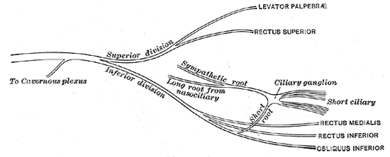
Plan of oculomotor nerve. (Short ciliary labeled at center right.)
Details
FromCiliary ganglion
Identifiers
Latinnervi ciliares breves
TA98A14.3.02.005
TA26664
FMA75474
Anatomical terms of neuroanatomy

The short ciliary nerves are nerves of the orbit around the eye. They are branches of the ciliary ganglion. They supply parasympathetic and sympathetic nerve fibers to the ciliary muscle, iris, and cornea. Damage to the short ciliary nerve may result in loss of the pupillary light reflex, or mydriasis.

Structure

[edit]

The short ciliary nerves are branches of the ciliary ganglion.[1] They arise from the forepart of the ganglion in two bundles connected with its superior and inferior angles. The lower bundle is larger than the upper bundle. These split into between 6 and 10 filaments.

They run forward with the ciliary arteries in a wavy course.[1] One bundle is set above the optic nerve, while the other bundle is set below it. They are accompanied by the long ciliary nerves from the nasociliary.

They pierce the sclera at the back part of the bulb of the eye, pass forward in delicate grooves on the inner surface of the sclera, and are distributed to the ciliary muscle, iris, and cornea.

Function

[edit]

The short ciliary nerves contain both parasympathetic and sympathetic nerve fibers. The parasympathetic fibers arise from the Edinger-Westphal nucleus and synapse in the ciliary ganglion via the oculomotor nerve, the postganglionic parasympathetics leave the ciliary ganglion in the short ciliary nerve and supply the ciliary body and iris. The sympathetic fibers are provided by the superior cervical ganglion and reach the ganglion either as branches of the nasociliary nerve or directly from the extension of the plexus on the ophthalmic artery (sympathetic branch to ciliary ganglion). The sympathetics are non-specific and provide vasoconstriction of the globe.

Clinical significance

[edit]

Damage to the short ciliary nerve may result in loss of the pupillary light reflex,[2] or mydriasis.

Additional images

[edit]

See also

[edit]

References

[edit]
[edit]
Revisions and contributorsEdit on WikipediaRead on Wikipedia
from Grokipedia
The short ciliary nerves are a group of approximately 10–20 small peripheral nerves that originate from the , located in the posterior to the eye, and provide mixed sensory, sympathetic, and parasympathetic innervation to the anterior segment of the eyeball, including the , , iris, and . These nerves arise as postganglionic branches from the , which receives preganglionic parasympathetic fibers via the (cranial nerve III), sensory fibers from the nasociliary branch of the (cranial nerve V1), and sympathetic fibers from the . They course anteriorly in a plexus around the , penetrating the near the in a radial pattern, before distributing within the uveal tract. Functionally, the short ciliary nerves play a critical role in ocular physiology: the parasympathetic components drive pupillary constriction () and accommodation of the lens by innervating the pupillae muscle and , respectively, in response to light or near-focus stimuli; sympathetic fibers regulate in the choroidal and ciliary vessels; and sensory fibers transmit pain, temperature, and touch sensations from the to the trigeminal nuclei. Disruption of these nerves, such as through trauma or compression at the orbital apex, can lead to , impaired accommodation, or sensory deficits in the eye, underscoring their importance in conditions like Adie's tonic pupil or orbital syndromes. Their anatomical proximity to short posterior ciliary arteries also facilitates coordinated neurovascular supply to the head and posterior .

Anatomy

Origin and Components

The short ciliary nerves are a group of delicate filaments, typically numbering 6 to 10, that arise from the anterior aspect of the in the . These nerves emerge in two bundles from the superior and inferior poles of the ganglion, with the inferior bundle generally being larger, and they exhibit bilateral symmetry across individuals, though the exact count can vary due to anatomical differences. These nerves are mixed in composition, carrying parasympathetic, sympathetic, and sensory fibers. The parasympathetic component consists of postganglionic fibers originating from preganglionic neurons in the Edinger-Westphal nucleus, which travel via the motor root of the (cranial nerve III) to synapse in the . The sensory fibers derive from the nasociliary branch of the ophthalmic division of the (cranial nerve V1), providing general afferent innervation. Sympathetic fibers are postganglionic, arising from the and reaching the via the . Microscopically, the short ciliary nerves comprise approximately 75% myelinated fibers and 25% unmyelinated fibers. The parasympathetic and sympathetic fibers are predominantly unmyelinated, while the sensory fibers are myelinated, reflecting their distinct functional roles and conduction properties.

Course and Distribution

The short ciliary nerves, carrying parasympathetic, sympathetic, and sensory fibers from the , emerge as 6 to 10 delicate filaments divided into superior and inferior bundles. These nerves travel anteriorly from the ganglion in a wavy course, accompanying the short posterior ciliary arteries, with bundles passing above and below the while running alongside the . Numbering typically between 8 and 10, they pierce the near the entry point at the posterior globe, posterior to the lamina cribrosa, and enter via the retrobulbar region. Upon penetrating the sclera, the short ciliary nerves follow an intrascleral path before transitioning to the suprachoroidal space, where they advance forward in grooves on the inner scleral surface toward the anterior uveal tract. In this pathway, larger nerves exhibit minimal branching, while smaller ones contribute to a suprachoroidal nerve plexus. The nerves distribute primarily to the choroid, ciliary body, and iris, with branches supplying the ciliary muscle and sphincter pupillae muscle. Sensory components provide innervation to the sclera, iris, and ciliary body. Along their course, the short ciliary nerves maintain close relations to the optic nerve, vortex veins, and long posterior ciliary arteries, positioning them in proximity to the orbital apex.

Physiology

Parasympathetic Role

The short ciliary nerves carry postganglionic parasympathetic fibers that originate from preganglionic neurons in the Edinger-Westphal nucleus of the (cranial nerve III). These preganglionic fibers travel via the inferior branch of the and the nerve to the before synapsing in the , located near the orbital apex. From the , the postganglionic parasympathetic fibers course through the short ciliary nerves to innervate the and the pupillae muscle of the iris. Activation of the causes its contraction, which relaxes the zonular fibers attached to the lens, allowing the lens to become more convex for accommodation during near vision. Innervation of the pupillae induces , or pupil constriction, which reduces the amount of light entering the eye and sharpens the . These parasympathetic pathways are integral to key ocular reflex arcs. In the pupillary light reflex, illumination of the retina triggers bilateral pupil constriction via the short ciliary nerves to protect the retina from excessive light. The near reflex involves coordinated accommodation (ciliary muscle contraction) and pupillary constriction (via sphincter pupillae), both mediated by postganglionic fibers in the short ciliary nerves, along with convergence of the eyes. The neurotransmitter released by these postganglionic parasympathetic fibers is , which binds to muscarinic receptors—predominantly the M3 subtype—on the cells of the and sphincter pupillae. This binding activates a protein-coupled pathway, increasing intracellular calcium levels and promoting .

Sympathetic and Sensory Roles

The short ciliary nerves carry postganglionic sympathetic fibers originating from the , which travel via the to the without synapsing. These fibers then pass through the short ciliary nerves to innervate the dilator pupillae muscle of the iris, facilitating pupil dilation () in response to dim light or as part of the low-light . Additionally, sympathetic fibers within the short ciliary nerves supply the choroidal blood vessels, contributing to regulation that modulates ocular blood flow and nutrient delivery to the and other intraocular structures. In contrast to the parasympathetic fibers that synapse in the ciliary ganglion, the sympathetic fibers traverse it without interruption, ensuring direct transmission to their targets. This arrangement supports efficient sympathetic control over pupillary dilation and vascular tone, distinct from the preganglionic-to-postganglionic relay observed in parasympathetic pathways. The sensory component of the short ciliary nerves consists of general somatic afferent fibers derived from the nasociliary branch of the ophthalmic division of the trigeminal nerve (CN V1), which also pass through the ciliary ganglion without synapsing. These fibers convey pain and temperature sensations from the cornea, iris, and sclera, playing a critical role in protective reflexes such as the corneal blink reflex. Furthermore, they contribute afferent input to the nasolacrimal reflex, where ocular irritation triggers tearing via connections to the parasympathetic system through the trigeminal pathway. This sensory integration helps maintain ocular surface integrity and responds to environmental threats without involving motor synapses in the ganglion.

Clinical Significance

Pathological Conditions

Trauma to the short ciliary nerves often results from orbital fractures or penetrating injuries, which can disrupt the parasympathetic fibers originating from the , leading to tonic pupil, also known as Adie's pupil. This condition manifests as a dilated with absent or sluggish light reflex but preserved accommodation due to denervation hypersensitivity and aberrant regeneration of postganglionic fibers. Inflammatory processes affecting the short ciliary nerves include herpes zoster ophthalmicus and ciliary ganglionitis, typically caused by varicella-zoster virus reactivation, which leads to , pupil irregularity, and potential tonic pupillary changes through direct or nerve damage. Neoplastic involvement arises from orbital tumors, such as meningiomas, that compress the or short ciliary nerves due to their proximity near the orbital apex, resulting in from impaired parasympathetic innervation and unequal pupil sizes. Unique symptoms of short ciliary nerve involvement include segmental paralysis, observed as vermiform iris movements indicating partial denervation, and isolated corneal hypoesthesia due to interruption of sensory fibers from the , without broader trigeminal sensory deficits.

Diagnostic and Therapeutic Implications

Diagnosis of short ciliary nerve dysfunction primarily involves assessing pupillary responses, as these nerves contribute to parasympathetic innervation of the iris sphincter and . Slit-lamp examination can reveal sectoral iris sphincter or vermiform movements indicative of tonic pupil, often resulting from postganglionic damage to the short ciliary nerves or . Pharmacological testing with dilute (0.125%) is a key diagnostic tool for conditions like Adie's tonic pupil, where the affected constricts more than the normal one due to denervation supersensitivity, confirming parasympathetic hypofunction within 30-60 minutes of instillation. This test is particularly useful in differentiating postganglionic lesions from preganglionic issues. (MRI) may visualize the to rule out compressive or inflammatory etiologies affecting the short ciliary nerves, though it is not routinely used for isolated nerve assessment. Electrophysiological studies, such as recordings of ciliary nerve fiber activity, are infrequently employed in clinical practice but have been utilized in research to evaluate sensory thresholds following corneal or orbital injuries. Therapeutic interventions target parasympathetic hypofunction or aim to prevent iatrogenic damage during procedures. For hypofunction in tonic pupil syndromes, low-dose topical or drops can alleviate symptoms like or accommodative by stimulating residual receptors in the short ciliary nerve pathways. In cases of trauma-induced damage, surgical decompression may be considered to relieve compression on the or short ciliary nerves, particularly in orbital fractures, though outcomes depend on the extent of . During intraocular surgeries such as extraction or procedures, regional anesthesia techniques like sub-Tenon's or peribulbar blocks are employed to transiently block short ciliary nerves, providing analgesia and akinesia while minimizing the risk of permanent damage through careful needle or placement to avoid globe . Prognostic factors for short ciliary nerve recovery vary by etiology, with post-viral damage often showing better outcomes than traumatic injuries. In post-viral cases, such as those associated with Adie's syndrome, accommodative paresis typically improves spontaneously over months, and pupillary light reactions may partially recover, leading to a benign, non-progressive course. Thermal or laser-induced damage, akin to minor trauma, frequently resolves within 6-12 weeks as parasympathetic function returns. Conversely, traumatic injuries causing structural disruption, like those from orbital surgery or , carry a poorer with potential for irreversible or persistent hypofunction due to axonal severance.

References

Add your contribution
Related Hubs
User Avatar
No comments yet.