Hubbry Logo
Strain (injury)Strain (injury)Main
Open search
Strain (injury)
Community hub
Strain (injury)
logo
8 pages, 0 posts
0 subscribers
Be the first to start a discussion here.
Strain (injury)
Strain (injury)
from Wikipedia
Strain
Other namesMuscle strain, pulled muscle, torn muscle
Two images of the same strain to the hamstring and associated bruising.
SpecialtyEmergency medicine
SymptomsBruise, swelling, redness and soreness
CausesExcessive stress and/or repeated injury on a muscle
3D Medical Animation Depicting Strain
3D animation depicting strain

A strain is an acute or chronic soft tissue injury that occurs to a muscle, tendon, or both. The equivalent injury to a ligament is a sprain.[1] Generally, the muscle or tendon overstretches and partially tears, under more physical stress than it can withstand, often from a sudden increase in duration, intensity, or frequency of an activity. Strains most commonly occur in the foot, leg, or back. Initial treatment typically includes rest, ice, compression, and elevation (RICE).[citation needed]

Signs and symptoms

[edit]

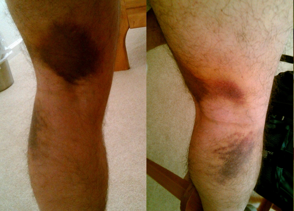
Typical signs and symptoms of a strain include pain, functional loss of the involved structure, muscle weakness, contusion, and localized inflammation.[2] A strain can range from mild overstretching to severe tears, depending on the extent of injury.[1]

Cause

[edit]

A strain can occur as a result of improper body mechanics with any activity (e.g., contact sports, lifting heavy objects) that can induce mechanical trauma or injury. Generally, the muscle or tendon overstretches and is placed under more physical stress than it can withstand.[1] Strains commonly result in a partial or complete tear of a tendon or muscle, or they can be severe in the form of a complete tendon rupture. Strains most commonly occur in the foot, leg, or back.[3] Acute strains are more closely associated with recent mechanical trauma or injury. Chronic strains typically result from repetitive movement of the muscles and tendons over a long period of time.[1]

Degrees of Injury (as classified by the American College of Sports Medicine):[4]

  • First degree (mildest) – little tissue tearing; mild tenderness; pain with full range of motion.
  • Second degree – torn muscle or tendon tissues; painful, limited motion; possibly some swelling or depression at the spot of the injury.
  • Third degree (most severe) – limited or no movement; severe acute pain, though sometimes painless straight after the initial injury

To establish a uniform definition amongst healthcare providers, in 2012 a Consensus Statement on suggested new terminology and classification of muscle injuries was published.[5]

The classifications suggested were:

The major difference suggested was the use of "indirect" muscle injury verse "grade 1" to provide subclassifications when advanced images were negative.

Indirect Muscle Injury FUNCTIONAL (Negative MSK US & MRI)[6]

  • Type 1: Overexertion-related Muscle Disorder
    • Type 1a: Fatigue induced
    • Type 1b: DOMS

• Type 2: Neuromuscular muscle disorder

    • Type 2a: Spine-Related
    • Type 2b: Muscle-Related

STRUCTURAL MUSCLE INJURY (Positive MSK US & MRI)[6] • Type 3: Partial Muscle Tear • Type 4: (Sub) total tear

DIRECT MUSCLE INJURY • Bump or Cut: Contact-related

Risk factors

[edit]

Although strains are not restricted to athletes and can happen while doing everyday tasks, people who play sports are more at risk for developing a strain, in particular sprinting or team sports.[7][8] It is common for an injury to develop when there is a sudden increase in duration, intensity, or frequency of an activity.[3]

Treatment

[edit]

The first-line treatment for a muscular strain in the acute phase include five steps commonly known as P.R.I.C.E.[9][10]

  • Protection: Apply soft padding to minimize impact with objects.
  • Rest: Rest is necessary to accelerate healing and reduce the potential for re-injury.
  • Ice: Apply ice to induce vasoconstriction, which will reduce blood flow to the site of injury. Never ice for more than 20 minutes at a time.
  • Compression: Wrap the strained area with a soft-wrapped bandage to reduce further diapedesis and promote lymphatic drainage.
  • Elevation: Keep the strained area as close to the level of the heart as is possible in order to promote venous blood return to the systemic circulation.

Immediate treatment is usually an adjunctive therapy of NSAIDs and Cold compression therapy. Cold compression therapy acts to reduce swelling and pain by reducing leukocyte extravasation into the injured area.[11][12] NSAIDs such as Ibuprofen/paracetamol work to reduce the immediate inflammation by inhibiting Cox-1 and Cox-2 enzymes, which are the enzymes responsible for converting arachidonic acid into prostaglandin. However, NSAIDs, including aspirin and ibuprofen, affect platelet function (this is why they are known as "blood thinners") and should not be taken during the period when tissue is bleeding because they will tend to increase blood flow, inhibit clotting, and thereby increase bleeding and swelling. After the bleeding has stopped, NSAIDs can be used with some effectiveness to reduce inflammation and pain.[13]

A new treatment for acute strains is the use of platelet rich plasma (PRP) injections which have been shown to accelerate recovery from non-surgical muscular injuries.[14]

It is recommended that the person injured should consult a medical provider if the injury is accompanied by severe pain, if the limb cannot be used, or if there is noticeable tenderness over an isolated spot. These can be signs of a broken or fractured bone, a sprain, or a complete muscle tear.[15]

See also

[edit]

References

[edit]
[edit]
Revisions and contributorsEdit on WikipediaRead on Wikipedia
from Grokipedia
A strain, commonly referred to as a pulled muscle, is an to a muscle or its —the fibrous that attaches muscle to —resulting from overstretching or tearing of the muscle fibers. Unlike a , which involves ligaments connecting bones, a strain affects muscles or tendons. These injuries range from mild overstretching with minimal fiber damage to severe cases involving partial or complete tears, often occurring during sudden movements, heavy lifting, or sports activities. Strains are among the most frequent soft tissue injuries, accounting for 10% to 55% of all acute sports-related injuries, with higher incidence in athletes involved in high-demand activities like running or contact sports. Symptoms typically include sudden sharp , swelling, bruising, weakness, limited , and spasms. Muscle strains are classified into three grades based on severity: grade 1 (mild, minor ), grade 2 (moderate, partial tears), and grade 3 (severe, complete tears). Initial management often follows the protocol (, , compression, ), with rehabilitation for recovery; severe cases may require . Prevention emphasizes warm-up, , and flexibility to reduce risk, as recurrence is common without proper measures.

Overview

Definition

A strain is an injury to the muscle fibers or the muscle-tendon junction resulting from overstretching or excessive contractile force, leading to partial or complete tearing of the affected tissues. This soft tissue damage typically occurs during sudden or forceful movements and is distinct from skeletal or joint injuries due to its localization in muscular structures. Unlike a sprain, which involves ligament damage around a joint, a strain primarily affects muscles or their tendinous attachments. The colloquial term "pulled muscle" has served as a longstanding lay descriptor for strains, reflecting their common occurrence in physical activities. Formal medical recognition of these injuries emerged in sports medicine during the early 20th century, with early investigations into muscle-related pain and trigger points documented in the 1930s. Pathophysiologically, strains initiate with microtears in myofibrils, often concentrated near the myotendinous junction due to mechanical overload during eccentric contractions. These disruptions cause localized hemorrhage and necrosis of muscle fibers, prompting an acute inflammatory response where phagocytic cells infiltrate the site to clear debris and form a hematoma that organizes into granulation tissue. This process impairs muscle contraction and elasticity until repair mechanisms, involving satellite cell proliferation, restore function.

Classification

Strains are classified into three grades based on the extent of muscle fiber damage and functional impairment. Grade 1 strains are mild, involving minor tearing of a small number of muscle fibers with less than 5% loss of strength or function, allowing most individuals to continue activity with minimal discomfort. Grade 2 strains are moderate, characterized by a partial tear affecting a notable portion of fibers, leading to noticeable weakness and limited . Grade 3 strains represent severe injuries with a complete rupture of the muscle or musculotendinous junction, resulting in significant functional loss, often including a palpable defect and inability to actively contract the muscle. Strains are further categorized by mechanism into direct and indirect subtypes. Direct strains occur due to or contusion, compressing the muscle against underlying bone and causing localized fiber disruption without . Indirect strains, more common in , result from excessive overstretching or eccentric loading of the muscle, typically at the musculotendinous . Additionally, strains are distinguished as acute or chronic; acute strains arise suddenly from a single event, while chronic strains develop gradually from repetitive overuse or microtrauma. The grading system carries important clinical implications for and management planning. Recovery from a Grade 1 strain typically takes 1 to 3 weeks with conservative care, enabling a swift return to normal activities. Grade 2 strains often require 4 to 8 weeks for healing, involving structured rehabilitation to restore strength. Grade 3 strains may demand 3 to 6 months or longer, sometimes necessitating surgical intervention for optimal outcomes. Representative examples illustrate these classifications. A hamstring strain during sprinting exemplifies an acute, indirect Grade 2 injury, where rapid eccentric contraction leads to partial tearing without external impact. In contrast, a direct strain might occur in the quadriceps from a contusion during contact sports, such as a knee strike in soccer, resulting in localized swelling and potential Grade 1 or 2 damage.

Causes and Risk Factors

Mechanisms of injury

Muscle strains typically result from excessive tensile forces applied to the muscle, often during sudden or deceleration movements that stretch the tissue beyond its capacity. These forces can lead to partial or complete tears in muscle fibers, particularly when the muscle is actively contracting against resistance. A primary mechanism involves eccentric contractions, where the muscle lengthens under load, generating higher forces than concentric or isometric actions and increasing vulnerability to . Direct impact from external trauma can also cause strains through contusion or laceration, compressing and disrupting muscle integrity. Common scenarios include sports activities such as sprinting, , or kicking, where rapid eccentric loading of muscles like the hamstrings occurs frequently. Occupational settings often involve lifting heavy loads or repetitive , which impose sustained tensile stress on back and lower extremity muscles. Accidental events, such as slips or trips, can trigger sudden corrective movements that stretch muscles abruptly, leading to strain. Biomechanically, injury arises when applied exceeds the muscle's tensile strength, typically ranging from 0.2 to 0.5 MPa (2 × 10^5 to 5 × 10^5 N/m²), resulting in disruption. Stress often concentrates at the muscle-tendon junction, a weak point where transmission is highest during . This site is particularly susceptible in biarticular muscles, amplifying the risk during multi-joint movements. Muscle physiology plays a key role, as fatigue diminishes fiber elasticity and impairs force absorption, predisposing the tissue to tears under peak loads. Poor conditioning can exacerbate this by reducing overall muscle resilience, though the immediate trigger remains the overload event.

Risk factors

Risk factors for muscle strains can be categorized as non-modifiable or modifiable, with certain demographic and environmental elements also contributing to increased susceptibility. Non-modifiable factors include age, history of previous injury, and sex. As individuals age, muscle elasticity decreases, leading to reduced tissue compliance and higher vulnerability to strain, particularly after the third decade of life when sarcopenia and connective tissue stiffening accelerate. Sex, with males generally exhibiting a higher risk of muscle strains compared to females, especially in athletic and high-impact activities. Previous injuries represent the strongest predictor of future strains, as scar tissue formation and neuromuscular adaptations from prior damage weaken the affected muscle's structural integrity and alter biomechanical loading patterns. Modifiable risk factors primarily involve training and conditioning deficits that can be addressed to mitigate likelihood. Poor flexibility, often manifested as muscle tightness, limits the and increases tensile stress on fibers during activity, elevating strain risk. Strength imbalances between and muscle groups, such as quadriceps-hamstring disparities, disrupt stability and predispose tissues to overload. Inadequate warm-up fails to elevate muscle and improve extensibility, leaving tissues more prone to acute failure under sudden demands. Overuse without sufficient recovery periods accumulates microtrauma, leading to and diminished force absorption capacity in muscles. Improper technique during sports or occupational tasks amplifies eccentric loading on vulnerable muscles, further heightening potential. Demographic factors highlight higher incidence among specific populations. Athletes in sports requiring explosive movements, such as runners and weightlifters, face elevated risks due to repetitive high-intensity demands on lower extremity muscles like the hamstrings and . Similarly, occupations involving manual labor, including and warehouse work, increase strain susceptibility through frequent heavy lifting and repetitive motions without ergonomic support. Environmental contributors include cold weather and , which impair muscle function. Exposure to cold temperatures reduces muscle pliability by constricting blood flow and increasing , making tissues stiffer and more susceptible to tears during exertion. compromises muscle hydration and balance, decreasing contractile efficiency and elevating the risk of cramping or straining under load.

Clinical Presentation

Signs and symptoms

Muscle strains typically present with sudden, sharp pain at the site of , often described as a tearing or pulling sensation, which is localized to the affected muscle and intensifies with movement, , or contraction of the muscle. This pain arises immediately upon during strenuous activity and can range from mild discomfort to severe, debilitating intensity depending on the extent of muscle fiber damage. Swelling and bruising commonly develop due to local hemorrhage and , usually becoming visible within 24 hours to 2 days after the injury, and may spread to adjacent areas such as the lower leg in strains. The injured area often feels tender to the touch, with possible redness or discoloration accompanying the bruising. Functional deficits include , reduced , and difficulty using the affected muscle, such as inability to bear weight or fully extend a limb, along with possible cramping or spasms that limit normal activity. These impairments stem from the partial or complete disruption of muscle fibers, making the muscle feel unstable or unusable. Severity of symptoms varies by grade: mild (grade I) strains cause minimal pain and slight discomfort with only a few fibers affected, allowing near-normal function; moderate (grade II) involve partial tears leading to noticeable weakness and limited motion; severe (grade III) result in complete rupture with intense pain, significant swelling, and total loss of muscle function. In the acute phase, symptoms such as and swelling typically peak within 24 to 48 hours post-injury before gradually subsiding with rest, though bruising may continue to evolve over several days. Common sites like the often exhibit these signs prominently due to the muscle's vulnerability during sudden movements.

Common locations

Muscle strains most frequently occur in the lower body, particularly among athletes involved in running, jumping, or explosive movements. The hamstrings, located in the posterior , are among the most commonly affected muscles, especially in sports like soccer where rapid acceleration and deceleration are involved. Hamstring strains account for approximately 19% of all reported injuries in professional soccer players. The calf muscles, specifically the gastrocnemius, are also prone to strain during push-off phases in activities such as sprinting or jumping. Strains behind the knee, often involving the hamstrings, calf, or gastrocnemius muscles, typically cause tightness and pain with movement or stretching, sometimes with localized swelling. The pain is often sharper or more activity-specific and improves with rest. strains involving the adductor muscles often result from twisting or sudden directional changes in sports like hockey or . strains, affecting the anterior , are typical in jumping sports such as or due to the high eccentric loads during landing. In the upper body, the lumbar paraspinal muscles in the lower back are a frequent site of strain, often from lifting or bending activities in both athletic and occupational settings. Lumbar strains represent about 70% of mechanical cases. Shoulder strains, particularly involving the muscles, commonly arise in overhead sports like or , where repetitive or reaching motions place repetitive stress on the supraspinatus and infraspinatus. strains, affecting the cervical paraspinals, occur in contact sports such as football or rugby from impacts or tackles that cause sudden hyperextension or flexion. Although less common overall, cervical muscle strains have a reported incidence of around 0.1 per 1,000 athlete-exposures in collegiate football. These sites highlight the role of biarticular muscles—those crossing two joints—in vulnerability to strain due to their involvement in multi-planar movements.

Diagnosis

History and physical examination

The diagnosis of a muscle strain begins with a thorough history to characterize the injury and guide the . Clinicians inquire about the onset of symptoms, distinguishing between acute sudden often associated with a specific inciting event, such as an eccentric during sports, and gradual onset linked to overuse or repetitive stress. Patients are asked to describe the pain's location, quality (e.g., sharp or aching), severity, and radiation, as well as any associated functional impacts like limping or inability to bear weight. Additional history includes prior episodes of similar injuries, which may indicate recurrent strains in vulnerable muscles like the hamstrings, and relevant risk factors such as recent activity levels or muscle imbalances. The systematically assesses the affected area through inspection, , (ROM) testing, strength evaluation, and specialized maneuvers. Inspection reveals asymmetry, swelling, or ecchymosis, while identifies localized tenderness, warmth, or a palpable gap in the muscle belly suggestive of partial or complete tears. Active and passive ROM is tested and compared to the contralateral side, using a for precision; restricted motion due to or guarding is common in moderate strains. Muscle strength is graded on the 0-5 Medical Research Council scale, with resistance applied against the patient's effort to detect weakness, such as inability to flex the against gravity in a quadriceps strain. Special tests, like the straight-leg raise for hamstring strains, involve passive flexion with extension to reproduce at the muscle's maximal length, or the active extension test at 90 degrees of flexion to assess flexibility and provoke symptoms. Red flags during examination warrant urgent evaluation to exclude severe pathology. A palpable defect, such as the "sulcus sign" in ruptures, indicates a complete rupture (grade III strain), while significant swelling, extensive bruising, or inability to voluntarily contract the muscle suggest high-grade injury or complications like . To differentiate from fractures, clinicians check for bony tenderness or ; absence of these, combined with soft-tissue findings, supports a strain diagnosis. is considered if there is tense swelling, disproportionate pain exacerbated by passive stretch, , or diminished pulses, prompting immediate intervention.

Imaging and tests

Imaging is typically reserved for cases where the clinical examination is inconclusive, such as suspected grade 2 or 3 strains, persistent symptoms beyond the initial acute phase, or atypical presentations that raise concern for complications like or associated fractures. In athletes or high-risk individuals, may also guide when return to activity timelines are critical. X-rays are not diagnostic for muscle strains themselves but are useful to exclude associated bony injuries, such as fractures or avulsions, particularly in high-impact trauma scenarios. They provide quick assessment of bone integrity, with normal findings supporting a . serves as a cost-effective, dynamic modality for evaluating muscle strains, allowing real-time assessment of , hematomas, and disruption. It is particularly advantageous for superficial muscles due to its portability and lack of , though operator dependence can affect accuracy. Typical findings include hypoechoic areas indicating or partial , with complete ruptures showing muscle discontinuity and possible retractions. Magnetic resonance imaging (MRI) is considered the gold standard for detailed evaluation of soft tissue injuries in muscle strains, offering superior visualization of edema, hemorrhage, and tear extent. On T2-weighted sequences, acute strains appear as hyperintense regions due to increased from inflammation and edema, while T1 sequences may highlight hemorrhage as hyperintense in subacute phases. Grade 2 strains often show perifascial fluid and partial fiber disruption, whereas grade 3 involves full-thickness tears with . Routine laboratory tests are not indicated for uncomplicated muscle strains, though they may be considered if infection or systemic involvement is suspected, such as elevated white blood cell count or in cases of atypical swelling. Despite their utility, modalities have limitations; overuse in mild strains, which comprise the majority of cases and are reliably diagnosed clinically, can escalate healthcare costs without altering treatment. may miss deep or small lesions, and MRI, while comprehensive, is more expensive and less accessible. Most strains can be managed initially without advanced , emphasizing conservative approaches.

Management

Acute treatment

The acute treatment of a muscle strain focuses on minimizing pain, swelling, and further tissue damage in the initial period following injury. Current guidelines recommend the PEACE & LOVE protocol as an evidence-based approach for managing soft tissue injuries like muscle strains, which has largely superseded the traditional protocol (rest, ice, compression, elevation). PEACE stands for protect the injured tissue from further damage by avoiding aggravating activities, such as halting sports or limiting for lower extremity injuries; elevate the limb above heart level when possible to reduce ; avoid anti-inflammatories in the first few days to allow the natural inflammatory response for healing; compress with an to control swelling without impeding circulation (loosen if numbness occurs); and educate on the and the benefits of active recovery to address factors and reduce fear of movement. For , acetaminophen is preferred in the acute phase as it provides relief without interfering with healing. Nonsteroidal anti-inflammatory drugs (NSAIDs), such as ibuprofen or naproxen, may be used cautiously after the initial 48-72 hours or if acetaminophen is insufficient, but under medical guidance to minimize gastrointestinal or renal risks and potential impact on tissue repair. (ice wrapped in cloth for 10-20 minutes as needed for pain) can be applied optionally during the first 48-72 hours to numb pain, but its routine use for swelling reduction is not strongly supported and should not delay early movement. Supportive measures include immobilization with aids like crutches for lower limb strains to offload weight in moderate to severe cases. Early gentle and optimal loading—light range-of-motion exercises once acute pain subsides—are encouraged to prevent and , transitioning into the LOVE components: load with progressive exercises, foster through education, promote vascularization via aerobic activity, and incorporate exercise for recovery. The acute phase typically lasts 48 to 72 hours, during which adherence to these principles is essential before advancing to rehabilitation. Treatment varies by strain grade: mild (Grade 1) strains allow quicker progression to loading, while moderate (Grade 2) and severe (Grade 3) strains require more protection initially.

Rehabilitation and surgical options

Rehabilitation for muscle strains typically progresses through three overlapping phases aimed at restoring function while minimizing reinjury risk. The initial protection phase, spanning 0-2 weeks post-injury, focuses on pain control and inflammation reduction through relative rest, gentle range-of-motion exercises, and modalities such as or to support early healing without excessive stress on the tissue. In the repair phase (2-6 weeks), gentle and strengthening begin once pain subsides, emphasizing controlled loading to promote alignment and tissue remodeling, with progression guided by pain-free tolerance. The remodeling phase (6+ weeks onward) incorporates sport- or activity-specific training, including dynamic exercises like and agility drills, to rebuild strength, power, and neuromuscular control for full return to function. Physical therapy modalities play a key role across phases to enhance recovery. Ultrasound therapy is commonly used in the repair phase to improve blood flow and reduce adhesions, while electrical stimulation, such as neuromuscular electrical stimulation (NMES), aids in muscle activation and prevention during immobilization. Progressive loading exercises form the core of rehabilitation, starting with isometric contractions in the protection phase to maintain muscle tension without movement, advancing to concentric and eccentric exercises in later phases to restore length-tension relationships and prevent weakness. These approaches are tailored to the strain grade, with grade 1 and 2 injuries often resolving fully through conservative means. Surgical options are reserved for severe cases, primarily grade 3 complete tears where conservative management fails. Indications include significant muscle retraction greater than 2 cm on imaging, large hematomas causing , or persistent symptoms after 3 months of rehabilitation, particularly in athletes requiring high performance. Techniques involve direct suture repair of the muscle-tendon unit or, in cases of extensive retraction, augmentation with grafts to restore and function. Outcomes for rehabilitation are generally favorable, with 80-90% of patients achieving return to pre-injury activity levels through structured programs, though recurrence rates can reach 30% in the first year without adequate progression. For complete tears treated surgically, success rates improve to approximately 95% return to sport in elite athletes, with reduced reinjury risk compared to non-operative approaches in select cases.

Prevention and Prognosis

Prevention strategies

Preventing muscle strains involves implementing evidence-based strategies that enhance muscle preparedness, strength, and overall physical resilience prior to engaging in activities. These approaches focus on preparation and habitual practices to mitigate overload and improve tissue tolerance. Warm-up routines, consisting of 5-10 minutes of dynamic and light aerobic activity, increase blood flow to muscles and enhance their elasticity, thereby reducing strain risk. Structured warm-up programs, such as the FIFA 11+ protocol, have been shown to decrease overall rates by up to 50% in soccer players, with particular benefits for lower extremity strains. Training principles emphasize balanced programs that incorporate strength and flexibility exercises, with gradual progression to prevent overload. Strength training reduces the overall risk of sports injuries by approximately 34% (relative risk 0.66), while proprioception and neuromuscular training further lower lower extremity injury incidence by 36-45%. For hamstring strains, eccentric exercises like the Nordic hamstring curl can reduce injury rates by 49-65% in athletes. Lifestyle factors play a supportive role in strain prevention through proper hydration, nutrition, and recovery practices. Maintaining adequate hydration prevents dehydration-related muscle and , which can contribute to strains during . Nutritional counseling, including balanced intake of proteins and micronutrients, supports muscle repair and resilience, with professionals identifying it as a key prevention strategy in elite athletes. Sufficient and rest periods facilitate recovery, reducing -induced injury risk. Targeted approaches address specific vulnerabilities, such as core strengthening exercises to prevent back strains. Core stability programs enhance spinal support and reduce low back recurrence by improving muscle endurance, with evidence indicating up to 62% risk reduction in low back and lower extremity injuries. In , protective gear like appropriate or supports can minimize strain by stabilizing vulnerable areas during high-impact activities.

Prognosis and complications

The prognosis for muscle strains varies significantly by injury grade, with milder strains generally resolving faster and with fewer long-term issues. Grade 1 strains, involving minimal fiber damage, typically achieve full recovery within 1 to 3 weeks through conservative management, with low recurrence rates of around 9%. Grade 2 strains, characterized by partial tears, often require 4 to 8 weeks for recovery, though some cases extend to 2 to 3 months, and carry a higher recurrence risk of approximately 20-24%. Grade 3 strains, featuring complete tears, demand 3 to 6 months for and may involve surgical intervention, which can elevate recurrence risks further due to potential complications like adhesions or incomplete restoration of function. Several factors influence recovery outcomes. Older age is associated with delayed and less efficient muscle repair, leading to prolonged recovery times compared to younger individuals. The injury site also plays a role; for instance, strains often heal more slowly—averaging 16 weeks for certain types—than calf strains, which typically resolve in 6 to 8 weeks. Adherence to rehabilitation protocols is crucial, as poor compliance increases the likelihood of suboptimal and reinjury. Potential complications arise particularly in moderate to severe cases or with inadequate management. These include from persistent or nerve involvement, scar tissue adhesions that limit mobility and strength, and re-injury rates reaching up to 30% among athletes due to incomplete rehabilitation. In rare severe instances, may develop, involving abnormal bone formation within the muscle that impairs function. Overall, 70-80% of strains resolve without significant long-term sequelae when treated promptly, but delayed intervention correlates with poorer prognosis, including higher rates of and functional deficits.

References

Add your contribution
Related Hubs
User Avatar
No comments yet.