Hubbry Logo
Coronary artery bypass surgeryCoronary artery bypass surgeryMain
Open search
Coronary artery bypass surgery
Community hub
Coronary artery bypass surgery
logo
8 pages, 0 posts
0 subscribers
Be the first to start a discussion here.
Coronary artery bypass surgery
from Wikipedia

Coronary artery bypass surgery
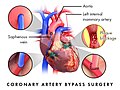
Early in a coronary artery bypass operation, during vein harvesting from the legs (left of image) and the establishment of cardiopulmonary bypass by placement of an aortic cannula (bottom of image). The perfusionist and heart-lung machine are on the upper right. The patient's head (not seen) is at the bottom.
Other namesCoronary artery bypass graft
ICD-10-PCS021209W
ICD-9-CM36.1
MeSHD001026
MedlinePlus002946

Coronary artery bypass surgery, also called coronary artery bypass graft (CABG /ˈkæbɪ/ KAB-ij, like "cabbage"), is a surgical procedure to treat coronary artery disease (CAD), the buildup of plaques in the arteries of the heart. It can relieve chest pain caused by CAD, slow the progression of CAD, and increase life expectancy. It aims to bypass narrowings in heart arteries by using arteries or veins harvested from other parts of the body, thus restoring adequate blood supply to the previously ischemic (deprived of blood) heart.

There are two main approaches. The first uses a cardiopulmonary bypass machine, a machine which takes over the functions of the heart and lungs during surgery by circulating blood and oxygen. With the heart in cardioplegic arrest, harvested arteries and veins are used to connect across problematic regions—a construction known as surgical anastomosis. In the second approach, called the off-pump coronary artery bypass (OPCAB), these anastomoses are constructed while the heart is still beating. The anastomosis supplying the left anterior descending branch is the most significant one and usually, the left internal mammary artery is harvested for use. Other commonly employed sources are the right internal mammary artery, the radial artery, and the great saphenous vein.

Effective ways to treat chest pain (specifically, angina, a common symptom of CAD) have been sought since the beginning of the 20th century. In the 1960s, CABG was introduced in its modern form and has since become the main treatment for significant CAD. Significant complications of the operation include bleeding, heart problems (heart attack, arrhythmias), stroke, infections (often pneumonia) and injury to the kidneys.

Three coronary artery bypass grafts, a pedicled LITA to LAD and two saphenous vein grafts – one to the right coronary artery system and one to the obtuse marginal system.

Uses

[edit]

Coronary artery bypass surgery aims to prevent death from coronary artery disease and improve quality of life by relieving angina, the associated feeling of chest pain.[1] The decision to perform surgery is informed by studies of CABG's efficacy in different patient subgroups, based on the lesions' anatomy or how well the heart is functioning. These results are compared with that of other strategies, most importantly percutaneous coronary intervention (PCI).[2][3]

Coronary artery disease

[edit]
With a coronary angiography, stenotic lesions of the coronary arteries are identified.

Coronary artery disease is caused when coronary arteries of the heart accumulate atheromatous plaques, causing stenosis (narrowing) in one or more arteries and risking myocardial infarction, the interruption of blood supply to the heart. CAD can occur in any of the major vessels of the coronary circulation: the left main stem, left ascending artery, circumflex artery, and right coronary artery, and branches thereof. CAD symptoms vary from none, to chest pain only when exercising (stable angina), to chest pain even at rest (unstable angina). It can even manifest as a myocardial infarction; if blood flow to the heart is not restored within a few hours, whether spontaneously or by medical intervention, the blood-deprived part of the heart becomes necrotic (dies) and is scarred. It may lead to other complications such as arrhythmias, rupture of the papillary muscles of the heart, or sudden death.[4]

There are various methods of detecting and assessing CAD. Apart from history and clinical examination, noninvasive methods include electrocardiography (ECG) at rest or during exercise, and X-ray of the chest. Echocardiography can quantify heart functioning by measuring, for example, enlargement of the left ventricle, the ejection fraction, and the situation of the heart valves. The most accurate ways to detect CAD are the coronary angiogram and the coronary CT angiography.[4] An angiogram can provide detailed anatomy of coronary circulation and lesions. The significance of each lesion is determined by the diameter loss. A diameter loss of 50% translates to a 75% cross-sectional area loss, considered moderate by most groups. Severe stenosis constitutes a diameter loss of 2/3 or more—a greater-than-90% loss of cross-sectional area.[5] To more accurately determine the severity of stenosis, interventional cardiologists may also employ intravascular ultrasound, which can determine the severity and provide information on the composition of the atheromatous plaque. With the technique of fractional flow reserve, the pressure after the stenosis is compared to mean aortic pressure. If the ratio is less than 0.80, then the stenosis is deemed significant.[5]

Indications for CABG

[edit]

Stable patients

[edit]

People with angina during exercise are usually first treated with medical therapy. Noninvasive tests help estimate which patients might benefit from undergoing coronary angiography. Generally, if portions of cardiac wall are receiving less blood than normal, coronary angiography is indicated; then, lesions are identified and inform a decision to undergo PCI or CABG.[6]

CABG is generally preferred over PCI when there is a significant burden of plaque on the coronary arteries, that is extensive and complex, due to survival benefit. Other indicators that a patient will benefit more from CABG rather than PCI include: decreased left-ventricle function; left main disease; diabetes; and complex triple system disease (including LAD, Cx and RCA), especially when the lesion in the LAD is at its proximal part.[2][3]

Acute coronary syndrome

[edit]

During an acute heart event, known as acute coronary syndrome, it is paramount to quickly restore blood flow to heart tissue. Typically, patients arrive at the hospital with chest pain. They are first treated with drugs, particularly the strongest drugs that prevent clots within vessels (dual anti-platelet therapy: aspirin and clopidogrel). Patients at risk of ongoing ischemia undergo PCI to restore blood flow and thus oxygen delivery to the struggling heart.[7] If PCI failed to restore blood flow because of anatomical considerations or other technical problems, urgent CABG is indicated to save heart tissue. The timing of the operation plays a role in survival: It is preferable to delay the surgery if possible (three days if the infarction affecting the total thickness of the cardiac muscle, and six hours if it does not).[2]

CABG is also indicated when there are mechanical complications of an infarction (ventricular septal defect, papillary muscle rupture or myocardial rupture).[8] There are no absolute contraindications of CABG, but severe disease of other organs such as the liver or brain, limited life expectancy, and patient fragility are considered.[8]

Other cardiac surgery

[edit]

CABG is also performed when a patient is to undergo another cardiac surgical procedure, most commonly for valve disease, and angiography reveals a significant lesion of the coronary arteries.[9] CABG can also address dissection of coronary arteries, where a rupture of the coronary layers creates a pseudo-lumen (cavity) and diminishes blood delivery to the heart. Such a dissection may be caused by pregnancy, tissue diseases like Ehlers–Danlos syndromes and Marfan syndrome, cocaine abuse, or PCI. A coronary aneurysm may also indicate CABG: A blood clot might develop within the vessel and travel downstream.[10]

CABG versus PCI

[edit]

CABG and percutaneous coronary intervention (PCI) are the two methods to restore blood flow caused by stenotic lesions of the coronary arteries. The choice of method is still a matter of debate, but it is clear that in the presence of complex lesions, significant left main disease, or diabetes, CABG yields better long-term survival and outcomes.[11][10] Strong indications for CABG also include symptomatic patients and impaired left ventricle function.[10] CABG offers better results than PCI in left main disease and in CAD that affects multiple vessels, because of the protection arterial conduits offer to the native arteries of the heart, by producing vasodilator factors and preventing the advancement of plaques. Studies published in 2023 show that CABG in patients with left main disease is associated with lower mortality and fewer adverse events compared to PCI.[12][13]

Patients with unprotected left main disease—when the runoff of the left main artery is not protected by a patent graft since a previous CABG operation—have been studied as a group. A 2016 European study found that in these patients, CABG outperforms PCI in the long run (5 years). Another 2016 study found that PCI has similar results to CABG at 3 years, but that CABG becomes better than PCI after 4 years.[14][15]

A 2012 trial and followup in diabetic patients demonstrated a significant advantage to CABG over PCI. The relative advantage remained evident at 3.8-year and 7.5-year follow ups, which found particular benefits in smokers and younger patients.[16] A 2015 trial compared CABG and the latest technological advancement of PCI, second-generation drug-eluting stents in multivessel disease. Their results indicated that CABG is a better option for CAD patients.[17] A trial published in 2021, comparing results after one year, also concluded that CABG is a safer option than PCI.[18] A large study published in 2023 showed that PCI patients had higher mortality than CABG patients with left main coronary artery disease.[19]

Procedure

[edit]
Illustration of a typical coronary artery bypass surgery. A vein from the leg is removed and grafted to the coronary artery to bypass a blockage at LAD.
Coronary artery bypass surgery during mobilization (freeing) of the right coronary artery from its surrounding adipose tissue (yellow). The tube visible at the bottom is the aortic cannula, which returns blood from the heart–lung machine. The tube above it (obscured by the surgeon on the right) is the venous cannula, which receives blood from the body. The patient's heart is stopped and the aorta is cross-clamped. The patient's head (not seen) is at the bottom.

Preoperative examination and strategy

[edit]

Routine preoperative examination aims to check the status of systems and organs besides the heart. The examination typically includes a chest X-ray to check the lungs, a complete blood count, and kidney and liver function tests. Physical examination to determine the quality of the grafts or the safety of removing them, such as varicosities in the legs, or the Allen test in the arm is performed to be sure that blood supply to the arm will not be critically disturbed.[20]

A patient taking anticoagulants—aspirin, clopidogrel, ticagrelol and others—will stop taking them several days before, to prevent excessive bleeding during and after the operation. Warfarin is also stopped for the same reason and the patient starts taking heparin products after the INR falls below 2.0.[20][21]

After the angiogram is reviewed by the surgical team, targets are selected (that is, which native arteries will be bypassed and where the anastomosis should be placed). Ideally, all major lesions in significant vessels should be addressed. Most commonly, the left internal thoracic artery (LITA; formerly, left internal mammary artery, LIMA) is anastomosed to the left anterior descending artery (LAD) because the LAD is the most significant artery of the heart and supplies blood to a larger portion of myocardium than other arteries.[21]

A conduit can be used to graft one or more native arteries. In the latter case, an end-to-side anastomosis is performed. In the former, using a sequential anastomosis, a graft can then deliver blood to two or more native vessels of the heart.[21] Also, the proximal part of a conduit can be anastomosed to the side of another conduit. It is preferred not to harvest too much conduit because it might necessitate re-operation.[21]

With cardiopulmonary bypass machine (on-pump)

[edit]

The intubated patient is brought to the operating theater. Lines (e.g., peripheral IV cannulae, central lines such as internal jugular cannulae) are inserted for drug administration and monitoring. A description of a traditional CABG follows.[21]

Harvesting

An incision in the sternum is made while vessels are being harvested, either from the arms or chest or from the leg, usually from the internal mammary artery or the saphenous vein. The LITA is harvested through the sternotomy. There are two common ways of mobilizing the LITA: the pedicle (i.e., a pedicle consisting of the artery plus surrounding fat and veins) and the skeletonized (i.e., freed of other tissues). Before the LITA is divided in its more distal part, the anticoagulant heparin is administered to the patient via a peripheral line, to prevent clots.[21]

Catheterization and establishment of cardiopulmonary bypass

After harvesting, the pericardium—the sac that surrounds the heart—is opened and stay sutures are placed to keep it open. Purse string sutures are placed in the aorta to prepare the insertions of the cannula into the aorta, and a catheter which temporarily arrests the heart using a solution high in potassium. Another purse string is placed in the right atrium for the venous cannula. Once the cannulas and the catheter are placed, cardiopulmonary bypass (CPB) is commenced. Deoxygenated blood arriving to the heart from veins is forwarded to the CPB machine to get oxygenated, then delivered to the aorta to keep the rest of the body saturated. The blood is often cooled to 32–34 °C (90–93 °F) to slow metabolism and minimize the demand for oxygen. A clamp is placed on the aorta between the cardioplegic catheter and aortic cannula, so that the flow of cardioplegic solution may be controlled by adjusting the clamp. Within minutes, the heart stops beating.[21][22]

Anastomosis (grafting)

With the heart still, the tip of the heart is taken out of pericardium so that native arteries lying on the posterior side of the heart are accessible. Usually, distal anastomoses are constructed first (first to the right coronary system, then to the circumflex) and then the sequential anastomosis if necessary. Surgeons check the anastomosis for patency (whether it is sufficiently open) or leaking. They then insert the graft within the pericardium, sometimes attached to the cardioplegic catheter. The anastomosis of the LIMA to the LAD is usually the last distal anastomosis to be constructed; while it is being constructed the blood rewarming process starts (by the CPB).[21] After the anastomosis is completed and checked for leaks, the proximal anastomoses of the conduits, if any, are next. They can be done either with the clamp still on, or after removing the aortic clamp and isolating a small segment of the aorta by placing a partial clamp. That said, aortas burdened by plaques might be damaged or release atheromatous debris by being overhandled.[21][23]

Weaning from cardiopulmonary bypass and closure

After the proximal anastomoses are done, the clamp is removed and the aorta and conduits de-aired. Pacing wires, which supply a current to assist the heartbeat, might be placed. If the heart and other systems are functioning, CPB is discontinued and cannulae are removed. Protamine is administered to reverse the effect of the anticoagulant heparin. After possible bleeding sites are checked, chest tubes are placed and the sternum is closed.[21][23]

Off-pump

[edit]

Off-pump coronary artery bypass (OPCAB) surgery avoids using the CPB machine by stabilizing small segments of the heart at a time. The surgical team and anesthesiologists must coordinate and take great care to not manipulate the heart too much, lest they compromise the stability of blood flow. Compromise should be detected immediately and appropriate action taken.[24]

Keeping a healthy heartbeat may involve maneuvers like placing atrial wires to protect from bradycardia, or by placing stitches or incisions into the pericardium to help exposure. Snares and tapes are used to facilitate exposure. The aim is to avoid distal ischemia caused by blockage of the vessel supplying distal portions of the left ventricle, so usually LITA to LAD is the first to be anastomosed and others follow. For the anastomosis, a fine tube blowing humidified CO2 keeps the surgical field clean of blood. Also, a shunt might be used so the blood can travel past the site of anastomosis. After the distal anastomoses are completed, proximal anastomoses to the aorta are constructed with a partially closed aortic clamp. The rest of the process is similar to on-pump CABG.[24]

Alternative approaches and special situations

[edit]

When CABG is performed as an emergency because of a myocardial infarction, the highest priority is to salvage the struggling myocardium. Before operation, an intra-aortic balloon pump (IABP) might be inserted to relieve some of the burden of pumping blood, effectively reducing the amount of oxygen needed by myocardium. During operation, the standard practice is to place the patient on CPB as soon as possible and revascularize the heart with three saphenous veins. A calcified aorta also poses a problem because it is very dangerous to clamp. In this case, the operation can be done as an off-pump CAB using both inferior mesenteric arteries (IMA) or Y, T and sequential grafts. Deep arrest may be induced with hypothermia, lowering the temperature of the body to slightly above 20 °C (68 °F).[25] In cases where a significant artery is totally blocked, it may be possible to remove the plaque and use the same hole in the artery to perform an anastomosis. This technique is called endarterectomy and is usually performed at the right coronary system.[26]

Re-operations of CABG (another CABG operation after a previous one) pose difficulties. The heart may be positioned too close to the sternum and thus at risk when cutting the sternum again, so an oscillating saw is used. The heart may be covered with strong adhesions to adjusting structures. Doctors must decide whether aging grafts should be replaced. Manipulation of vein grafts is avoided because it risks dislodgement of plaque.[27]

Minimally invasive direct coronary artery bypass (MIDCAB) strives to avoid a large incision in the sternum. It utilizes off-pump techniques to place a graft, usually of the LIMA at the LAD. The LIMA is freed through an incision between the left ribs (thoractomy), or even using an endoscope placed in the left chest.[28] Robot-assisted coronary revascularization, which is not yet widely used, avoids the sternum incision to prevent infections and bleeding. Both conduit harvesting and the anastomosis are performed with the aid of a robot, through a thoracotomy. Usually, the procedure is combined with hybrid coronary revascularization, in which methods of CABG and PCI are both employed. Anastomosis of the LIMA to the LAD is performed in the operating theater and other lesions are treated with PCI—either at the operating room immediately following the anastomosis, or several days later.[29]

Post-operative care

[edit]

After the procedure, the patient is usually transferred to the intensive care unit (ICU), where intubations are removed if not already done in the operating theater. They usually exit the ICU by the following day, and four days later, if no complications occur, the patient is discharged from the hospital.[30]

A series of drugs are commonly used in early post-operative care. Dobutamine, a beta agent, can increase the cardiac output and is administered some hours after the operation. Beta blockers are used to prevent atrial fibrillation and other supraventricular arrhythmias. Pacing wires attached to both atria, inserted during the operation, may help prevent atrial fibrillation. Aspirin (80 mg) is used to prevent graft failure.[30] Angiotensin-converting enzyme (ACE) inhibitors and angiotensin receptor blockers (ARBs) are used to control blood pressure, especially in patients with low cardiac function (<40%). Amlodipine, a calcium channel blocker, is used for patients whose radial artery was used as a graft.[2]

After the discharge, patients may experience insomnia, low appetite, decreased sex drive, and memory problems. This effect is usually transient and lasts 6 to 8 weeks.[30] A tailored exercise plan is usually beneficial.[30]

Results

[edit]

CABG is the best procedure to reduce mortality from severe CAD and improve quality of life.[31][32]: 153  Operative mortality strongly relates to the patient's age. According to a study by Eagle et al., patients 50–59 years old have an operative mortality rate of 1.8%, while patients older than 80 have a rate of 8.3%.[33] Other factors that increase mortality are being female, re-operation, dysfunction of the left ventricle, and left main disease.[33] CABG usually relieves angina, but in some patients it reoccurs. Around 60% of patients will be angina-free 10 years after their operation.[33] Myocardial infarction is rare five years after a CABG, but its risk increases with time.[34] The risk of sudden death for CABG patients is low.[34] Quality of life is also high for at least five years, then can slowly start to decline.[35] However, the use of bilateral mammary artery in patients of younger age and those without specific comorbidities (diabetes, obesity, steroid use) can provide excellent long-term survival and quality of life.[36]

The beneficial effects of CABG are clear at the cardiac level. Left-ventricle function is improved and malfunctioning segments of the heart—dyskinetic (moving inefficiently) or even akinetic (not moving)—can show signs of improvement. Both systolic and diastolic functions are improved and keep improving for up to five years in some cases.[37] Left-ventricle function and myocardial perfusion during exercise also improves after CABG. When the left ventricle is severely impaired before operation (ejection fraction below 30%), however, benefits are less impressive in terms of segmental wall movement but still significant because other parameters might improve as LV function improves; the pulmonary hypertension might be relieved and lengthen survival.[37][38]

Determining the total risk of the procedure is difficult because of the diversity of patients undergoing CABG; different subgroups have different risk, but younger patients see better results than older ones. A CABG using two, rather than one, internal mammary arteries (IMAs) may offer greater protection from CAD, but results are not yet conclusive.[33][39]

Grafts

[edit]

Conduits that can be used for CABG may be arteries or veins. Arteries have a superior long-term patency (expandedness), but veins are more commonly used due to their practicality.[40]

Arterial grafts originate from the part of the internal thoracic artery (ITA) that runs near the edge of sternum, and can easily be mobilized and anastomosed to the native target vessel of the heart. The left artery is most often used because it is closer to the heart, but the right artery is sometimes used, depending on patient and surgeon preferences. The ITAs are advantageous because of their endothelial cells, which produce endothelium-derived relaxing factor and prostacyclin, protecting the artery from atherosclerosis and thus stenosis or occlusion. Disadvantages include a high rate of complications, such as deep sternal wound infections, in some subgroups of patients—mainly obese and diabetic ones. The left radial artery and left gastroepiploic artery can be also used. Long-term patency is influenced by the type of artery used and intrinsic factors of the cardiac arterial circulation.[41]

Veins used are mostly great saphenous veins and, in some cases, the lesser saphenous vein. Their patency rate is lower than that of arteries. Aspirin protects grafts from occlusion; adding clopidogrel does not improve rates.[41]

Compared to PCI

[edit]

CABG and PCI are the two methods to revascularize stenotic lesions of the cardiac arteries. The preferences for each patient is still a matter of debate but in the presence of complex lesions and significant Left Main Disease, and in diabetic patients, CABG seems to offer better results in patients than PCI.[11][10] Strong indications for CABG also include symptomatic patients and those with impaired LV function.[10]

Complications

[edit]

The most common complications of CABG are postoperative bleeding, heart failure, atrial fibrillation (a form of arrhythmia), stroke, kidney dysfunction, and infection of the wound near the sternum.[38]

Postoperative bleeding occurs in 2–5% of cases and may require returning to the operating room;[42] the most common indicator is the amount of blood being drained by chest tubes, which are inserted during the operation to drain fluid or air from the chest. Bleeding may originate from the aorta, the anastomosis, an insufficiently sealed branch of the conduit, or from the sternum. Other causes include platelet abnormalities or their failure to clot—perhaps due to the bypass or to the rebound heparin effect, which occurs when the anti-coagulant heparin is administered at the beginning of surgery and reappears in the blood after its neutralization by protamine.[43]

Low cardiac output syndrome (LCOS) can occur in up to 14% of CABG patients. According to its severity, LCOS is treated with inotropes, an intra-aortic balloon pump (IABP), optimization of pre-load and afterload, or correction of blood gauzes and electrolytes. The aim is to maintain a systolic blood pressure above 90 mmHg and a cardiac index of more than 2.2 L/min/m2.[38] LCOS is often transient.[42] Myocardial infarction can occur after the operation because of either technical or patient-specific factors. Its incidence is difficult to estimate due to varying definitions, but most studies place its occurrence at between 2% and 5%. The incidence is also dependent on whether it is isolated CABG (average, 4%, range, 0.3%–10%) or a combined operation (average, 2.0%, range, 0.7%–12%).[44] New electrocardiogram features, such as Q waves or ultrasound-documented alternation of cardiac wall motions, are indicative. Ongoing ischemia might prompt emergency angiography and PCI or re-operation.[45][42] Immediate coronary angiography offers the most expeditious modality not only for diagnosis but also for potential reintervention.[46] Echocardiography is less valuable for the detection or confirmation of postoperative myocardial ischemia.[47] Arrhythmias can also occur, most-commonly atrial fibrillation (incidence of 20–40%) that is treated with correcting electrolyte balance, and rate and rhythm control.[42][38] However, arrhythmia such as ventricular tachycardia or fibrillation can be a sign of postoperative myocardial ischemia that is treated depending on the cause.[48]

Adverse neurological effects occur after CABG in about 1.5% of patients.[42] They can manifest as type-1 deficits—focal deficits such as stroke or coma—or type-2 global deficits such as delirium caused by CPB, hypoperfusion, or cerebral embolism.[38] Cognitive impairment has been reported in up to 80% cases after CABG at discharge and lasts for a year in up to 40% of cases. The cause remains unclear; CPB is an unlikely cause because even in CABG patients without CPB, as in off-pump CABG, and PCI patients, the incidence is the same.[38][39]

Infections, such as wound infections in the sternum (superficial or deep) are most commonly caused by Staphylococcus aureus, and may complicate the post-operation process. The harvesting of both two thoracic arteries is a risk factor because it significantly impairs the perfusion of blood through the sternum.[38] Pneumonia can also occur.[42] Complications in the gastrointestinal tract have been described and are most commonly due to medications administered during the operation.[39]

History

[edit]

Pre-CABG

[edit]
René Gerónimo Favaloro was an Argentine cardiac surgeon and educator best known for his pioneering work on coronary artery bypass surgery using the great saphenous vein.[49]

In the early 20th century, surgical interventions aiming to relieve angina and prevent death were either sympathectomy — a cut on the sympathetic chain that supplies the heart—or pericardial abrasion, with the hope adhesions would create significant collateral circulation. Sympathectomy produced disappointing and inconsistent results.[50] French surgeon Alexis Carrel was the first to anastomose a vessel—a branch of the carotid artery—to a native artery in the heart of a dog, but the experiment could not be reproduced.[51] In the mid-20th century, revascularization efforts continued. Beck C. S. used a carotid conduit to connect the descending aorta to the coronary sinus, the biggest vein of the heart. In the "Vineberg Procedure", Arthur Vineberg used skeletonized LITA, placing it in a small tunnel he created next to the LAD and hoping spontaneous collateral circulation would form. This occurred in canine experiments but not in humans. Goetz RH was the first to perform an anastomosis of the ITA to LAD in the 1960 using a sutureless technique.[50]

The development of coronary angiography in 1962 by Mason Sones helped medical doctors to identify patients in need of operation, and which native heart vessels should be bypassed.[52] In 1964, Soviet cardiac surgeon Vasilii Kolesov performed the first successful internal thoracic artery–coronary artery anastomosis, followed by Michael DeBakey in the United States. Argentine surgeon René Favaloro standardized the procedure. Their advances made CABG the standard of care of CAD patients.[53]

The CABG era

[edit]

The modern era of the CABG began in 1964 when Soviet cardiac surgeon Vasilii Kolesov performed the first successful internal thoracic artery–coronary artery anastomosis. The same year, American surgeon Michael DeBakey used a saphenous vein to create an aorta-coronary artery bypass. Argentinean surgeon René Favaloro advanced and standardized the CABG technique using the patient's saphenous vein.[53]

The introduction of arresting the heart during operation (cardioplegia) made CABG much less risky. A major obstacle of CABG was ischemia and infarction occurring while the heart was stopped to allow surgeons to construct the distal anastomosis. In the 1970s, potassium-based cardioplegia was used. Cardioplegia minimized the oxygen demands of the heart, thus reducing the effects of ischemia. Refinement of cardioplegia in the 1980s made CABG less risky, lowering mortality during operation.[54]

In the late 1960s, after the work of René Favaloro, the operation was performed in only a few centers, but was anticipated to more broadly change the outcome of coronary artery disease. By 1979, there were 114,000 procedures per year in the US. The introduction of percutaneous coronary intervention (PCI) did not obsolesce CABG; rates of both procedures continued to increase, but PCIs grew more rapidly. In the following decades, CABG was extensively studied and compared to PCI. The absence of a clear advantage of CABG over PCI led to a small decrease in numbers of CABGs in some countries (like the US) by 2000. In Europe—mainly Germany—CABG was increasingly performed. As of 2023, research comparing the two techniques is continuing.[55] Meta-analysis published in 2023 suggests that CABG provides a consistent survival benefit over PCI with drug-eluting stents (DES).[56]

Favaloro's work is fundamental to the history of graft selection. He established the use of bilateral ITAs as superior to vein grafts. Surgeons examined the use of other arterial grafts—splenic, gastroepiploic mesenteric, subscapular and others—but none matched the patency rates of ITA. In 1971, Carpentier introduced the use of the radial artery, which was initially prone to failure, but the development of harvesting techniques in the following 20 years significantly improved patency.[57]

CABG In Animals

[edit]

Pigs, sheep, and dogs have been used as experimental models, for the development of CABG.[58] Performing CABG to treat a sick animal though is extremely rare.[59]

See also

[edit]

References

[edit]

Sources

[edit]
[edit]
Revisions and contributorsEdit on WikipediaRead on Wikipedia
from Grokipedia
Coronary artery bypass (CABG), commonly known as coronary artery bypass surgery, is a surgical procedure designed to improve blood flow to the heart muscle by creating new pathways that blocked or narrowed sections of the . This is achieved by harvesting a healthy , typically from the chest (internal mammary ), leg (saphenous vein), or arm (), and it to connect the to the coronary artery beyond the obstruction. The surgery addresses coronary heart disease, a condition where plaque buildup restricts blood supply, potentially leading to (), heart attacks, or . CABG is primarily indicated for patients with severe that cannot be adequately managed with medications or less invasive procedures like and stenting. It is often recommended when there is significant blockage in the left main coronary artery, multiple vessels are affected, or in cases involving , reduced heart function, or post-heart attack scenarios where emergency intervention is needed. Unlike treatments that open narrowed arteries, CABG provides a durable alternative route for blood flow, though it does not cure the underlying and requires ongoing lifestyle modifications and medications to prevent further disease progression. The procedure can be performed as an or emergently during a heart attack to restore circulation and reduce mortality risk. The surgery typically lasts 3 to 6 hours under general anesthesia and involves two main approaches: traditional on-pump CABG, where the heart is stopped and a heart-lung machine maintains circulation, or off-pump (beating-heart) CABG, which avoids the bypass machine for select patients to potentially reduce complications. In the , the makes a long incision along the breastbone to access the heart, spreads the , and attaches the grafts while the heart is temporarily halted. Minimally invasive variants use smaller incisions between the ribs, often for fewer bypasses, leading to shorter recovery times. Multiple grafts may be needed depending on the number of blockages; terms such as double bypass (two grafts), triple bypass (three grafts), or quadruple bypass (four grafts) are commonly used, with one to four being typical and arterial grafts preferred for better long-term patency. Postoperative recovery involves 1 to 2 days in the , a total hospital stay of about one week, and full recuperation over 6 to 12 weeks, supported by programs that emphasize exercise, diet, and risk factor management. While CABG effectively relieves symptoms and improves long-term survival in appropriately selected patients, it carries risks such as , , , arrhythmias, and problems, which are higher in cases or those with comorbidities like or lung disease. Advances in surgical techniques, including robotic assistance and "no-touch" vein harvesting, continue to enhance outcomes and minimize complications.

Medical background

Coronary artery disease

Coronary artery disease (CAD), also known as coronary heart disease, is a pathological condition characterized by the accumulation of atherosclerotic plaques within the walls of the , leading to narrowing () and potential obstruction of blood flow to the heart muscle (myocardium). This buildup primarily results from , a process involving the deposition of , inflammatory cells, and fibrous elements that form plaques. CAD manifests in various types, including stable CAD with chronic plaque progression causing predictable symptoms, and acute forms such as unstable CAD where plaques rupture or erode, triggering formation and sudden events. Stable plaques feature a thick fibrous cap and smaller cores, providing relative structural integrity, whereas unstable plaques have thin fibrous caps, large necrotic cores rich in , and high inflammatory activity, increasing the risk of rupture. Epidemiologically, CAD represents the leading cause of cardiovascular mortality worldwide, accounting for approximately 9.1 million deaths in 2019 as part of ischemic heart disease. Global prevalence is influenced by rising rates in low- and middle-income countries due to , dietary shifts toward high-fat processed foods, and sedentary lifestyles, with projections indicating a 90% increase in cardiovascular prevalence by 2050. Key risk factors include nonmodifiable elements such as advancing age (incidence rises sharply after 45 years in men and 55 in women), male sex, and genetic predispositions like family history, alongside modifiable factors including (affecting an estimated 1.4 billion adults aged 30–79 years worldwide as of 2024), diabetes mellitus, cigarette smoking (responsible for approximately 20% of deaths from coronary heart disease), and with elevated cholesterol. These risks synergistically promote , accelerating plaque formation and progression to myocardial ischemia, where oxygen demand exceeds supply. The of CAD centers on chronic inflammation and lipid accumulation in the arterial intima, initiating endothelial injury and recruitment that evolves into foam cell-laden plaques. As plaques enlarge, they reduce luminal diameter, impairing coronary blood flow and causing myocardial ischemia—particularly during increased demand such as exercise—manifesting as pectoris, a substernal chest pressure or tightness often radiating to the jaw, neck, or arms. Severe or prolonged ischemia can lead to (heart attack) if a plaque ruptures, forming an occlusive that fully blocks , resulting in cardiomyocyte . In advanced cases, chronic ischemia contributes to and . The arise from the aortic root and form a network essential for myocardial , delivering oxygenated blood to sustain cardiac contraction. The left main originates from the left and bifurcates into the left anterior descending (LAD) , which supplies the anterior left ventricle and two-thirds of the , and the left circumflex (LCx) , which the lateral and posterior left ventricle and left atrium. The (RCA) emerges from the right , providing blood to the right ventricle, right atrium, , and often the and posterior descending for inferior myocardial regions. Dominance varies, with right-dominant circulation (RCA supplies posterior descending) in about 85% of individuals, left-dominant (LCx) in 10%, and codominant in the rest, influencing patterns and ischemia risks. Common symptoms of CAD include stable angina, triggered by exertion and relieved by rest or , reflecting reversible ischemia, as well as atypical presentations like , dyspnea, or , particularly in women, diabetics, and the elderly. signals acute plaque instability with rest pain or escalating episodes, heralding potential infarction. Diagnosis typically begins with clinical history and , followed by noninvasive assessments such as to detect ischemic changes and (exercise or pharmacologic) to provoke and evaluate myocardial oxygen supply-demand mismatch, confirming CAD in symptomatic patients.

Revascularization principles

Coronary encompasses surgical and procedures designed to restore blood flow to ischemic myocardium in patients with obstructive by bypassing or opening occluded coronary arteries. The primary goals of these interventions include relieving myocardial ischemia to alleviate symptoms such as , preventing , improving long-term survival, and enhancing overall . Revascularization strategies are broadly classified into surgical approaches, such as coronary artery bypass grafting (CABG), which involves creating a conduit to detour blood around the blockage using harvested arterial or venous grafts, and methods, such as (PCI) with stenting, which mechanically dilate the stenotic lesion to reopen the vessel lumen. These mechanisms differ fundamentally: CABG reroutes flow externally via grafts, while PCI directly addresses the intraluminal obstruction without altering the native vessel's overall architecture. Physiologically, the choice of graft material significantly influences long-term outcomes, with arterial conduits like the internal mammary artery exhibiting superior rates—often exceeding 90% at 10 years—compared to venous grafts such as the saphenous vein, due to their endothelial properties and resistance to progression. In bypassed vessels, blood flow dynamics are predominantly diastolic, mirroring native coronary patterns to optimize myocardial oxygen delivery during ventricular relaxation, though competitive flow from partially native arteries can compromise graft durability. Landmark studies, such as the Coronary Artery Surgery Study (CASS), have established the foundational role of in reducing mortality among patients with severe , particularly those with multivessel involvement or left main stenosis, by demonstrating improved survival through surgical bypass compared to medical therapy alone.

Indications and patient selection

Stable ischemic heart disease

Stable ischemic heart disease (SIHD) refers to a clinical characterized by myocardial ischemia due to a mismatch between coronary blood supply and demand, typically presenting as stable angina or equivalent symptoms without evidence of or hemodynamic instability. This condition arises from chronic (CAD) and is managed initially with optimal medical therapy (OMT), including antianginal medications, risk factor modification, and lifestyle interventions. Coronary artery bypass grafting (CABG) is indicated in SIHD for patients with significant left main coronary artery stenosis (≥50% diameter reduction), where it is recommended to improve survival compared to medical therapy alone (Class 1 recommendation, Level of Evidence B-R). CABG is also recommended for patients with multivessel CAD involving the proximal left anterior descending (LAD) artery, particularly in those with diabetes, to reduce long-term adverse cardiac events (Class 1, Level of Evidence A). In cases of three-vessel disease without left ventricular dysfunction, CABG may be reasonable to enhance survival, especially when anatomical complexity favors surgical over percutaneous approaches (Class 2b, Level of Evidence B-R). Ideal patient profiles for CABG in SIHD include those with persistent or refractory despite guideline-directed OMT, such as beta-blockers, nitrates, and , or evidence of significant ischemia on noninvasive testing. These patients often have multivessel involvement or high-risk features like diabetes mellitus, where surgical addresses diffuse or complex lesions not amenable to stenting. Selection emphasizes a multidisciplinary Heart Team approach to weigh symptom burden, functional status, and comorbidities. Prognostic benefits of CABG in SIHD are most pronounced in subgroups with left main disease, three-vessel CAD, or , where it confers improved long-term survival and reduced rates of compared to medical management. For instance, in diabetic patients with multivessel disease, CABG demonstrates superior outcomes in preventing major adverse cardiac events over a decade of follow-up. These advantages stem from complete of multiple territories, mitigating ischemia-related risks. Decision-making incorporates anatomical scoring systems like the SYNTAX score to assess lesion complexity, with scores >22 generally favoring CABG over for better outcomes in complex cases. Surgical risk is evaluated using models such as the Society of Thoracic Surgeons (STS) score or EuroSCORE II, which predict and morbidity to guide patient optimization in stable settings. Comorbidities conferring high bleeding risk, such as recent cerebral hemorrhage, may favor percutaneous coronary intervention over CABG due to lower perioperative bleeding risks and the option for shorter dual antiplatelet therapy durations with modern drug-eluting stents. In less complex anatomy, may serve as an alternative to CABG for symptom relief.

Acute coronary syndromes

Acute coronary syndromes (ACS) represent a spectrum of clinical conditions arising from acute myocardial ischemia due to coronary artery occlusion or reduced perfusion, encompassing , non–ST-segment elevation (NSTEMI), and ST-segment elevation (STEMI). is characterized by new-onset or worsening angina at rest or with minimal exertion without biomarker elevation, while NSTEMI involves myocardial necrosis evidenced by elevated cardiac biomarkers alongside ischemic symptoms or electrocardiographic changes; STEMI features transmural ischemia with persistent ST-segment elevation and biomarker rise. These presentations necessitate urgent to restore coronary blood flow and limit myocardial damage. In ACS, coronary artery bypass grafting (CABG) is indicated for patients with complex multivessel or left main unsuitable for (PCI), particularly when anatomy involves high-risk features such as with proximal left anterior descending involvement or significant left ventricular dysfunction. CABG is also recommended following failed PCI with ongoing ischemia, in refractory to medical therapy or mechanical support, and for mechanical complications post-myocardial , including ventricular septal rupture, acute from papillary muscle dysfunction, or free wall rupture. Guideline-directed referral to a Heart Team for CABG consideration is emphasized in high-risk ACS, defined by the GRACE risk score exceeding 140, which integrates age, , systolic , , at admission, ST-segment deviation, and elevated biomarkers to predict in-hospital and 6-month mortality. Timing of CABG in ACS is guided by hemodynamic stability and ischemic burden, with emergent surgery (within 6 hours) reserved for life-threatening complications like mechanical rupture or refractory shock to prevent further deterioration. For post-STEMI patients with multivessel disease and ongoing ischemia but without instability, delayed CABG (3–7 days after the index event) allows to resolve and reduces operative mortality compared to immediate intervention. Special considerations in ACS patients selected for CABG include meticulous perioperative management of antithrombotic therapy to balance thrombotic and risks. Dual antiplatelet therapy (aspirin plus a inhibitor like clopidogrel or ) initiated for ACS should be interrupted preoperatively—typically 5 days for clopidogrel, 7 days for , and 3–5 days for in elective cases, or at least 24 hours in urgent scenarios—with resumption 24–72 hours postoperatively once is achieved. Parenteral anticoagulation, such as , is continued until surgery with dosing adjustments to avoid excess . In select cases with hybrid anatomy (e.g., isolated non-left anterior descending culprit lesions amenable to stenting), staged hybrid revascularization—combining initial PCI on the culprit lesion followed by CABG 10–14 days later—offers a safe alternative to standalone CABG, potentially reducing transfusion needs while achieving comparable long-term outcomes.

Concomitant cardiac procedures

Concomitant cardiac procedures involve performing coronary artery bypass grafting (CABG) alongside other cardiac surgeries in patients with severe (CAD) requiring non-coronary interventions. Common combinations include CABG with valve repair or replacement, such as for or , ascending aortic aneurysm repair, and left ventricular aneurysm reconstruction or closure following . These combined approaches address multivessel CAD in patients who would otherwise face high ischemic risk during isolated non-coronary procedures. Indications for concomitant CABG arise when preoperative angiography reveals significant CAD (typically ≥50-70% stenosis in major vessels, or ≥70% in non-left main epicardial arteries and ≥50% in left main per ACC/AHA guidelines) in patients undergoing elective or urgent cardiac surgery for valvular, aneurysmal, or structural heart disease. In octogenarians undergoing surgical aortic valve replacement (SAVR), the prevalence of CAD necessitating CABG is approximately 50-60%. For aneurysmectomy, concomitant CABG is indicated in 20-40% of cases with concomitant CAD to prevent perioperative myocardial ischemia, while ventricular reconstruction often pairs with CABG in post-infarction patients with akinetic segments and viable myocardium or mechanical complications like ventricular septal defects. These combinations are prioritized in guidelines for patients with left ventricular dysfunction or angina, ensuring comprehensive revascularization. Surgical sequencing typically prioritizes CABG before other procedures to optimize myocardial protection, with distal graft anastomoses performed first under cardiopulmonary bypass and cardioplegic arrest, followed by valve intervention or aneurysm repair. This order minimizes ischemic time by restoring coronary flow early, using antegrade or retrograde cold blood cardioplegia to maintain myocardial viability during subsequent steps. For arterial grafts like the left internal mammary artery, proximal anastomosis is deferred until after the primary procedure to avoid tension or kinking. Combined procedures increase operative complexity, with reported in-hospital mortality rates of 4.8-6.8% for CABG plus aortic or surgery, higher than isolated CABG (2-3%) or (3-5%) but comparable to age-matched controls when multivessel CAD is addressed. Society of Thoracic Surgeons (STS) data indicate unadjusted operative mortality around 4.4-5% for elective SAVR with CABG (vs. 2.3% isolated SAVR), though emergency operations elevate risks to 15-20%. Despite added morbidity, long-term survival benefits are evident, with 5-year rates of 85-92% in combined CABG-valve cohorts, attributed to reduced recurrent ischemia in multivessel disease. Registries like STS highlight improved outcomes with experienced centers, supporting concomitant CABG for survival advantage in high-risk anatomies.

Comparison with alternative treatments

Versus percutaneous coronary intervention

(PCI) involves balloon angioplasty to dilate narrowed coronary arteries, often followed by deployment of stents, including drug-eluting stents to reduce restenosis, but it faces limitations in complex anatomy such as multivessel disease or left main involvement due to challenges in achieving complete and higher rates of restenosis or . In contrast, coronary artery bypass grafting (CABG) provides more durable in such cases by bypassing multiple lesions with grafts, leading to superior long-term vessel patency. Key randomized trials have informed the comparative efficacy of CABG and PCI. The SYNTAX trial demonstrated that CABG reduced major adverse cardiac or cerebrovascular events (MACCE) compared to PCI at 5 years, particularly in patients with high SYNTAX scores (≥33) indicating complex coronary anatomy (29.7% vs. 46.5% MACCE rate). The FREEDOM trial in patients with diabetes and multivessel disease showed CABG superiority over PCI with drug-eluting stents, with lower rates of death (16.3% vs. 10.9%) and myocardial infarction at 5 years. Conversely, the EXCEL trial in left main disease with low-to-intermediate SYNTAX scores found PCI noninferior to CABG for the composite outcome of death, stroke, or myocardial infarction at 5 years (22.0% vs. 19.2%). Overall outcomes favor CABG for long-term survival and reduced repeat in multivessel or left main disease, with meta-analyses showing improved 10-year survival ( 0.82) and lower rates compared to PCI. PCI, however, is preferred for single-vessel disease or patients at high surgical risk, including those with recent cerebral hemorrhage indicating high bleeding risk, due to lower periprocedural rates (0.6% vs. 1.2% in SYNTAX), reduced perioperative bleeding risk from its less invasive nature, ability to shorten dual antiplatelet therapy to 1-3 months with modern drug-eluting stents, and faster recovery. Regarding cost and recovery, PCI typically involves shorter hospital stays (average 1-2 days vs. 5-7 days for CABG) and lower initial procedural costs ($17,000 less per hospitalization), but higher reintervention rates (e.g., 12-20% at 5 years vs. 5-8% for CABG) increase long-term expenses. Guideline consensus, including the 2018 ESC/EACTS guidelines and 2021 ACC/AHA/SCAI guidelines, recommends a hybrid approach: CABG as class I for complex multivessel disease (SYNTAX score >22) or diabetes, PCI as class I or IIa for simpler anatomy or left main with low SYNTAX scores (<22), factoring in comorbidities, surgical risk, and patient preference.

Versus optimal medical therapy

Optimal medical therapy (OMT) for coronary artery disease typically includes anti-ischemic agents such as beta-blockers and nitrates, lipid-lowering drugs like statins, antiplatelet therapy, and lifestyle modifications including smoking cessation, diet, and exercise. This approach aims to reduce ischemic symptoms, prevent plaque progression, and lower cardiovascular risk without invasive intervention. In patients with stable ischemic heart disease and low-risk anatomy, trials like COURAGE demonstrated no survival benefit from revascularization added to OMT, with similar rates of death or myocardial infarction between groups over five years. However, in subgroups with diabetes and more severe disease, the BARI 2D trial showed that CABG superior to OMT in reducing major adverse cardiac events, primarily driven by a lower incidence of nonfatal myocardial infarction (7.4% vs. 14.6% at five years), though all-cause mortality was comparable. The ISCHEMIA trial further indicated that an invasive strategy, including CABG in about 25% of cases, reduced angina symptoms compared to OMT alone but did not lower the primary composite endpoint of cardiovascular death or myocardial infarction in patients with moderate or severe ischemia. CABG is indicated over OMT in patients with high-risk anatomy, such as left main disease, proximal left anterior descending artery stenosis, or three-vessel disease, particularly when combined with diabetes or reduced left ventricular function, as well as in those with refractory symptoms despite OMT. Long-term data from meta-analyses of randomized trials reveal that CABG reduces the risk of myocardial infarction and death in severe coronary artery disease, with relative risk reductions of approximately 20-30% at 10 years compared to OMT. Following the ISCHEMIA trial, clinical decision-making has shifted toward a more symptom-driven approach, reserving CABG for patients with persistent angina or high-risk features unresponsive to OMT, while emphasizing guideline-directed medical management as the initial strategy in stable cases.

Preoperative evaluation

Risk assessment and optimization

Risk assessment prior to coronary artery bypass grafting (CABG) involves validated predictive models to estimate perioperative mortality and morbidity, enabling informed decision-making and patient stratification. The European System for Cardiac Operative Risk Evaluation II (EuroSCORE II), developed in 2012, incorporates patient-related factors such as age, comorbidities, and surgical details to predict in-hospital mortality after cardiac procedures including CABG, with demonstrated good discriminative accuracy in validation studies across diverse populations. Similarly, the Society of Thoracic Surgeons (STS) score, updated periodically based on national database analyses, assesses operative mortality and major morbidity risks for CABG by integrating variables like ejection fraction, renal function, and procedural complexity, achieving high predictive performance in contemporary cohorts. Institution-specific tools, such as those from the , further refine these predictions by incorporating local outcome data alongside national models like STS. Comorbidity assessment is essential for identifying modifiable risks that can be optimized preoperatively to improve outcomes. Diabetes requires glycemic control, with guidelines recommending preoperative hemoglobin A1c below 8% to reduce infection and wound complications, achieved through multidisciplinary management including endocrinologists. Renal failure, often quantified by estimated glomerular filtration rate, heightens stroke and dialysis risks; optimization strategies include hydration protocols and nephrology consultation to mitigate acute kidney injury. Chronic obstructive pulmonary disease (COPD) assessment via spirometry identifies patients at risk for prolonged ventilation, with smoking cessation at least four weeks prior to surgery recommended to lower respiratory complications. Frailty evaluation, using tools like the Fried phenotype, predicts functional decline; preoperative interventions such as nutritional support and physical therapy can enhance resilience in frail elderly patients. Neurological risk stratification complements standard models and may include preoperative brain magnetic resonance imaging (MRI) in select cases or institutions, although it is not routinely recommended in major guidelines. Brain MRI can detect preexisting abnormalities such as silent cerebral ischemia, lacunar infarcts, white matter hyperintensities, or intracranial arterial stenosis, which are more prevalent in CABG candidates due to shared atherosclerotic risk factors. These findings are associated with increased postoperative risks of complications including delirium, stroke, and poorer long-term neurological outcomes. In certain settings, such as some Japanese institutions, preoperative brain MRI is performed routinely to enhance risk stratification beyond tools like EuroSCORE II and STS score, aiding patient selection and potential perioperative management adjustments. Cardiac evaluation focuses on left ventricular function and myocardial viability to determine revascularization benefit. Ejection fraction, measured via echocardiography, stratifies patients with reduced function (≤35%) as higher risk, influencing surgical planning per guidelines. Viability testing, particularly with positron emission tomography (PET) using 18F-fluorodeoxyglucose, identifies hibernating myocardium—dysfunctional but viable tissue that may recover post-revascularization—supporting CABG in ischemic cardiomyopathy to improve survival. A multidisciplinary heart team approach, comprising cardiologists, surgeons, and allied specialists, is recommended by guidelines for complex cases to integrate risk assessments, optimize comorbidities, and align with evidence-based revascularization strategies. Patient counseling emphasizes informed consent, discussing personalized risks derived from models; for low-risk patients (e.g., STS-predicted mortality <2%), overall in-hospital mortality is approximately 1-2%, balancing benefits against potential complications like stroke or renal failure.

Diagnostic imaging and planning

Coronary angiography remains the gold standard for preoperative diagnostic imaging in coronary artery bypass grafting (CABG), providing detailed visualization of coronary artery anatomy to identify lesion locations, assess stenosis severity—typically considering lesions greater than 70% as hemodynamically significant—and evaluate overall disease complexity. This invasive procedure enables precise mapping of target vessels for grafting, guiding surgeons in selecting optimal anastomosis sites and avoiding unsuitable segments. Additionally, it facilitates calculation of the SYNTAX score, an angiographic tool that quantifies coronary lesion complexity based on factors such as bifurcation involvement, calcification, and total occlusion length, thereby informing revascularization strategy and predicting procedural risks. Higher SYNTAX scores, particularly above 33, often favor CABG over percutaneous intervention due to anatomical challenges. Non-invasive imaging modalities complement angiography by offering preoperative planning insights without procedural risks. Computed tomography (CT) angiography provides high-resolution three-dimensional reconstructions of coronary anatomy, aiding in graft trajectory planning, assessment of vessel tortuosity, and evaluation of potential complications like atherosclerosis in native vessels or prior stents. It is particularly valuable for complex cases, such as redo surgeries, where it delineates retrosternal adhesions and internal mammary artery positions to enhance surgical safety. Stress echocardiography and cardiac magnetic resonance (CMR) imaging assess the extent of myocardial ischemia by detecting wall motion abnormalities or perfusion defects under stress, helping delineate ischemic territories that would benefit from revascularization and prioritizing high-risk regions like the left anterior descending (LAD) artery territory. These techniques improve patient selection by quantifying viable ischemic myocardium, ensuring grafts target areas with potential functional recovery. Functional assessments refine lesion significance and guide graft placement decisions. Fractional flow reserve (FFR), measured during angiography via a pressure wire, evaluates the hemodynamic impact of stenoses by comparing distal to proximal coronary pressure under hyperemia, identifying functionally significant lesions (FFR ≤0.80) that warrant grafting while allowing deferral of non-ischemic ones to simplify procedures and potentially enhance arterial graft patency. Myocardial viability imaging, often via CMR with late gadolinium enhancement or positron emission tomography, distinguishes hibernating myocardium from scar tissue, informing whether revascularization will restore function in akinetic segments and optimizing graft allocation to viable areas. Such assessments reduce unnecessary grafts, minimizing operative time and competitive flow risks. Although not routinely recommended in major guidelines such as the 2021 ACC/AHA/SCAI Guideline for Coronary Artery Revascularization, preoperative brain magnetic resonance imaging (MRI) may be performed in specific cases or institutions to detect preexisting silent cerebral ischemia, lacunar infarcts, white matter hyperintensities, or other brain abnormalities. These findings are more prevalent in patients undergoing CABG due to shared atherosclerotic risk factors and can predict higher risks of postoperative complications such as delirium, stroke, or poorer long-term outcomes. In some settings, particularly certain Japanese institutions, preoperative brain MRI is routine for risk stratification in cardiac surgery. These imaging modalities directly influence surgical strategy, including the determination of graft number and type. Prioritizing the left internal mammary artery (LIMA) to the LAD anastomosis is standard due to its superior long-term patency rates exceeding 90% at 10 years, driven by endothelial function and resistance to atherosclerosis, which imaging confirms by verifying LAD suitability and proximal disease extent. Angiography and CT help calculate required grafts—typically 2-4 based on multi-vessel involvement—by mapping all significant stenoses, while assessing off-pump feasibility through evaluation of target vessel accessibility, hemodynamic stability, and absence of diffuse disease that might necessitate cardiopulmonary bypass. Recent advances in the 2020s have integrated three-dimensional (3D) imaging with robotic systems for enhanced CABG planning, allowing virtual simulations of graft paths and anastomotic sites to improve precision in minimally invasive approaches. CT-derived 3D models fused with intraoperative fluoroscopy enable real-time navigation, reducing conversion rates to open surgery and optimizing outcomes in complex anatomies. This uptake reflects broader adoption of hybrid imaging-robotics platforms, particularly for off-pump and robotic-assisted procedures.

Surgical techniques

Graft harvesting and selection

In coronary artery bypass grafting (CABG), grafts or conduits are selected from arterial or venous sources to bypass coronary artery blockages, with arterial conduits generally preferred for their superior long-term patency rates exceeding 90% at 10 years compared to venous options. The left internal mammary artery (LIMA) is the most commonly used arterial graft, routinely anastomosed to the left anterior descending (LAD) artery due to its excellent endothelial function and resistance to atherosclerosis, achieving patency rates of over 90% at 10 years. Other arterial options include the right internal mammary artery (RIMA) and radial artery, which are recommended for additional targets when feasible, particularly in patients under 75 years old, women, or those with preserved renal function, as multiple arterial grafts improve mid- and long-term outcomes over single arterial use. Venous grafts, primarily the great saphenous vein, are employed for multiple distal targets or when arterial conduits are limited, though they exhibit lower patency of approximately 50-60% at 10 years due to accelerated intimal hyperplasia. Harvesting techniques vary by conduit type to minimize trauma and preserve vessel integrity. For the saphenous vein, open harvesting involves a continuous leg incision, while endoscopic methods use small ports and carbon dioxide insufflation for minimally invasive extraction, reducing incision length and wound exposure. Endoscopic harvesting is associated with fewer leg wound complications, such as infections or hematomas (rates of 3-7% versus 20-30% for open techniques), though early concerns about higher graft failure rates have been debated in recent studies showing comparable patency when performed by experienced surgeons. Arterial grafts like the LIMA and RIMA are typically harvested via median sternotomy as pedicled (with surrounding tissue) or skeletonized (dissected free) preparations; skeletonization preserves sternal blood supply and reduces wound infection risk in bilateral mammary use. Radial artery harvesting employs an open longitudinal forearm incision or endoscopic approaches, preferentially from the non-dominant arm after confirming ulnar collateral flow via a modified Allen test with plethysmography to prevent hand ischemia. Selection criteria for grafts prioritize patient-specific factors, target vessel characteristics, and procedural goals to optimize durability and minimize risks. Arterial conduits are favored for proximal, high-flow targets like the LAD or right coronary artery in patients with longer life expectancy (>10 years) and severe stenosis (>70%) to leverage their vasoreactivity and reduce competitive flow issues. Radial arteries are contraindicated in cases of inadequate palmar arch patency, peripheral , or prior transradial catheterization, which can compromise graft quality. Saphenous veins are selected for peripheral or sequential grafting in diabetic patients or those with calcified arteries, where multiple bypasses are needed, but avoidance is advised in smokers or those with due to poorer conduit quality. Total arterial revascularization is a guiding principle when permits, as it enhances survival benefits and symptom relief, though hybrid arterial-venous strategies balance operative time and complexity. Harvesting-specific complications include wound infections (1-5% overall, higher with open saphenous techniques at up to 28%), leg edema, and hematomas, which are mitigated by endoscopic approaches but may increase operative time. For arterial grafts, radial harvesting carries risks of damage (affecting 15-20% with ) and reduced strength (5-6%), while mammary dissection can lead to or sternal complications in bilateral use (2-5% deep infections in diabetics). Proper sizing during selection—matching graft diameter to target vessel (typically 2-4 mm)—and gentle handling during harvest are essential to prevent or kinking.

On-pump coronary artery bypass grafting

On-pump coronary artery bypass grafting (CABG) is the conventional technique for myocardial revascularization, involving the use of to temporarily take over the heart's pumping and oxygenation functions while the heart is arrested. The procedure begins with a , a longitudinal incision along the to access the heart and facilitate graft harvesting, such as the left internal mammary artery. Cannulation follows, with tubes inserted into the for arterial return and into the right atrium or vena cava for venous drainage to connect to the heart-lung machine, also known as the circuit. An is then applied to isolate the heart from systemic circulation, allowing for a bloodless and motionless operative field. Once on , systemic is often induced to reduce metabolic demand, and the heart is arrested using solutions administered via the aortic root (antegrade) or (retrograde) to prevent myocardial ischemia during the ischemic period. The heart-lung machine maintains circulation by oxygenating and pumping blood throughout the body, typically at a flow rate adjusted to the patient's needs. Distal anastomoses are created by the ends of harvested grafts—such as saphenous veins or arterial conduits—to the beyond the stenoses, followed by proximal anastomoses connecting the graft to the . Multiple grafts are fashioned as needed for multivessel , with the procedure lasting 3 to 6 hours depending on the number of bypasses. This approach offers a stable operative field, enabling precise suturing and complete , particularly beneficial for complex multivessel or left main disease, and accounts for approximately 80% of CABG procedures in Western countries. The sequence is strategically planned to optimize myocardial preservation, often prioritizing less critical territories first—such as the right coronary or systems—before the (LAD) to minimize ischemia time to the most vital vessel. After completing the anastomoses, the cross-clamp is removed, the heart is reperfused and restarted, either spontaneously or with electrical , and the patient is gradually weaned from bypass.

Off-pump coronary artery bypass grafting

Off-pump coronary artery bypass grafting (OPCABG) is a surgical technique that performs coronary artery bypasses on a beating heart without the use of , allowing the heart to continue ejecting blood throughout the procedure. This approach relies on mechanical stabilization devices, such as the Octopus tissue stabilizer, to temporarily immobilize the specific epicardial area around the target , creating a relatively bloodless and steady field for construction. Surgeons typically perform sequential , connecting grafts to multiple in a stepwise manner to achieve complete while minimizing hemodynamic instability. One key advantage of OPCABG is the reduction in perioperative stroke risk, with studies showing up to a 50% decrease compared to on-pump methods due to less aortic manipulation and . It also lowers the need for blood transfusions, with transfusion rates as low as 50.7% versus 63.3% in on-pump cases, alongside shorter times and stays. This technique is particularly beneficial for high-risk patients, such as those with calcified aortas or advanced age, where avoiding mitigates inflammatory responses and end-organ injury like acute kidney damage. Despite these benefits, OPCABG presents technical challenges, including the need for precise heart positioning and stabilization, which demands advanced surgical expertise and can lead to hemodynamic fluctuations during manipulation. A notable drawback is the risk of incomplete revascularization, with conversion rates to on-pump surgery ranging from 5% to 10% in experienced centers, often due to poor target vessel exposure or instability. To address transient ischemia during clamping, alternatives like intracoronary shunts may be used briefly to maintain distal perfusion. Patient selection is crucial for optimal outcomes in OPCABG, with ideal candidates typically having accessible anterior vessels like the , where stabilization is more straightforward. For patients requiring grafts to posterior or lateral vessels, a hybrid approach combining off-pump for anterior targets and on-pump for posterior ones may be employed to balance feasibility and completeness. Evidence from randomized trials, including updates from the ROOBY trial, indicates that mid-term outcomes such as survival and major adverse cardiac events are generally similar between OPCABG and on-pump CABG, though off-pump may show slightly lower graft patency rates at one year (82.6% versus 87.8%). Systematic reviews confirm comparable early and late survival across both techniques, supporting OPCABG's role in select populations without compromising overall efficacy.

Minimally invasive and hybrid approaches

Minimally invasive coronary artery bypass grafting (CABG) techniques aim to reduce surgical trauma by avoiding full sternotomy and cardiopulmonary bypass, often performed on a beating heart to facilitate faster recovery in selected patients. These approaches, including minimally invasive direct CABG (MIDCAB) and robotic-assisted procedures, are particularly suited for single-vessel disease involving the left anterior descending (LAD) artery, where the left internal mammary artery (LIMA) provides durable revascularization. Hybrid procedures integrate CABG with percutaneous coronary intervention (PCI) to address multivessel disease, combining surgical precision for proximal lesions with catheter-based treatment for distal ones.00299-6/fulltext) MIDCAB involves a small left anterior thoracotomy incision (5-6 cm) for direct LIMA-to-LAD without , typically reserved for isolated proximal LAD when PCI is unsuitable due to anatomy or prior failure. Indications include high-risk patients such as those with , , , or prior sternotomy, where traditional CABG poses excessive morbidity. The procedure offers advantages like reduced postoperative pain, shorter stays (often 2-3 days), and lower infection rates compared to conventional CABG. Outcomes demonstrate low (0-4.9%) and high graft patency (95.5%), with 5-year survival rates around 88% in experienced centers, though a steep limits widespread use. Robotic-assisted CABG, using systems like the da Vinci platform, enables endoscopic harvesting of the and through small ports, supporting both minimally invasive direct (RA-MIDCAB) and totally endoscopic (TECAB) variants, often off-pump. This approach is indicated for single- or multivessel disease in patients seeking cosmetic benefits and rapid return to activity, including those at risk for sternotomy complications like reoperative cases. Perioperative outcomes include mortality rates of 0.9-1.5%, conversion to open below 3%, and reduced length of stay versus conventional methods. Long-term graft patency reaches 96% at 5 years, with freedom from major adverse cardiac and cerebrovascular events (MACCE) at 83-92%, though high costs and a prolonged (initially requiring 20-50 cases) hinder broader adoption despite technological improvements post-2020. Hybrid coronary revascularization (HCR) combines minimally invasive CABG, usually LIMA-to-LAD via MIDCAB or robotic assistance, with PCI for remaining vessels, performed in staged (e.g., surgery first, PCI within days) or simultaneous fashion in a . It is indicated for multivessel with low-to-intermediate SYNTAX scores, particularly in high-risk patients with left main or proximal LAD involvement unsuitable for full PCI, such as those with , , or . Benefits include shorter recovery times (hospital stays of 5-6 days), fewer wound complications, and improved compared to traditional CABG. Recent trials, including the HYBRID and MERGING studies, report comparable 5-year outcomes to CABG in mortality, , and repeat , with increasing adoption after 2020 driven by evidence of safety in multivessel cases and reduced transfusions.00299-6/fulltext)

Postoperative management

Immediate recovery and monitoring

Following coronary artery bypass grafting (CABG), patients are transferred to the (ICU) for initial stabilization and close monitoring, typically lasting 1-2 days. Hemodynamic parameters are assessed using continuous (ECG) and, in select high-risk cases, invasive tools such as catheters (Swan-Ganz) to guide fluid therapy and detect issues like hypoperfusion or right ventricular failure. Ventilation weaning is prioritized through fast-track protocols, aiming for extubation within 6 hours postoperatively using short-acting anesthetics and low-dose s to minimize respiratory complications. Pain control employs multimodal strategies, including acetaminophen, gabapentinoids, and , to reduce opioid requirements and facilitate early recovery. Common challenges in this phase include postoperative , occurring in 20-40% of patients, often peaking on the second postoperative day and managed with beta-blockers or to restore . complications necessitate vigilant chest tube drainage monitoring, with re-exploration required in 2-5% of cases to address excessive output or . Active clearance of retained blood via chest tubes is recommended to mitigate risks of and . Standard protocols emphasize glycemic control with continuous intravenous insulin infusion targeting 140-180 mg/dL to limit hyperglycemia-related morbidity, particularly in diabetic patients. Deep vein thrombosis prophylaxis combines mechanical compression with pharmacologic agents starting after postoperative day 1, alongside early mobilization to promote circulation and prevent . Enhanced recovery after surgery (ERAS) protocols, updated through 2025, integrate these elements with opioid-sparing analgesia to accelerate extubation, reduce ICU length of stay, and enhance overall outcomes. Discharge from occurs once criteria are met, including stable cardiac rhythm, absence of ischemia on ECG, adequate oral intake, and ability to ambulate independently, typically 5-7 days postoperatively. Prior to discharge, aspirin is initiated within 24 hours to support graft patency, with education on wound care and activity limitations. Wound care education provided prior to discharge includes instructions to keep the surgical incisions clean and dry while monitoring for signs of infection such as redness, swelling, drainage, or fever. Non-absorbable sutures or staples used for skin closure are typically removed 7 to 14 days after surgery, commonly around 8 to 10 days, during a follow-up visit by a healthcare provider such as a nurse or doctor. Some surgeons use absorbable sutures that dissolve naturally and do not require removal.

Long-term rehabilitation and follow-up

Following discharge from the hospital, patients transitioning from immediate postoperative care engage in structured long-term rehabilitation to optimize recovery and minimize recurrent cardiovascular events. Cardiac rehabilitation programs, particularly phases II and III, form the cornerstone of this process, emphasizing supervised exercise training, risk factor education, and psychosocial support. Phase II typically involves outpatient sessions lasting 6 to 12 weeks, with progressive aerobic and resistance exercises tailored to individual fitness levels, alongside counseling on nutrition and stress management. These programs have been shown to reduce hospital readmissions by approximately 20% to 30% through improved adherence to healthy behaviors and enhanced physical capacity. Phase III extends into community-based or home maintenance, focusing on sustained independent activity to promote long-term cardiovascular health. Lifelong pharmacotherapy is essential for secondary prevention, including aspirin (81-325 mg daily) indefinitely for all patients to prevent graft thrombosis; dual antiplatelet therapy (aspirin plus a P2Y12 inhibitor such as clopidogrel 75 mg daily) for 12 months in patients with acute coronary syndrome or high on-treatment platelet reactivity. High-intensity statin therapy is recommended indefinitely to achieve low-density lipoprotein cholesterol levels below 70 mg/dL, reducing atherosclerosis progression in native vessels and grafts. Beta-blockers (e.g., metoprolol or carvedilol) are recommended for at least 1-3 years post-CABG to reduce early mortality and arrhythmia risk, with continuation lifelong only if ongoing indications exist, such as heart failure, hypertension, or persistent arrhythmias. Renin-angiotensin-aldosterone system inhibitors, such as ACE inhibitors (e.g., lisinopril) or ARBs (e.g., losartan), are recommended indefinitely for patients with diabetes, heart failure, reduced left ventricular ejection fraction (<40%), or chronic kidney disease to further reduce cardiovascular risk. Routine surveillance ensures early detection of issues, with exercise recommended around 6 months post-surgery to assess functional capacity and ischemia, particularly in symptomatic patients. Annual lipid panel monitoring is standard to guide adjustments and maintain target levels. Coronary is reserved for patients with recurrent symptoms such as or evidence of ischemia on noninvasive tests. Lifestyle modifications are integrated throughout rehabilitation, with programs achieving abstinence rates up to 30% higher when combined with like . A heart-healthy diet emphasizing fruits, , whole grains, and lean proteins supports and lipid control, while regular adherence monitoring via patient diaries or apps reinforces compliance. goals include at least 150 minutes of moderate weekly, progressively building from rehabilitation sessions. In special populations, such as patients with , tighter glycemic control targeting hemoglobin A1c below 7% is prioritized through intensified monitoring and insulin adjustments to mitigate vascular complications. The rise of tele-rehabilitation in the 2020s, accelerated by the , has integrated virtual platforms for remote exercise supervision and education, improving access for those unable to attend in-person sessions while maintaining efficacy comparable to traditional programs.

Clinical outcomes

Graft patency and durability

Graft patency refers to the ability of bypass conduits to remain open and functional, allowing adequate blood flow to the myocardium, while durability encompasses long-term resistance to occlusion. Arterial grafts, particularly the left internal mammary (LIMA), demonstrate superior patency rates of 85–95% at 10 years post-surgery, attributed to their endothelial properties and resistance to changes. In contrast, saphenous vein grafts (SVGs) exhibit lower patency of 50–60% at 10 years, primarily due to accelerated that leads to luminal narrowing and occlusion. Failure of grafts occurs through distinct temporal mechanisms. Early failure within the first month is dominated by , resulting from endothelial injury, platelet activation, and technical factors such as size mismatch or poor distal runoff. Subacute failure between 1 and 12 months involves , driven by cell proliferation in response to arterial pressure and on the wall. Late failure beyond 1 year stems from progression of native coronary disease and accelerated in the graft, characterized by diffuse, concentric plaques that promote and secondary . Several factors influence graft patency, with arterial conduits inherently outperforming venous ones due to better vasoreactivity and lower susceptibility to . Continued post-surgery triples the risk of graft occlusion by exacerbating and promoting thrombotic events, underscoring the importance of cessation for longevity. Surgical techniques, such as graft harvesting methods, also play a role in selection and preparation to optimize durability. Patency is assessed through imaging surveillance, with invasive coronary serving as the gold standard for confirming occlusion and evaluating flow dynamics. Multidetector computed (CT) angiography has emerged as a reliable noninvasive alternative, offering high (≥96%) for detecting graft patency across early and late postoperative periods, with advantages in volumetric assessment of anastomoses. Advances in SVG handling, such as the no-touch harvesting technique—which preserves perivascular tissue and minimizes endothelial trauma—have improved vein graft outcomes, achieving patency rates of 83% at 16 years compared to 64% with conventional methods in randomized trials from the 2010s. This approach reduces early and , bringing SVG durability closer to that of arterial grafts.

Survival benefits and symptom relief

Coronary artery bypass grafting (CABG) provides substantial survival benefits, particularly in patients with severe (CAD), as demonstrated by landmark randomized trials. In the Coronary Artery Surgery Study (CASS), patients with three-vessel disease and reduced (≤50%) experienced significantly lower 7-year mortality with CABG compared to medical therapy (21% vs. 39%; p=0.01), reflecting reduced cardiac death rates in this high-risk subgroup. Similarly, the Veterans Affairs Cooperative Study showed that CABG reduced post-myocardial infarction mortality by 35% at 10 years (p<0.001) in patients with stable and severe disease, underscoring its role in preventing fatal events. For low-risk patients undergoing elective CABG, 10-year survival rates typically range from 85% to 90%, with excellent outcomes in those without significant comorbidities. In patients with left main , CABG offers a clear advantage over optimal medical therapy (OMT), with studies reporting an absolute benefit of approximately 15-25% in long-term . For instance, a of randomized trials indicated 4-year of 88% with CABG versus 63% with medical therapy (p<0.001), driven by lower rates of cardiac death and . These benefits are most pronounced in subgroups such as younger patients (<65 years) and those with multivessel disease, where CABG reduces all-cause mortality by up to 20% at 10 years compared to OMT, based on prognostic modeling from large registries. Recent data from the 2020s, including the FAME 3 trial, further affirm CABG's efficacy in complex multivessel CAD, with 1-year mortality as low as 0.9% and a primary event rate of 6.9% in the CABG arm, supporting its use in anatomically challenging cases. Regarding symptom relief, CABG effectively alleviates in the majority of patients, with 80-90% achieving freedom from at 5 years post-surgery. In a cohort of patients with , 81% reported substantial relief persisting at 5 years, alongside reduced need for anti-anginal medications. This durable symptom improvement contributes to lower rates of recurrent , with freedom from major ischemic events approximately 75% at 5 years in multivessel disease patients. Graft patency plays a key role in these outcomes, as high-quality conduits maintain myocardial and support long-term survival and symptom control.

Quality of life and functional improvements

Coronary artery bypass grafting (CABG) significantly enhances patient-reported quality of life (QoL), particularly through improvements in physical functioning, reduced dyspnea, and increased exercise capacity, as measured by validated instruments like the Short Form-36 (SF-36) Health Survey. Studies demonstrate substantial gains in SF-36 domain scores post-surgery, with physical component summaries often rising by 10-20 points from baseline levels typically below population norms, reflecting better daily activities and vitality. These enhancements build upon symptom relief from angina, providing a foundation for broader functional gains. Adherence to cardiac rehabilitation plays a crucial role in maximizing these QoL benefits, as consistent participation correlates with greater improvements in exercise tolerance and overall compared to non-adherent patients. Approximately 70-80% of eligible patients return to work within three months post-CABG, with rates influenced by preoperative status, age under 65, and absence of comorbidities; for instance, one study reported 81% resumption by this timeframe among working-age individuals. These returns to activity underscore CABG's role in restoring productive lifestyles. Long-term QoL advantages persist for up to 10 years after CABG, with sustained elevations in both physical and mental components relative to preoperative states, though gradual declines may occur in the presence of comorbidities such as or renal . Subgroup analyses from trials like indicate superior QoL outcomes with CABG versus (PCI) in patients with multivessel and , particularly in disease-specific measures like frequency and general perceptions at intermediate follow-up. Emerging research as of 2025 highlights a growing focus on aspects of post-CABG recovery, with depression affecting 20-30% of patients in the initial months, often linked to surgical stress and preexisting vulnerabilities; this prevalence underscores the need for integrated screening and interventions to preserve holistic QoL gains.

Complications and risks

Perioperative adverse events

Perioperative adverse events in coronary bypass grafting (CABG) encompass a range of complications arising during or within the immediate postoperative period, typically the first 30 days, with overall mortality rates ranging from 1% to 3% depending on patient characteristics and surgical factors. These events contribute significantly to morbidity, often requiring intensive monitoring in the immediate recovery phase to detect and manage issues such as hemodynamic instability or . Major cardiovascular events include and (MI). Perioperative , primarily due to emboli from aortic manipulation or , occurs in approximately 1% to 3% of cases, with higher rates in patients undergoing combined procedures. Similarly, perioperative MI, often classified as type 5 under universal definitions and linked to graft failure or incomplete , affects 2% to 5% of patients, though incidence varies with diagnostic criteria like elevation. Renal complications, manifesting as (AKI), are common, with incidence rates of 20% to 30% across all stages, and 1% to 2% progressing to dialysis-requiring failure, driven by factors like cardiopulmonary bypass-induced and . Bleeding remains a frequent issue, leading to in 0.2% to 1% of cases, often necessitating reoperation, while transfusion requirements occur in 20% to 50% of patients due to factors such as anticoagulation reversal and surgical challenges. Infectious complications include sternal wound infections, affecting 1% to 5% of patients and classified as superficial or deep (mediastinitis), with the latter carrying up to 25% mortality, and , occurring in 5% to 10% of cases, predominantly in the first postoperative week due to ventilation and immobility. Arrhythmias, particularly , develop in 20% to 40% of patients, typically 2 to 3 days postoperatively, and are managed with agents like to restore and prevent . Key risk factors amplifying these events include advanced age over 75 years and low , each approximately doubling the odds of major adverse outcomes and mortality.

Long-term sequelae

Long-term sequelae of coronary artery bypass grafting (CABG) primarily involve progressive complications that manifest months to years after , often stemming from graft degeneration and ongoing processes. Graft failure, particularly in saphenous vein grafts (SVGs), is a leading issue, with failure rates estimated at 40-50% by 10 years post-procedure due to intimal , , and . This degeneration can lead to recurrent ischemia, with approximately 10-20% of patients requiring reintervention, such as (PCI) or repeat CABG, within 10 years. Furthermore, graft failure is associated with an elevated risk of and subsequent progression to heart failure, as occluded grafts contribute to and reduced over time. Neurological complications in the long term include cognitive decline and late-onset . Postoperative cognitive dysfunction (POCD) persists or emerges in 10-30% of patients beyond 6 months, manifesting as mild impairments in , executive function, and , potentially linked to microemboli, , and hypoperfusion during surgery. Late , occurring more than 30 days post-CABG, affects 1-4% of patients within the first year and up to 7-11% by 5 years, driven by from aortic manipulation or progression of , with cumulative freedom from at 89% after 5 years. Additional long-term issues encompass morbidity from saphenous vein harvest sites and recurrent due to new development. Leg harvest complications, such as healing disturbances, , and neuropathy, occur in up to 20% of cases with traditional open techniques, though endoscopic methods reduce acute risks but may not eliminate late sensory or vascular deficits. Recurrent from progression of native coronary s or incomplete arises in 20-40% of patients by 5-10 years, often necessitating further diagnostic and intervention. Overall reoperation rates for any cause hover at 5-10% within a decade, reflecting the cumulative burden of these sequelae. Management of these late complications focuses on and supportive care. Repeat CABG is reserved for complex multivessel failures but carries higher mortality (3-7%) compared to primary procedures, while PCI offers a less invasive option for targeted lesions in failed grafts or native vessels. Emerging in the , bioresorbable scaffolds show promise for treating late graft failures via PCI, providing temporary support without permanent metallic implants, though their application in post-CABG settings remains investigational with ongoing trials assessing patency and resorption.

Historical development

Early innovations and pre-CABG era

The escalating incidence of (CAD) in the early , driven by lifestyle and demographic shifts, prompted initial surgical efforts to address myocardial ischemia beyond conservative measures. From the 1900s through the 1940s, experimental approaches focused on indirect revascularization, culminating in the Vineberg procedure developed by Arthur Vineberg. In 1946, Vineberg implanted the internal mammary artery directly into the left ventricular myocardium to foster collateral vessel growth and improve in ischemic regions, marking an early milestone in surgical myocardial revascularization. This technique, first applied clinically in humans around 1950, represented a foundational indirect method but faced limitations in consistent efficacy. The 1950s brought transformative technological progress with the invention of the cardiopulmonary bypass (CPB) machine by John H. Gibbon Jr., which was successfully employed for the first time in open-heart surgery on May 6, 1953, during an atrial septal defect repair. This innovation enabled safer exposure of the heart, facilitating early revascularization attempts such as further refinements in internal mammary artery implantation into the myocardium to enhance blood supply to ischemic areas. Concurrently, alternatives like coronary endarterectomy gained traction; first described by Charles P. Bailey in 1956, this procedure involved incising the artery and extracting atherosclerotic plaques to reopen luminal flow, offering a direct but technically challenging option for select cases. Despite these advances, medical management dominated CAD treatment, emphasizing bed rest, digitalis, diuretics, and lifestyle modifications due to the prohibitive risks and variable outcomes of surgical interventions. Pivotal contributions in the early bridged toward modern bypass concepts, exemplified by Robert H. Goetz's first successful clinical coronary artery bypass on May 2, 1960, using a nonsuture technique to connect the internal mammary artery to the . Additionally, C. Sabiston's landmark procedure on April 4, 1962—the first planned coronary artery bypass using a saphenous graft to an occluded —though the patient died three days postoperatively from a , underscoring the era's high mortality rates exceeding 30%. The also witnessed the conceptualization of the left internal mammary artery () for direct coronary , evolving from Vineberg's indirect implantation to targeted end-to-side connections, which promised superior long-term patency and set the stage for standardized grafting.

Evolution of modern CABG techniques

The modern era of coronary artery bypass grafting (CABG) began in 1967 with pivotal innovations that established foundational techniques for . Argentine surgeon performed the first successful CABG using an interposed saphenous vein graft on May 9, 1967, at the , marking a shift toward direct coronary artery bypass with venous conduits. Concurrently, Soviet surgeon Vasilii Ivanovich Kolesov conducted the first end-to-end between the left internal mammary artery () and the left anterior descending (LAD) artery on a beating heart in 1967, pioneering arterial grafting without . These advancements built on earlier experimental work, transitioning CABG from isolated procedures to a viable clinical option for multivessel . During the 1970s and 1980s, CABG procedures standardized around on-pump techniques using , which allowed for a motionless surgical field and improved precision in multi-vessel grafting. This era saw widespread adoption, with saphenous vein grafts becoming the primary conduit due to their availability and ease of harvest. Multi-center randomized trials, such as the Coronary Artery Surgery Study (CASS) published in 1983, provided robust evidence validating CABG's survival benefits over medical therapy alone in patients with significant left main or three-vessel , solidifying its role in clinical guidelines. The 1990s introduced refinements aimed at reducing procedural invasiveness and enhancing long-term patency. Off-pump CABG (OPCAB), performed on a beating heart without bypass, experienced a resurgence with the development of stabilizers and exposure techniques, enabling safer in select patients and potentially lowering risks of and renal dysfunction. Simultaneously, there was a strong push toward arterial , including bilateral internal mammary arteries and radial arteries, driven by superior patency rates compared to s—arterial grafts demonstrated over 90% patency at 10 years in observational studies—prompting efforts to minimize vein use for better durability. From the 2000s to the 2020s, technological and procedural evolutions further minimized invasiveness and optimized outcomes. Minimally invasive direct CABG (MIDCAB) and port-access techniques reduced incision size, shortening recovery times, while the first robotic-assisted totally endoscopic CABG (TECAB) on a beating heart was performed in 2000 using the da Vinci system, enabling precise multi-vessel through small ports. Guideline updates, such as the 2011 ACCF/AHA recommendations, emphasized patient selection for these approaches, recommending arterial grafts as the default for improved longevity and integrating hybrid strategies for complex cases. In recent years, hybrid revascularization has gained prominence, combining minimally invasive CABG (typically LIMA-to-LAD) with (PCI) for non-LAD vessels, offering reduced morbidity in high-risk s with multivessel disease; randomized trials like HYBRID (2016) and observational through 2024 show comparable mid-term outcomes to conventional CABG with fewer complications. Emerging AI-assisted planning tools, including predictive models for graft selection and risk stratification, have entered clinical trials from 2023 to 2025, enhancing preoperative by analyzing and to optimize strategies.

References

Add your contribution
Related Hubs
User Avatar
No comments yet.