
Community hub
Recent from talks
Contribute something
Nothing was collected or created yet.
Cryopreservation
View on Wikipedia
Cryopreservation or cryoconservation is a process where biological material - cells, tissues, or organs - are frozen to preserve the material for an extended period of time.[1] At low temperatures (typically −80 °C (−112 °F) or −196 °C (−321 °F) using liquid nitrogen) any cell metabolism which might cause damage to the biological material in question is effectively stopped. Cryopreservation is an effective way to transport biological samples over long distances, store samples for prolonged periods of time, and create a bank of samples for users.
Molecules, referred to as cryoprotective agents (CPAs), are added to reduce the osmotic shock and physical stresses cells undergo in the freezing process.[2] Some cryoprotective agents used in research are inspired by plants and animals in nature that have unique cold tolerance to survive harsh winters, including: trees,[3][4] wood frogs,[5] and tardigrades.[6]
The first human corpse to be frozen with the hope of future resurrection was James Bedford's, a few hours after his cancer-caused death in 1967.[7]
Natural cryopreservation
[edit]Tardigrades, microscopic animals sometimes known as water bears, can survive freezing by replacing most of their internal water with a sugar called trehalose, preventing it from crystallization that otherwise damages cell membranes. Mixtures of solutes can achieve similar effects. Some solutes, including salts, have the disadvantage that they may be toxic at intense concentrations. Wood frogs can also tolerate the freezing of their blood and other tissues. Urea is accumulated in tissues in preparation for overwintering, and liver glycogen is converted in large quantities to glucose in response to internal ice formation. Both urea and glucose act as "cryoprotectants" to limit the amount of ice that forms and to reduce osmotic shrinkage of cells. Frogs can survive many freeze/thaw events during winter if no more than about 65% of the total body water freezes. Research exploring the phenomenon of "freezing frogs" has been performed primarily by the Canadian researcher, Dr. Kenneth B. Storey.[citation needed]
Freeze tolerance, in which organisms survive the winter by freezing solid and ceasing life functions, is known in a few vertebrates: five species of frogs (Rana sylvatica, Pseudacris triseriata, Hyla crucifer, Hyla versicolor, Hyla chrysoscelis), one of salamanders (Salamandrella keyserlingii), one of snakes (Thamnophis sirtalis) and three of turtles (Chrysemys picta, Terrapene carolina, Terrapene ornata).[8] Snapping turtles Chelydra serpentina and wall lizards Podarcis muralis also survive nominal freezing but it has not been established to be adaptive for overwintering. In the case of Rana sylvatica one cryopreservant is ordinary glucose, which increases in concentration by approximately 19 mmol/L when the frogs are cooled slowly.[8]
History
[edit]
One early theoretician of cryopreservation was James Lovelock. In 1953, he suggested that damage to red blood cells during freezing was due to osmotic stress,[9] and that increasing the salt concentration in a dehydrating cell might damage it.[10][11] In the mid-1950s, he experimented with the cryopreservation of rodents, determining that hamsters could be frozen with 60% of the water in the brain crystallized into ice with no adverse effects; other organs were shown to be susceptible to damage.[12]
Cryopreservation was applied to human materials beginning in 1954 with three pregnancies resulting from the insemination of previously frozen sperm.[13] Fowl sperm was cryopreserved in 1957 by a team of scientists in the UK directed by Christopher Polge.[14] During 1963, Peter Mazur, at Oak Ridge National Laboratory in the U.S., demonstrated that lethal intracellular freezing could be avoided if cooling was slow enough to permit sufficient water to leave the cell during progressive freezing of the extracellular fluid. That rate differs between cells of differing size and water permeability: a typical cooling rate around 1 °C/minute is appropriate for many mammalian cells after treatment with cryoprotectants such as glycerol or dimethyl sulphoxide, but the rate is not a universal optimum.[15]
On April 22, 1966, the first human cadaver was frozen—it had been embalmed for two months—by being placed in liquid nitrogen and stored at just above freezing. The cadaver was that of an elderly woman from Los Angeles, whose name is unknown, and was soon thawed out and buried by relatives. The first human corpse to be frozen with the hope of future resurrection was James Bedford's, a few hours after his cancer-caused death in 1967.[7] Bedford's is the only cryonics corpse frozen before 1974 still frozen today.[16]
Risks
[edit]Phenomena which can cause damage to cells during cryopreservation mainly occur during the freezing stage, and include solution effects, extracellular ice formation, dehydration, and intracellular ice formation. Many of these effects can be reduced by cryoprotectants. Once the preserved material has become frozen, it is relatively safe from further damage.[17]
- Solution effects
- As ice crystals grow in freezing water, solutes are excluded, causing them to become concentrated in the remaining liquid water. High concentrations of some solutes can be very damaging.
- Extracellular ice formation
- When tissues are cooled slowly, water migrates out of cells and ice forms in the extracellular space. Too much extracellular ice can cause mechanical damage to the cell membrane due to crushing.
- Dehydration
- Migration of water, causing extracellular ice formation, can also cause cellular dehydration. The associated stresses on the cell can cause damage directly.
- Intracellular ice formation
- While some organisms and tissues can tolerate some extracellular ice, any appreciable intracellular ice is almost always fatal to cells.
Main methods to prevent risks
[edit]The main techniques to prevent cryopreservation damages are a well-established combination of controlled rate and slow freezing and a newer flash-freezing process known as vitrification.
Slow programmable freezing
[edit]
Controlled-rate and slow freezing, also known as slow programmable freezing (SPF),[18] is a technique where cells are cooled to around -196 °C over the course of several hours.
Slow programmable freezing was developed during the early 1970s, and eventually resulted in the first human frozen embryo birth in 1984. Since then, machines that freeze biological samples using programmable sequences, or controlled rates, have been used for human, animal, and cell biology – "freezing down" a sample to better preserve it for eventual thawing, before it is frozen, or cryopreserved, in liquid nitrogen. Such machines are used for freezing oocytes, skin, blood products, embryos, sperm, stem cells, and general tissue preservation in hospitals, veterinary practices and research laboratories around the world. As an example, the number of live births from frozen embryos 'slow frozen' is estimated at some 300,000 to 400,000 or 20% of the estimated 3 million in vitro fertilization (IVF) births.[19]
Lethal intracellular freezing can be avoided if cooling is slow enough to permit sufficient water to leave the cell during progressive freezing of the extracellular fluid. To minimize the growth of extracellular ice crystals and recrystallization,[20] biomaterials such as alginates, polyvinyl alcohol or chitosan can be used to impede ice crystal growth along with traditional small molecule cryoprotectants.[21] That rate differs between cells of differing size and water permeability: a typical cooling rate of about 1 °C/minute is appropriate for many mammalian cells after treatment with cryoprotectants such as glycerol or dimethyl sulfoxide (DMSO), but the rate is not a universal optimum. The 1 °C / minute rate can be achieved by using devices such as a rate-controlled freezer or a benchtop portable freezing container.[22]
Several independent studies have provided evidence that frozen embryos stored using slow-freezing techniques may in some ways be 'better' than fresh in IVF. The studies indicate that using frozen embryos and eggs rather than fresh embryos and eggs reduced the risk of stillbirth and premature delivery though the exact reasons are still being explored.
Vitrification
[edit]Vitrification is a flash-freezing (ultra-rapid cooling) process that helps to prevent the formation of ice crystals and helps prevent cryopreservation damage.
Researchers Greg Fahy and William F. Rall helped to introduce vitrification to reproductive cryopreservation in the mid-1980s.[23] As of 2000, researchers claim vitrification provides the benefits of cryopreservation without damage due to ice crystal formation.[24] The situation became more complex with the development of tissue engineering as both cells and biomaterials need to remain ice-free to preserve high cell viability and functions, integrity of constructs and structure of biomaterials. Vitrification of tissue engineered constructs was first reported by Lilia Kuleshova,[25] who also was the first scientist to achieve vitrification of oocytes, which resulted in live birth in 1999.[26] For clinical cryopreservation, vitrification usually requires the addition of cryoprotectants before cooling. Cryoprotectants are macromolecules added to the freezing medium to protect cells from the detrimental effects of intracellular ice crystal formation or from the solution effects, during the process of freezing and thawing. They permit a higher degree of cell survival during freezing, to lower the freezing point, to protect cell membrane from freeze-related injury. Cryoprotectants have high solubility, low toxicity at high concentrations, low molecular weight and the ability to interact with water via hydrogen bonding.
Instead of crystallizing, the syrupy solution becomes an amorphous ice—it vitrifies. Rather than a phase change from liquid to solid by crystallization, the amorphous state is like a "solid liquid", and the transformation is over a small temperature range described as the "glass transition" temperature.
Vitrification of water is promoted by rapid cooling, and can be achieved without cryoprotectants by an extremely rapid decrease of temperature (megakelvins per second). The rate that is required to attain glassy state in pure water was considered to be impossible until 2005.[27]
Two conditions usually required to allow vitrification are an increase of viscosity and a decrease in the freezing temperature. Many solutes do both, but larger molecules generally have a larger effect, particularly on viscosity. Rapid cooling also promotes vitrification.
For established methods of cryopreservation, the solute must penetrate the cell membrane in order to achieve increased viscosity and decrease the freezing temperature inside the cell. Sugars do not readily permeate through the membrane. Those solutes that do, such as DMSO, a common cryoprotectant, are often toxic in intense concentration. One of the difficult compromises of vitrifying cryopreservation concerns limiting the damage produced by the cryoprotectant itself due to cryoprotectant toxicity. Mixtures of cryoprotectants and the use of ice blockers have enabled the 21st Century Medicine company to vitrify a rabbit kidney to −135 °C with their proprietary vitrification mixture. Upon rewarming, the kidney was transplanted successfully into a rabbit, with complete functionality and viability, able to sustain the rabbit indefinitely as the sole functioning kidney.[28] In 2000, FM-2030 became the first person to be successfully vitrified posthumously.[29]
Persufflation
[edit]Blood can be replaced with inert noble gases and/or metabolically vital gases like oxygen, so that organs can cool more quickly and less antifreeze is needed. Since regions of tissue are separated by gas, small expansions do not accumulate, thereby protecting against shattering.[30] A small company, Arigos Biomedical, "has already recovered pig hearts from the 120 degrees below zero",[31] although the definition of "recovered" is not clear. Pressures of 60 atm can help increase heat exchange rates.[32] Gaseous oxygen perfusion / persufflation can enhance organ preservation relative to static cold storage or hypothermic machine perfusion, since the lower viscosity of gases, may help reach more regions of preserved organs and deliver more oxygen per gram tissue.[33]
Freezable tissues
[edit]Generally, cryopreservation is easier for thin samples and suspended cells, because these can be cooled more quickly and so require lesser doses of toxic cryoprotectants. Therefore, cryopreservation of human livers and hearts for storage and transplant is still impractical.
Nevertheless, suitable combinations of cryoprotectants and regimes of cooling and rinsing during warming often allow the successful cryopreservation of biological materials, particularly cell suspensions or thin tissue samples. Examples include:
- Semen in semen cryopreservation
- Blood
- Special cells for transfusion like platelets (Thrombosomes by Cellphire)
- Stem cells. It is optimal in high concentration of synthetic serum, stepwise equilibration and slow cooling.[34]
- Genetic Material Additionally, cryopreservation is used for gene therapy treatments e. g. for cancer patients suffering from leukemia or lymphoma. The genetic materials used for gene therapy have to be modified in vivo or ex vivo. In order to do that they need to be kept viable during transport and storage. With cryopreservation they are brought to ultra-low temperatures and thawed when needed.[35]
- Umbilical cord blood in a Cord blood bank
- Tissue samples like tumors and histological cross sections
- Eggs (oocytes) in oocyte cryopreservation
- Embryos at cleavage stage (that are 2, 4, 8 or 16 cells) or at early blastocyst stage, in embryo cryopreservation
- Ovarian tissue in ovarian tissue cryopreservation
- Plant seeds, callus, shoots tips or dormant buds are cryopreserved for conservation purposes.[36][37][38]
Embryos
[edit]Cryopreservation for embryos is used for embryo storage, e.g., when IVF has resulted in more embryos than is currently needed.
One pregnancy and resulting healthy birth has been reported from an embryo stored for 27 years, after the successful pregnancy of an embryo from the same batch three years earlier.[39] Many studies have evaluated the children born from frozen embryos, or "frosties". The result has been uniformly positive with no increase in birth defects or development abnormalities.[40] A study of more than 11,000 cryopreserved human embryos showed no significant effect of storage time on post-thaw survival for IVF or oocyte donation cycles, or for embryos frozen at the pronuclear or cleavage stages.[41] Additionally, the duration of storage did not have any significant effect on clinical pregnancy, miscarriage, implantation, or live birth rate, whether from IVF or oocyte donation cycles.[41] Rather, oocyte age, survival proportion, and number of transferred embryos are predictors of pregnancy outcome.[41]
Ovarian tissue
[edit]Cryopreservation of ovarian tissue is of interest to women who want to preserve their reproductive function beyond the natural limit, or whose reproductive potential is threatened by cancer therapy,[42] for example in hematologic malignancies or breast cancer.[43] The procedure is to take a part of the ovary and perform slow freezing before storing it in liquid nitrogen whilst therapy is undertaken. Tissue can then be thawed and implanted near the fallopian, either orthotopic (on the natural location) or heterotopic (on the abdominal wall),[43] where it starts to produce new eggs, allowing normal conception to occur.[44] The ovarian tissue may also be transplanted into mice that are immunocompromised (SCID mice) to avoid graft rejection, and tissue can be harvested later when mature follicles have developed.[45]
Oocytes
[edit]Human oocyte cryopreservation is a new technology in which a woman's eggs (oocytes) are extracted, frozen and stored. Later, when she is ready to become pregnant, the eggs can be thawed, fertilized, and transferred to the uterus as embryos. Since 1999, when the birth of the first baby from an embryo-derived from vitrified-warmed woman's eggs was reported by Kuleshova and co-workers in the journal of Human Reproduction,[25] this concept has been recognized and widespread. This breakthrough in achieving vitrification of a woman's oocytes made an important advance in our knowledge and practice of the IVF process, as the clinical pregnancy rate is four times higher after oocyte vitrification than after slow freezing.[46] Oocyte vitrification is vital for preserving fertility in young oncology patients and for individuals undergoing IVF who object, for either religious or ethical reasons, to the practice of freezing embryos.
Semen
[edit]Semen can be used successfully almost indefinitely after cryopreservation. The longest reported successful storage is 22 years.[47] It can be used for sperm donation where the recipient wants the treatment in a different time or place or as a means of preserving fertility for men undergoing vasectomy or treatments that may compromise their fertility, such as chemotherapy, radiation therapy or surgery.
Testicular tissue
[edit]Cryopreservation of immature testicular tissue is a developing method to avail reproduction to young boys who need to have gonadotoxic therapy. Animal data are promising since healthy offspring have been obtained after transplantation of frozen testicular cell suspensions or tissue pieces. However, none of the fertility restoration options from frozen tissue, i.e. cell suspension transplantation, tissue grafting and in vitro maturation has proved efficient and safe in humans as yet.[48]
Moss
[edit]
Cryopreservation of whole moss plants, especially Physcomitrella patens, has been developed by Ralf Reski and coworkers[49] and is performed at the International Moss Stock Center. This biobank collects, preserves, and distributes moss mutants and moss ecotypes.[50]
Mesenchymal stromal cells (MSCs)
[edit]MSCs, when transfused immediately within a few hours post-thawing, may show reduced function or show decreased efficacy in treating diseases as compared to those MSCs which are in log phase of cell growth (fresh). As a result, cryopreserved MSCs should be brought back into the log phase of cell growth in in vitro culture before these are administered for clinical trials or experimental therapies. Re-culturing of MSCs will help in recovering from the shock the cells get during freezing and thawing. Various clinical trials on MSCs have failed which used cryopreserved products immediately post-thaw as compared to those clinical trials which used fresh MSCs.[51]
Seed
[edit]Plant cryopreservation is becoming vital for its biodiversity value. Seeds are often considered as an important delivery system of genetic information. Cryopreservation of recalcitrant seed is the hardest due to intolerance to low temperature and low water content.[52] However, plant vitrification solution can solve the problem and help recalcitrant seed (Nymphaea caerulea) cryopreserve.[53]
Preservation of microbiology cultures
[edit]Bacteria and fungi can be kept short-term (months to about a year, depending) refrigerated, however, cell division and metabolism is not completely arrested and thus is not an optimal option for long-term storage (years) or to preserve cultures genetically or phenotypically, as cell divisions can lead to mutations or sub-culturing can cause phenotypic changes. A preferred option, species-dependent, is cryopreservation. Nematode worms are the only multicellular eukaryotes that have been shown to survive cryopreservation.[54][55]
Fungi
[edit]Fungi, notably zygomycetes, ascomycetes, and higher basidiomycetes, regardless of sporulation, are able to be stored in liquid nitrogen or deep-frozen. Cryopreservation is a hallmark method for fungi that do not sporulate (otherwise other preservation methods for spores can be used at lower costs and ease), sporulate but have delicate spores (large or freeze-dry sensitive), are pathogenic (dangerous to keep metabolically active fungus) or are to be used for genetic stocks (ideally to have an identical composition as the original deposit). As with many other organisms, cryoprotectants like DMSO or glycerol (e.g. filamentous fungi 10% glycerol or yeast 20% glycerol) are used. Differences between choosing cryoprotectants are species (or class) dependent, but generally for fungi penetrating cryoprotectants like DMSO, glycerol or polyethylene glycol are most effective (other non-penetrating ones include sugars mannitol, sorbitol, dextran, etc.). Freeze-thaw repetition is not recommended as it can decrease viability. Back-up deep-freezers or liquid nitrogen storage sites are recommended. Multiple protocols for freezing are summarized below (each uses screw-cap polypropylene cryotubes):[56]
Bacteria
[edit]Many common culturable laboratory strains are deep-frozen to preserve genetically and phenotypically stable, long-term stocks.[57] Sub-culturing and prolonged refrigerated samples may lead to loss of plasmid(s) or mutations. Common final glycerol percentages are 15, 20, and 25. From a fresh culture plate, one single colony of interest is chosen and liquid culture is made. From the liquid culture, the medium is directly mixed with an equal amount of glycerol; the colony should be checked for any defects like mutations. All antibiotics should be washed from the culture before long-term storage. Methods vary, but mixing can be done gently by inversion or rapidly by vortex and cooling can vary by either placing the cryotube directly at −50 to −95 °C, shock-freezing in liquid nitrogen or gradually cooling and then storing at −80 °C or cooler (liquid nitrogen or liquid nitrogen vapor). Recovery of bacteria can also vary, namely, if beads are stored within the tube then the few beads can be used to plate or the frozen stock can be scraped with a loop and then plated, however, since only little stock is needed the entire tube should never be completely thawed and repeated freeze-thaw should be avoided. 100% recovery is not feasible regardless of methodology.[58][59][60]
Freeze tolerance in animals
[edit]Worms
[edit]The microscopic soil-dwelling nematode roundworms Panagrolaimus detritophagus and Plectus parvus are the only eukaryotic organisms that have been proven to be viable after cryopreservation for many years (30,000 to 40,000 years). In this case, the preservation was natural rather than artificial, due to permafrost. They came alive when warmed up.
Vertebrates
[edit]Several animal species, including fish, amphibians, and reptiles have been shown to tolerate freezing. At least four species of frogs (Pseudacris crucifer, Hyla versicolor, Pseudacris triseriata, Lithobates sylvaticus) and several species of turtles (Terrapene carolina, hatchling Chrysemys picta), lizards, and snakes are freeze tolerant and have developed adaptations for surviving freezing. While some frogs hibernate underground or in water, body temperatures still drop to −5 to −7 °C, causing them to freeze. The wood frog (Lithobates sylvaticus) can withstand repeated freezing, during which about 65% of its extracellular fluid is converted to ice.[57]
Anthropological perspective on cryopreservation
[edit]Based on a speculative science, cryonics is controversial in scientific debate and can be better understood as an emergent death ritual along the social evolution of human culture and technology.[citation needed] Belief in an afterlife, or second life, where the phenomenological body endures a transition or resurrection is recurrent across ancient tradition, religion and science fiction. However, the increasingly socialised language of cryotechnology in health and wellness treatments, recontextualises waking of the un/dead into the biosocial sphere, framing mortality as something akin to illness which can be controlled or cured. Cryonics draws into question the boundaries of the sovereign self [61] and the individual body, challenging legal definitions of personhood.[62] These boundaries, however, are not universal and ideas which limit the self within the dichotomy of Cartesian dualism are defined through Western philosophy and law.
To understand the imprint of cryonics on the body politic,[63] it is useful to apply the Foucauldian definition of biopower. Ability to access and harness forms of cryotechnology (from cryostorage of food, blood or sperm) is historically bound to class, wealth and power. It is central to fertility, health and death and in this sense, cryonics is a mechanism in the 'cold chain' [61] power structure with potential to produce, preserve, and/or restrict life.
Power imbalance
[edit]Cryopreservation requires substantial financial and medical resources in order for its potential success. Therefore, suggesting those able to access cryonics must descend from significant wealth or power. This modern form of biopower is integrated into society as a new method of dictating power over the individual or phenomenological body when determining life or death outcomes. Considering the cyclical nature of wealth and power in society already (systemically undercut by race, gender, class, and religion), it is likely that the use of cryonics in the future will have a self-perpetuating influence on these structures. Hence, there is further potential to amplify already existing power imbalances as implications from a legal, financial, and socio-cultural perspective will contribute to sustaining the cryonic practice, excluding most members of society in order to benefit an already dominant group. Ultimately, cryonics reinforces hegemonic inequalities already existing in society today in which few will benefit and calls into question the ethical ambiguity of individual bodily autonomy in the pursuit of self-preservation or survival.[64][65][66][67]
Issues relative to the body politic
[edit]Cryopreservation has long been an issue of the individual body against the body politic. Those seeking to expand their lifespan in spite of death through preservation suffer from chronic, incurable, and/or degenerative conditions, having to overcome numerous legalities regarding body disposal, human tissue storage, the rights of minors, and in some cases medically assisted suicide.[68][69] In 1993, Thomas Donaldson, suffering from a brain tumour, requested a medically assisted death.[70] Due to the tumor, he was denied and his body was cryopreserved after the tumour had so devastated the surrounding brain tissue that Donaldson passed.[70] It was not until 25 years later in 2018 that the first person, Norman Hardy, was successfully cryopreserved after being allowed a medically aided death.[70][71] In 2016, a fourteen-year-old girl won the legal right to have her corpse cryogenically frozen, becoming a landmark case in the United Kingdom.[72] In that same year, it was confirmed by Cryonics UK that their youngest member was just 7 years old.[73]
See also
[edit]References
[edit]- ^ Hunt, Charles J. (2017). "Cryopreservation: Vitrification and Controlled Rate Cooling". Stem Cell Banking. Methods in Molecular Biology. Vol. 1590. pp. 41–77. doi:10.1007/978-1-4939-6921-0_5. ISBN 978-1-4939-6919-7. PMID 28353262.
- ^ Gurruchaga, H; Saenz del Burgo, L; Hernandez, R.M; Orive, G; Selden, C; Fuller, B; Ciriza, J; Pedraz, J.L (July 2018). "Advances in the slow freezing cryopreservation of microencapsulated cells". Journal of Controlled Release. 281: 119–138. doi:10.1016/j.jconrel.2018.05.016. PMID 29782945.
- ^ "How do trees survive the winter?". www.nationalforests.org. Retrieved 2023-01-08.
- ^ Cavender-Bares, Jeannine (2005). "Impacts of Freezing on Long Distance Transport in Woody Plants". Vascular Transport in Plants. pp. 401–424. doi:10.1016/B978-012088457-5/50021-6. ISBN 978-0-12-088457-5.
- ^ "Antifreeze-Like Blood Lets Frogs Freeze and Thaw With Winter's Whims". National Geographic. 2007-02-20. Archived from the original on March 2, 2021. Retrieved 2023-01-08.
- ^ Mayer-Grenu, rea; Stuttgart, University of. "How tardigrades survive freezing temperatures". phys.org. Retrieved 2023-01-08.
- ^ a b "Dear Dr. Bedford (and those who will care for you after I do)". Cryonics. July 1991. Archived from the original on 2012-09-16. Retrieved 2009-08-23.
- ^ a b Costanzo JP, Lee RE, Wright MF (December 1991). "Glucose loading prevents freezing injury in rapidly cooled wood frogs" (PDF). The American Journal of Physiology. 261 (6 Pt 2): R1549–53. doi:10.1152/ajpregu.1991.261.6.R1549. PMID 1750578.
- ^ Lovelock JE (March 1953). "The haemolysis of human red blood-cells by freezing and thawing". Biochimica et Biophysica Acta. 10 (3): 414–26. doi:10.1016/0006-3002(53)90273-X. PMID 13058999.
- ^ Fuller BJ, Lane N, Benson EE, eds. (2004). Life in the Frozen State. CRC Press. p. 7. ISBN 978-0-203-64707-3.
- ^ Mazur P (May 1970). "Cryobiology: the freezing of biological systems". Science. 168 (3934): 939–49. Bibcode:1970Sci...168..939M. doi:10.1126/science.168.3934.939. PMID 5462399.
- ^ "The Cryobiological Case for Cryonics" (PDF). Cryonics. Vol. 9, no. 3. Alcor Life Extension Foundation. March 1988. p. 27. Issue #92. Archived from the original (PDF) on 2020-04-17. Retrieved 2018-10-03.
- ^ "Fatherhood After Death Has Now Been Proved Possible". Cedar Rapids Gazette. April 9, 1954.
- ^ Polge C (December 1957). "Low-temperature storage of mammalian spermatozoa". Proceedings of the Royal Society of London. Series B, Biological Sciences. 147 (929): 498–508. Bibcode:1957RSPSB.147..498P. doi:10.1098/rspb.1957.0068. PMID 13494462.
- ^ Mazur P (July 1963). "Studies on rapidly frozen suspensions of yeast cells by differential thermal analysis and conductometry". Biophysical Journal. 3 (4): 323–53. Bibcode:1963BpJ.....3..323M. doi:10.1016/S0006-3495(63)86824-1. PMC 1366450. PMID 13934216.
- ^ Perry RM (October 2014). "Suspension Failures – Lessons from the Early Days". ALCOR: Life Extension Foundation. Archived from the original on April 16, 2020. Retrieved August 29, 2018.
- ^ Mazur, P. (1984). "Freezing of living cells: Mechanisms and implications". American Journal of Physiology. Cell Physiology. 247 (3): C125 – C142. doi:10.1152/ajpcell.1984.247.3.C125. PMID 6383068.
- ^ Vutyavanich T, Piromlertamorn W, Nunta S (April 2010). "Rapid freezing versus slow programmable freezing of human spermatozoa". Fertility and Sterility. 93 (6): 1921–8. doi:10.1016/j.fertnstert.2008.04.076. PMID 19243759.
- ^ "dead link". Retrieved 2020-07-26.[dead link]
- ^ Deller RC, Vatish M, Mitchell DA, Gibson MI (February 3, 2014). "Synthetic polymers enable non-vitreous cellular cryopreservation by reducing ice crystal growth during thawing". Nature Communications. 5 3244. Bibcode:2014NatCo...5.3244D. doi:10.1038/ncomms4244. PMID 24488146.
- ^ Sambu S (June 25, 2015). "A Bayesian approach to optimizing cryopreservation protocols". PeerJ. 3 e1039. doi:10.7717/peerj.1039. PMC 4485240. PMID 26131379.
- ^ Thompson M, Nemits M, Ehrhardt R (May 2011). "Rate-controlled Cryopreservation and Thawing of Mammalian Cells". Protocol Exchange. doi:10.1038/protex.2011.224.
- ^ Rall WF, Fahy GM (February 14–20, 1985). "Ice-free cryopreservation of mouse embryos at -196 degrees C by vitrification". Nature. 313 (6003): 573–5. Bibcode:1985Natur.313..573R. doi:10.1038/313573a0. PMID 3969158.
- ^ "Alcor: The Origin of Our Name" (PDF). Alcor Life Extension Foundation. Winter 2000. Retrieved August 25, 2009.
- ^ a b Kuleshova LL, Wang XW, Wu YN, Zhou Y, Yu H (2004). "Vitrification of encapsulated hepatocytes with reduced cooling and warming rates". Cryo Letters. 25 (4): 241–54. PMID 15375435.
- ^ Kuleshova L, Gianaroli L, Magli C, Ferraretti A, Trounson A (December 1999). "Birth following vitrification of a small number of human oocytes: case report". Human Reproduction. 14 (12): 3077–9. doi:10.1093/humrep/14.12.3077. PMID 10601099.
- ^ Bhat SN, Sharma A, Bhat SV (December 2005). "Vitrification and glass transition of water: insights from spin probe ESR". Physical Review Letters. 95 (23) 235702. arXiv:cond-mat/0409440. Bibcode:2005PhRvL..95w5702B. doi:10.1103/PhysRevLett.95.235702. PMID 16384318.
- ^ Fahy GM, Wowk B, Pagotan R, Chang A, Phan J, Thomson B, Phan L (July 2009). "Physical and biological aspects of renal vitrification". Organogenesis. 5 (3): 167–75. doi:10.4161/org.5.3.9974. PMC 2781097. PMID 20046680.
- ^ Chamberlain, Fred (Winter 2000). "A Tribute to FM-2030" (PDF). Cryonics. 21 (4): 11. Archived (PDF) from the original on November 19, 2010. Retrieved 2009-08-25.
- ^ Geddes, Linda (11 September 2013). "Heart of glass could be key to banking organs". New Scientist.
- ^ Flynn M (Oct 10, 2018). "Heart of Ice". BOSS Magazine.
- ^ US 9314015, Van Sickle, Stephen & Jones, Tanya, "Method and apparatus for prevention of thermo-mechanical fracturing in vitrified tissue using rapid cooling and warming by persufflation", published 2016-04-19, assigned to Arigos Biomedical Inc.
- ^ Suszynski TM, Rizzari MD, Scott WE, Tempelman LA, Taylor MJ, Papas KK (June 2012). "Persufflation (or gaseous oxygen perfusion) as a method of organ preservation". Cryobiology. 64 (3): 125–43. doi:10.1016/j.cryobiol.2012.01.007. PMC 3519283. PMID 22301419.
- ^ Lee JY, Lee JE, Kim DK, Yoon TK, Chung HM, Lee DR (February 2010). "High concentration of synthetic serum, stepwise equilibration and slow cooling as an efficient technique for large-scale cryopreservation of human embryonic stem cells". Fertility and Sterility. 93 (3): 976–85. doi:10.1016/j.fertnstert.2008.10.017. PMID 19022437.
- ^ Fischer, Barbara. "Cryopreservation: What you need to know about cryogenic freezing". www.susupport.com. Retrieved 3 August 2022.
- ^ Panis B, Nagel M, Van den houwe I (November 2020). "Challenges and prospects for the conservation of crop genetic resources in field genebanks, in in vitro collections and/or in liquid nitrogen". Plants. 9 (12): 1634. Bibcode:2020Plnts...9.1634P. doi:10.3390/plants9121634. PMC 7761154. PMID 33255385.
- ^ Malek Zadeh S (2009). "ICryopreservation of the axial meristem of Crocus sativus L.". Cryobiology. 59 (3): 412. doi:10.1016/j.cryobiol.2009.10.163.
- ^ Mikuła, Anna; Chmielarz, Paweł; Hazubska-Przybył, Teresa; Kulus, Dariusz; Maślanka, Małgorzata; Pawłowska, Bożena; Zimnoch-Guzowska, Ewa (14 December 2022). "Cryopreservation of Plant Tissues in Poland: Research Contributions, Current Status, and Applications". Acta Societatis Botanicorum Poloniae. 91 9132. Bibcode:2022AcSBP..91.9132M. doi:10.5586/asbp.9132.
- ^ Cramer, Maria (3 December 2020). "Girl Is Born in Tennessee From Embryo Frozen for 27 Years". The New York Times.
- ^ "Genetics & IVF Institute". Givf.com. Archived from the original on December 6, 2012. Retrieved July 27, 2009.
- ^ a b c Riggs R, Mayer J, Dowling-Lacey D, Chi TF, Jones E, Oehninger S (January 2010). "Does storage time influence postthaw survival and pregnancy outcome? An analysis of 11,768 cryopreserved human embryos". Fertility and Sterility. 93 (1): 109–15. doi:10.1016/j.fertnstert.2008.09.084. PMID 19027110.
- ^ Isachenko V, Lapidus I, Isachenko E, Krivokharchenko A, Kreienberg R, Woriedh M, et al. (August 2009). "Human ovarian tissue vitrification versus conventional freezing: morphological, endocrinological, and molecular biological evaluation". Reproduction. 138 (2): 319–27. doi:10.1530/REP-09-0039. PMID 19439559.
- ^ a b Oktay K, Oktem O (February 2010). "Ovarian cryopreservation and transplantation for fertility preservation for medical indications: report of an ongoing experience". Fertility and Sterility. 93 (3): 762–8. doi:10.1016/j.fertnstert.2008.10.006. PMID 19013568.
- ^ Donnez, J; Dolmans, Mm; Demylle, D; Jadoul, P; Pirard, C; Squifflet, J; Martinez-Madrid, B; Van Langendonckt, A (October 2004). "Livebirth after orthotopic transplantation of cryopreserved ovarian tissue". The Lancet. 364 (9443): 1405–1410. doi:10.1016/S0140-6736(04)17222-X. PMID 15488215.
- ^ Lan C, Xiao W, Xiao-Hui D, Chun-Yan H, Hong-Ling Y (February 2010). "Tissue culture before transplantation of frozen-thawed human fetal ovarian tissue into immunodeficient mice". Fertility and Sterility. 93 (3): 913–9. doi:10.1016/j.fertnstert.2008.10.020. PMID 19108826.
- ^ Glujovsky D, Riestra B, Sueldo C, Fiszbajn G, Repping S, Nodar F, Papier S, Ciapponi A (2014). "Vitrification versus slow freezing for women undergoing oocyte cryopreservation". Cochrane Database of Systematic Reviews (9) CD010047. doi:10.1002/14651858.CD010047.pub2. PMC 11246547. PMID 25192224.
- ^ Planer NEWS and Press Releases > Child born after 22-year semen storage using Planer controlled rate freezer Archived 2012-09-08 at archive.today 14/10/2004
- ^ Wyns C, Curaba M, Vanabelle B, Van Langendonckt A, Donnez J (2010). "Options for fertility preservation in prepubertal boys". Human Reproduction Update. 16 (3): 312–28. doi:10.1093/humupd/dmp054. PMID 20047952.
- ^ Schulte J, Reski R (2004). "High throughput cryopreservation of 140,000 Physcomitrella patens mutants". Plant Biology. 6 (2). Plant Biotechnology, Freiburg University, Freiburg, Germany: 119–27. Bibcode:2004PlBio...6..119S. doi:10.1055/s-2004-817796. PMID 15045662.
- ^ "Mosses, deep-frozen". ScienceDaily (Press release). Albert-Ludwigs-Universität Freiburg. 6 March 2010.
- ^ François M, Copland IB, Yuan S, Romieu-Mourez R, Waller EK, Galipeau J (February 2012). "Cryopreserved mesenchymal stromal cells display impaired immunosuppressive properties as a result of heat-shock response and impaired interferon-γ licensing". Cytotherapy. 14 (2): 147–52. doi:10.3109/14653249.2011.623691. PMC 3279133. PMID 22029655.
- ^ Roque-Borda, Cesar Augusto; Kulus, Dariusz; Vacaro de Souza, Angela; Kaviani, Behzad; Vicente, Eduardo Festozo (7 June 2021). "Cryopreservation of Agronomic Plant Germplasm Using Vitrification-Based Methods: An Overview of Selected Case Studies". International Journal of Molecular Sciences. 22 (11): 6157. doi:10.3390/ijms22116157. PMC 8201202. PMID 34200414.
- ^ 李崇豪 (2016). 埃及藍睡蓮種子的冷凍保存 — 使用添加穀胱甘肽的植物抗凍配方 [Cryopreservation of seeds of blue waterlily (Nymphaea caerulea) using glutathione adding plant vitrification solution, PVS+] (Thesis) (in Chinese). hdl:11296/477356. OCLC 1009363362.[page needed]
- ^ Weisberger, Mindy (27 July 2018). "Worms Frozen for 42,000 Years in Siberian Permafrost Wriggle to Life". Live Science.
- ^ Shatilovich AV, Tchesunov AV, Neretina TV, Grabarnik IP, Gubin SV, Vishnivetskaya TA, Onstott TC, Rivkina EM (May 2018). "Viable Nematodes from Late Pleistocene Permafrost of the Kolyma River Lowland". Doklady Biological Sciences. 480 (1): 100–102. doi:10.1134/S0012496618030079. PMID 30009350.
- ^ Nakasone, Karen K.; Peterson, Stephen W.; Jong, Shung-Chang (2004). "Preservation and Distribution of Fungal Cultures". Biodiversity of Fungi. pp. 37–47. doi:10.1016/B978-012509551-8/50006-4. ISBN 978-0-12-509551-8.
- ^ a b Vitt, Laurie J.; Caldwell, Janalee P. (2014). Herpetology. doi:10.1016/C2010-0-67152-5. ISBN 978-0-12-386919-7.[page needed]
- ^ Freeze-Drying and Cryopreservation of Bacteria
- ^ "Addgene: Protocol - How to Create a Bacterial Glycerol Stock". Addgene.org. Retrieved 9 September 2015.
- ^ "Growth of Bacterial Cultures". Archived from the original on 2013-09-07. Retrieved 2014-05-15.
- ^ a b Friedrich, Alexander (2017). "The Rise of Cryopower: Biopolitics in the Age of Cryogenic Life". Cryopolitics. The MIT Press. pp. 59–70. doi:10.7551/mitpress/10456.003.0006. ISBN 978-0-262-33869-1. JSTOR j.ctt1n2ttqh.6.
- ^ Falconer, Kate (October 2024). "Cryopreservation and the death of legal personhood". Mortality. 29 (4): 762–777. doi:10.1080/13576275.2023.2230906.
- ^ Scheper-Hughes, Nancy; Lock, Margaret M. (March 1987). "The Mindful Body: A Prolegomenon to Future Work in Medical Anthropology". Medical Anthropology Quarterly. 1 (1): 6–41. doi:10.1525/maq.1987.1.1.02a00020.
- ^ Foucault, Michel (1991). "Right to Death and Power over Life". In Rabinow, Paul (ed.). The Foucault Reader: An Introduction to Foucault's Thought. Penguin. pp. 258–272. ISBN 978-0-14-012486-6.
- ^ Perry, R. Michael (October 2014). "Suspension Failures: Lessons from the Early Years". Cryonics Archive.
- ^ Scheper-Hughes, Nancy; Lock, Margaret M. (March 1987). "The Mindful Body: A Prolegomenon to Future Work in Medical Anthropology". Medical Anthropology Quarterly. 1 (1): 6–41. doi:10.1525/maq.1987.1.1.02a00020.
- ^ Umeh, George-Franklin; Umeh, Ann C. (22 March 2024). "Cryonics and the Dignity of Human Life: an Ethical Approach". Nnadiebube Journal of Languages and Literatures. 2 (1).
- ^ Dein, Simon (August 2022). "Cryonics: Science or Religion". Journal of Religion and Health. 61 (4): 3164–3176. doi:10.1007/s10943-020-01166-6. PMID 33523374.
- ^ Stan, Oana-Mara (2016). "Cryonics suspension – debating life finitude, extending time capital and cancelling death". Journal of Comparative Research in Anthropology and Sociology. 7 (2): 71–91.
- ^ a b c Cohen, Jeremy (5 November 2020). "Frozen Bodies and Future Imaginaries: Assisted Dying, Cryonics, and a Good Death". Religions. 11 (11): 584. doi:10.3390/rel11110584.
- ^ Romain, Tiffany (18 May 2010). "Extreme Life Extension: Investing in Cryonics for the Long, Long Term". Medical Anthropology. 29 (2): 194–215. doi:10.1080/01459741003715391. PMID 20455144.
- ^ Conway, Heather (January 2018). "Frozen Corpses and Feuding Parents: Re JS (Disposal of Body)". The Modern Law Review. 81 (1): 132–141. doi:10.1111/1468-2230.12319.
- ^ Leake, Jon Ungoed-Thomas and Jonathan (20 November 2016). "Child of 7 signed up for body freezing". The Sunday Times.
Further reading
[edit]- Engelmann F, Dulloo ME, Astorga C, Dussert S, Anthony F, eds. (2007). Conserving coffee genetic resources. Bioversity International, CATIE, IRD. p. 61. Archived from the original on 2007-12-04. Retrieved 2007-12-12.
- Panis B (2009). Cryopreservation of Musa germplasm: 2nd Edition (PDF). Montpellier, France: Bioversity International. p. 51. ISBN 978-2-910810-86-3.
- ReproTech Limited (2012). "Fertility Preservation". ReproTech Limited. Archived from the original on 2012-09-04.
- Nakasone KK, Peterson SW, Jong SC (2004). "Preservation and distribution of fungal cultures.". Biodiversity of fungi: inventory and monitoring methods. Amsterdam: Elsevier Academic Press. pp. 37–47. ISBN 978-0-12-509551-8.
- Perry SF (1995). "Freeze-drying and cryopreservation of bacteria". Cryopreservation and Freeze-Drying Protocols. Methods in Molecular Biology. Vol. 38. Clifton, N.J. pp. 21–30. doi:10.1385/0-89603-296-5:21. ISBN 0-89603-296-5. PMID 7647859.
{{cite book}}: CS1 maint: location missing publisher (link)
| International | |
|---|---|
| National | |
| Other | |
Cryopreservation
View on GrokipediaFundamentals
Definition and Core Principles
Cryopreservation is the process of cooling biological materials, including cells, tissues, organs, or entire organisms, to cryogenic temperatures below -130°C, typically -196°C in liquid nitrogen, to preserve their structural integrity and potential viability by suspending metabolic activity and biochemical degradation.[1] This technique relies on the thermodynamic principle that at such low temperatures, molecular motion and reaction rates approach negligible levels, effectively halting decay processes that occur at physiological conditions.[10] The goal is to enable indefinite storage without loss of functionality upon thawing, though revival success varies by material type and method.[2] Central to cryopreservation are the biological challenges posed by water's phase transition during cooling, where intracellular and extracellular ice formation causes mechanical damage through crystal growth, osmotic stress from solute concentration, and denaturation of proteins and membranes due to eutectic salt precipitation.[2] Cryoprotective agents (CPAs), such as glycerol or dimethyl sulfoxide (DMSO), are essential additives that mitigate these effects by lowering the freezing point, inhibiting ice nucleation via colligative properties, and stabilizing cellular components through direct molecular interactions like hydrogen bonding or vitrification promotion.[10] Non-permeating CPAs, like sugars (e.g., trehalose), extracellularly prevent dehydration shrinkage, while permeating ones penetrate cells to replace water and reduce intracellular ice.[11] Optimal CPA concentrations balance protection against toxicity, which arises from high molarity disrupting cellular homeostasis.[1] Core protocols diverge on freezing dynamics: slow freezing induces controlled extracellular ice formation to dehydrate cells osmotically, minimizing intracellular ice while requiring precise cooling rates (typically 1°C/min) to avoid supercooling hazards, whereas vitrification employs ultra-rapid cooling (>10^3°C/min) with high CPA concentrations to achieve a glass-like amorphous state, bypassing crystalline ice entirely by surpassing the critical cooling velocity for nucleation.[1] Thermodynamically, vitrification exploits the glass transition temperature (Tg), where viscosity exceeds 10^12 Pa·s, kinetically trapping the solution in a non-crystalline solid; this demands uniform heat extraction to prevent devitrification upon warming.[12] Biologically, efficacy hinges on species- and cell-specific factors like membrane permeability to water and CPAs, with sensitive structures (e.g., oocytes) favoring vitrification for higher post-thaw viability rates, often exceeding 90% in optimized human embryo protocols.[13] Unprotected freezing remains lethal due to these biophysical insults, underscoring cryopreservation's reliance on engineered interventions over natural cold adaptation.[10]Thermodynamic and Biological Mechanisms
Cryopreservation achieves long-term storage of biological materials by cooling them to cryogenic temperatures, typically -196°C using liquid nitrogen, where kinetic energy is minimized and metabolic rates approach zero, preserving structural integrity without ongoing biochemical activity.[14] Thermodynamically, the process involves heat transfer governed by Fourier's law, with cooling rates determining phase transitions: slow cooling promotes extracellular ice nucleation at around -5°C to -15°C due to supercooling limits, while rapid cooling exceeds the critical rate to induce vitrification, bypassing crystallization by forming a glass-like amorphous solid below the glass transition temperature (Tg), often -120°C to -140°C for aqueous solutions with cryoprotectants.[15] In vitrification, the system's viscosity increases exponentially near Tg, trapping molecules in a non-equilibrium, kinetically arrested state that resists devitrification upon rewarming if rates are sufficiently high.[16] Biologically, ice crystal formation during slow freezing causes mechanical damage through intracellular ice (IIF) piercing cell membranes and organelles or extracellular ice inducing hyperosmotic stress via solute exclusion, leading to cellular dehydration and shrinkage as water effluxes to form ice.[17] This osmotic disequilibrium concentrates ions and proteins, denaturing macromolecules and disrupting membrane fluidity via phase separation into gel and liquid crystalline domains.[18] Cryoprotective agents (CPAs) like dimethyl sulfoxide (DMSO) or glycerol mitigate these effects by lowering the freezing point, increasing solution viscosity to inhibit nucleation, and permeating cells to equalize osmotic gradients, though high concentrations (>2-5 M) can introduce chemical toxicity through reactive oxygen species generation or protein unfolding.[19] During thawing, additional injury arises from recrystallization, where small ice crystals merge into larger ones due to thermodynamic favorability of minimizing surface energy, exacerbating mechanical rupture, particularly if warming is slow.[2] In vitrification, biological preservation hinges on avoiding IIF entirely, as the absence of ice prevents osmotic and mechanical insults, but requires ultra-high cooling rates (>10^3–10^6 °C/min) to prevent heterogeneous nucleation, with success dependent on sample size and CPA penetration. Overall, cellular viability post-thaw reflects a balance between thermodynamic control of phase behavior and biological tolerance to stresses, with empirical data showing survival rates dropping below 50% for many cell types without optimized protocols due to cumulative membrane lipid peroxidation and apoptotic signaling triggered by freeze-thaw cycles.[1]History
Early Observations and Experiments (Pre-20th Century)
In the 18th century, natural philosophers began systematically investigating cryptobiosis, the reversible suspension of metabolism in simple organisms through cooling or desiccation, laying foundational observations for later cryopreservation concepts. Researchers documented cases where microorganisms and small invertebrates, such as rotifers and tardigrades, entered dormancy-like states under subzero conditions and revived upon thawing, attributing survival to minimized cellular activity rather than active preservation techniques.[20] These studies, often conducted via early microscopy, highlighted ice formation's disruptive effects on cellular structures but noted sporadic viability in hardy species exposed to natural winter freezes.[21] By the late 19th century, initial experimental attempts focused on freezing spermatozoa and red blood cells at temperatures around −20°C, achieving partial post-thaw functionality without chemical protectants. These efforts demonstrated that certain isolated cells could endure ice crystal formation and osmotic stress during slow cooling, though viability rates remained low due to intracellular ice damage and dehydration.[22] Pioneering work in low-temperature storage of tissues originated around this period, driven by advances in refrigeration and interest in blood banking precursors, yet lacked controlled variables like cooling rates, foreshadowing 20th-century breakthroughs.[2] Such experiments underscored the thermodynamic challenges of phase transitions in aqueous biological systems, where water's expansion upon freezing caused mechanical rupture, limiting success to resilient cell types.[20]20th Century Developments
In the early 20th century, initial experiments focused on freezing simple biological materials, with limited success due to ice crystal damage. By 1938, researchers achieved the first reported cryopreservation of frog spermatozoa using rapid cooling techniques, though revival rates were low.[23] That same year, Jahnel demonstrated partial viability in frozen human sperm, marking an early step toward preserving reproductive cells.[23] A pivotal breakthrough occurred in 1949 when Christopher Polge, Audrey U. Smith, and Alan S. Parkes accidentally discovered glycerol's cryoprotective properties while attempting to freeze fowl spermatozoa; the addition of 15% glycerol enabled over 50% revival post-thawing at -79°C, revolutionizing low-temperature storage by mitigating intracellular ice formation.[24] This finding, published in Nature, shifted cryopreservation from empirical trials to a scientifically grounded practice, enabling routine semen banking for artificial insemination in livestock by the early 1950s; by 1953, cryopreserved bovine sperm achieved fertilization rates comparable to fresh samples.[25] The 1950s and 1960s saw expansion to other cell types, with dimethyl sulfoxide (DMSO) introduced in 1959 by James Lovelock and M.W.H. Bishop as an alternative cryoprotectant for erythrocytes, protecting red blood cells during freezing by reducing solute concentration effects.[26] The Society for Cryobiology was established in 1964 to advance research into freezing injuries and protective agents, fostering systematic studies on thermodynamic mechanisms like supercooling and devitrification.[27] Bone marrow cryopreservation became viable in the late 1950s, supporting early hematopoietic stem cell therapies for leukemia patients. Embryo cryopreservation advanced in the 1970s, with David Whittingham, Ian Wilmut, and Peter Mazur reporting the first successful freezing of eight-cell mouse embryos in 1972 using DMSO and controlled slow cooling to -196°C in liquid nitrogen, yielding live births upon transfer.[28] Human applications followed: in 1983, Alan Trounson and Linda Mohr cryopreserved eight-cell embryos with propanediol, resulting in the first live birth from a thawed human embryo in 1984.[29] The 1980s introduced vitrification, a glass-like freezing method avoiding ice crystals; William F. Rall and Gregory M. Fahy vitrified mouse embryos in 1985 using high-concentration cryoprotectants like VS1, achieving 65-80% survival rates superior to slow freezing.[30] Oocyte cryopreservation lagged due to chilling sensitivity but saw its first clinical success in 1986, when Chen reported a pregnancy from a thawed human oocyte frozen via slow cooling with DMSO.[31] By the 1990s, protocols refined for complex tissues, though organ-scale cryopreservation remained elusive, limited by vascular damage and cryoprotectant toxicity; rabbit kidneys were perfused with cryoprotectants and cooled to -140°C in 1995, but rewarming viability was not achieved.[32]Key Milestones Post-2000
In the early 2000s, vitrification protocols for human oocytes were refined, enabling higher survival rates and clinical pregnancies compared to slow freezing; by 2005, multiple live births had been reported using these methods, marking a shift toward routine application in assisted reproduction.[29] In 2009, researchers at 21st Century Medicine achieved the first successful vitrification of a rabbit kidney to −130 °C, followed by rewarming via magnetic nanoparticle heating and transplantation into a recipient rabbit that demonstrated functional urine production and indefinite survival without immediate rejection.[33] This represented a critical advance in organ-scale cryopreservation, overcoming ice formation challenges through high-concentration cryoprotectants.[34] In 2016, aldehyde-stabilized cryopreservation (ASC) was introduced as a technique for preserving mammalian brain ultrastructure; applied to rabbit brains, it combined chemical fixation with vitrification, enabling electron microscopy verification of synaptic integrity after freezing to −135 °C and rewarming, and earning the Brain Preservation Prize for demonstrating connectome-level preservation.[35] This method addressed fracturing and cryoprotectant toxicity issues in neural tissue cryopreservation.[36] Supercooling emerged as a complementary strategy in the late 2010s; in 2019, human donor livers were preserved at subzero temperatures (−4 °C to −6 °C) without freezing using cryoprotectants and machine perfusion, extending viable storage from 9 hours to 27 hours while maintaining metabolic function and bile production comparable to conventional hypothermic storage.[37] In 2023, vitrification combined with nanowarming via iron oxide nanoparticles enabled cryopreservation of whole rat kidneys to −150 °C; post-rewarming transplants restored renal function in recipients, sustaining life for up to 30 days with normal glomerular filtration and minimal fibrosis.[38] These developments highlight progress toward scalable organ banking, though scalability to human-sized organs remains limited by cryoprotectant distribution and vascular rewarming uniformity.[39]Preservation Methods
Conventional Slow Freezing
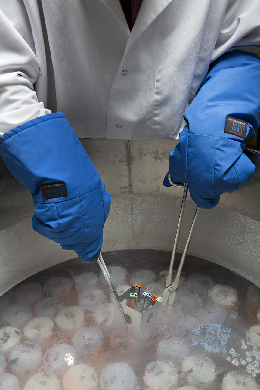
Conventional slow freezing, also known as equilibrium freezing, is a cryopreservation technique that involves the controlled, gradual cooling of biological samples—typically at rates of 0.3–1°C per minute—in the presence of cryoprotective agents (CPAs) to facilitate primarily extracellular ice formation while minimizing lethal intracellular ice crystals.[2] This method relies on the principle that slow cooling allows time for water to exit cells osmotically as extracellular ice nucleates, concentrating solutes outside the cells and dehydrating the intracellular environment to prevent ice nucleation within the cytoplasm.[2] CPAs such as dimethyl sulfoxide (DMSO) at concentrations of 1–2 M, glycerol, or propylene glycol are added prior to cooling to lower the freezing point, reduce ice crystal size, and stabilize biomolecules against denaturation, though their toxicity necessitates careful equilibration to avoid osmotic stress.[2][40] The process begins with sample preparation: cells or tissues are suspended in a CPA solution, such as those containing DMSO or glycerol, and equilibrated at 4°C for 10–30 minutes to achieve uniform penetration. Cooling proceeds in a programmable freezer: the sample is chilled to approximately -5 to -7°C (above the freezing point to induce supercooling), followed by manual or automatic ice seeding to initiate extracellular crystallization, often with a 5–10 minute hold at -7°C to enhance dehydration. Subsequent cooling at -1°C/min continues to -30 to -80°C, after which samples are plunged into liquid nitrogen at -196°C for storage, ensuring vitrification of residual unfrozen solution; alternatively, in standard laboratory protocols, samples pre-cooled to -80°C may be held for 1-24 hours in a -80°C freezer to gradually lower temperature and avoid ice crystal damage, then transferred using long tongs or specialized tools in sealed cryotubes directly into liquid nitrogen or vapor phase storage to prevent cross-contamination.[2][41] Thawing samples frozen in liquid nitrogen requires performing quick operations while wearing full protective equipment to ensure safety and minimize contamination risks, typically in a 37°C water bath for rapid rewarming at rates of 45–70°C/min to limit ice recrystallization and avoid damage from slow thawing, followed by stepwise CPA removal via dilution to prevent osmotic shock.[2] This protocol, optimized through studies on cooling rates by Peter Mazur in the 1960s–1970s, balances dehydration against solution effects like elevated intracellular salts that can harm cells.[2] Developed as the foundational cryopreservation approach, slow freezing gained prominence after Christopher Polge's 1949 discovery of glycerol's protective role for fowl sperm, enabling post-thaw viability exceeding 50% where unprotected freezing yielded none.[42] Subsequent refinements, including DMSO's introduction in 1959 by James Lovelock and others for red blood cells, established it as the standard for sperm, embryos, and hematopoietic stem cells by the 1980s, with survival rates often 70–90% for robust cell types like spermatozoa.[2][42] Advantages include compatibility with larger volumes via cost-effective controlled-rate freezers, lower CPA concentrations (reducing toxicity), and applicability to suspension cultures or tissues where vitrification's high CPA demands pose risks.[40] However, challenges persist: suboptimal rates can lead to intracellular ice (causing mechanical rupture) or excessive dehydration (elevating solute toxicity), yielding lower recoveries—e.g., 60–75% for oocytes—compared to vitrification in sensitive applications.[2][40] Ongoing advances focus on CPA alternatives like trehalose for enhanced stability and hybrid protocols for tissues, but slow freezing remains prevalent for microbial stocks, plant germplasm, and cell therapies requiring scalable, "on-demand" storage at -150°C to -196°C.[40] Post-thaw assessments, including viability assays (e.g., trypan blue exclusion) and functional tests, confirm efficacy, with meta-analyses indicating equivalence to vitrification for primordial follicles in ovarian tissue but inferiority for blastocyst implantation rates (e.g., 25–35% vs. 40–50%).[2][43]Vitrification
Vitrification is a cryopreservation technique that transforms aqueous solutions containing biological materials into a stable, amorphous glass-like solid by ultra-rapid cooling, preventing the formation of damaging ice crystals.[2] This method relies on high concentrations of cryoprotective agents (CPAs), such as dimethyl sulfoxide (DMSO), ethylene glycol (EG), or propylene glycol, combined with rapid cooling rates exceeding 10,000°C per minute, often achieved by direct immersion in liquid nitrogen at -196°C.[44] Unlike conventional freezing, vitrification avoids phase separation of water into ice, minimizing intracellular dehydration, osmotic imbalances, and mechanical disruption from crystal growth.[1] The foundational principles were advanced in the 1980s by Gregory Fahy, who demonstrated that sufficiently high CPA concentrations and cooling velocities could induce vitrification in complex solutions without ice nucleation.[23] The first practical success occurred in 1985 when William F. Rall and Fahy vitrified eight-cell mouse embryos using a mixture of DMSO, acetamide, and propylene glycol, achieving post-thaw viability and live births upon transfer.[45] Subsequent refinements in the 1990s and 2000s optimized CPA formulations, reducing equilibration times and introducing carriers like straws or open-pulled straws to enhance cooling efficiency for small volumes.[46] Compared to slow freezing, vitrification offers superior post-thaw survival rates, particularly for sensitive cells like human oocytes and embryos, with meta-analyses reporting survival exceeding 90% versus 50-70% for slow methods due to eliminated ice-related damage.[47] It also shortens procedural time, lowers equipment costs, and preserves follicular integrity better in ovarian tissue, as evidenced by reduced DNA strand breaks in primordial follicles.[13] However, challenges persist, including CPA toxicity from high molarities (often 4-6 M), which can cause chemical damage to membranes and proteins during exposure, and risks of fracturing in larger samples due to thermal stresses or devitrification upon rewarming. Rewarming samples frozen in liquid nitrogen requires performing quick operations while wearing full protective equipment, typically in a 37°C water bath or controlled methods for rapid thawing to minimize devitrification risks and avoid damage from slow thawing.[48] Strategies to mitigate toxicity include stepwise CPA loading, use of non-permeating sugars like trehalose for osmotic balance, and novel mixtures that lower concentrations while maintaining glass stability.[7] Despite these hurdles, vitrification has become the standard for gamete and embryo banking since the early 2010s, with ongoing research targeting scalability for organs.[49]Novel and Experimental Techniques
One promising experimental approach involves nanowarming, which addresses uneven rewarming in vitrified large tissues by using magnetic nanoparticles (such as iron oxide) infused with cryoprotective agents; these particles enable rapid, uniform heating via alternating magnetic fields, minimizing thermal fractures and devitrification. In 2023, researchers demonstrated successful vitrification, 100-day cryogenic storage, and nanowarming of whole pig kidneys, followed by transplantation into pigs with 72-hour function recovery, outperforming conventional conductive rewarming.[38] Similar techniques applied to rat hearts in 2021 showed preserved contractility post-perfusion with magnetic agents and nanowarming, suggesting potential for scaling to human organs despite challenges like nanoparticle toxicity and field uniformity.[50] A 2024 study further validated physical feasibility of nanowarming human-scale organs (e.g., 0.5-1 L volumes for kidneys) without ice formation, though biological viability remains unproven at that scale.[51] Aldehyde-stabilized cryopreservation (ASC) combines chemical fixation with vitrification to preserve neural ultrastructure for potential connectome mapping or revival, targeting applications in brain banking where traditional freezing causes synaptic loss. Developed by 2015, ASC perfuses tissue with glutaraldehyde to stabilize proteins and cytoskeleton, followed by high-concentration ethylene glycol for vitrification at -135°C, enabling electron microscopy-quality preservation of rabbit brains, including glial morphology and nanoscale details.[36] In 2018, the method earned the Brain Preservation Prize for demonstrating synaptic preservation in a postmortem pig brain, though critics note fixation renders tissue non-viable for biological revival, limiting it to informational preservation rather than functional recovery.[35] Ongoing refinements focus on minimizing shrinkage and optimizing aldehyde concentrations, but human-scale application awaits perfusion efficacy trials.[52] Other experimental methods include supercooling with cryoprotectants to enable subzero non-freezing preservation of organs, extending static storage beyond conventional 4-8°C limits by inhibiting nucleation at temperatures like -4°C to -6°C using polyethylene glycol or antifreeze proteins. A 2019 study supercooling human livers to -6°C preserved viability for up to 27 hours (tripling standard times) with immediate post-thaw function in machine perfusion models, reducing ischemic injury.[53] For vascularized composites like rat limbs, 2024 isochoric supercooling (constant-volume systems) maintained tissue integrity for days at -2°C to -4°C without ice, outperforming hypothermic storage in ATP levels and histology.[54] These techniques bridge toward full cryopreservation but face risks of heterogeneous supercooling and ice propagation upon rewarming, with clinical translation pending large-animal validation.[55] Emerging efforts also explore ice-blocking agents and miniaturized constructs for 3D bioprinted tissues, where trehalose or nanoparticles prevent intracellular ice in scaffolds, achieving higher post-thaw viability in ovarian or cartilage models as of 2025.[56] Directional freezing variants, incorporating nanowarming, have shown promise in preventing cracking during organ-scale thawing, as in 2025 Texas A&M trials targeting subzero storage without phase change damage.[57] These remain preclinical, with viability metrics (e.g., <50% cell survival in complex tissues) underscoring the need for integrative perfusion and agent optimization.[58]Biological Applications
Reproductive Cells and Tissues
Cryopreservation of human sperm, established since the 1950s, employs slow freezing with permeable cryoprotectants such as glycerol at concentrations of 5-10%, achieving post-thaw motility rates of 40-60% depending on initial semen quality and protocol.[59] This technique supports fertility preservation for men facing gonadotoxic treatments, with intracytoplasmic sperm injection (ICSI) fertilization rates comparable to fresh sperm, often exceeding 70% in clinical settings.[60] DNA integrity post-thaw remains high in fertile donors but declines more in infertile samples due to osmotic stress and ice crystal formation.[61] Oocyte cryopreservation relies predominantly on vitrification, a rapid cooling method using high concentrations of cryoprotectants like ethylene glycol and dimethyl sulfoxide to form a glass-like state, yielding survival rates over 90% and warming survival exceeding 95% in optimized protocols.[62] For elective preservation, cryopreserving at least 20 mature oocytes before age 38 correlates with a 70% live birth rate per patient via subsequent IVF.[63] Clinical pregnancy rates per transfer reach 38.5% with vitrified oocytes, matching fresh counterparts, though outcomes diminish with advanced maternal age at freezing due to inherent oocyte aneuploidy risks.[62][64] Embryo cryopreservation, integral to in vitro fertilization (IVF), utilizes vitrification for blastocysts, attaining thaw survival rates of 90-95% and implantation rates often surpassing fresh transfers by 5-10% due to optimized endometrial receptivity in frozen cycles.[65] Long-term storage beyond 10 years maintains viability, with reported embryo survival at 74% after such durations and subsequent live births.[66] Utilization rates for cryopreserved embryos in fertility preservation average 25.5% over 10-year follow-ups, yielding clinical pregnancies in line with age-matched fresh IVF benchmarks.[67] Ovarian tissue cryopreservation (OTC), typically via slow freezing of cortical strips, preserves primordial follicles for transplantation in patients with cancer or premature ovarian insufficiency, restoring ovarian function in 70-95% of cases post-autotransplantation and enabling spontaneous pregnancies.[68] As of 2024, OTC has resulted in over 200 live births globally, with live birth rates per patient around 27-30% in large cohorts, though lower than oocyte methods at 8.76% per cycle due to variable follicle yield and ischemia risks during grafting.[69][70] Vitrification of tissue fragments shows promise for improved follicular survival over traditional slow freezing.[71] Testicular tissue cryopreservation offers the sole fertility preservation avenue for prepubertal boys at risk of sterility from chemotherapy or radiation, involving enzymatic digestion or xenotransplantation to derive spermatogonial stem cells post-thaw.[72] Over 3,000 boys have undergone this procedure worldwide by 2024, primarily via slow freezing, with animal models demonstrating spermatogenesis restoration, though human applications remain experimental without confirmed live births due to ethical and technical hurdles in maturation.[73] Post-thaw tissue viability exceeds 80% in optimized protocols, preserving stem cell potential for future in vitro gametogenesis.[74]Microbial and Plant Materials
Cryopreservation of microorganisms, including bacteria, fungi, and yeast, employs controlled slow freezing with cryoprotective agents such as glycerol (typically 10-20%) or dimethyl sulfoxide (DMSO) to mitigate ice crystal formation and osmotic stress, followed by storage at -196°C in liquid nitrogen.[75] [76] This approach enables long-term viability, often exceeding several decades, supporting microbial biobanks for research, diagnostics, and biotechnology applications.[77] For bacterial strains like Lactobacillus rhamnosus, post-thaw survival rates range from 70% to 90% under optimized freezing conditions, preserving metabolic and genetic stability.[78] Fungal cultures across 61 genera demonstrate sustained viability and functionality post-cryopreservation, with protocols emphasizing reproducible quality control to ensure strain authenticity.[79] [80] In plant materials, cryopreservation facilitates the conservation of genetic resources, particularly for orthodox seeds via direct immersion after desiccation and for recalcitrant or intermediate seeds through vitrification of excised embryos, shoot tips, or cell suspensions using solutions like plant vitrification solution 2 (PVS2).[81] [82] This method prevents intracellular ice formation by achieving a glass-like state, allowing indefinite storage without loss of regenerative capacity, critical for biodiversity preservation in gene banks such as the USDA National Plant Germplasm System.[81] Recovery rates vary by species and protocol; for instance, a study of 164 medicinal plant seeds reported viable regrowth post-cryopreservation under standardized conditions, while potato germplasm accessions achieved success ratios dependent on explant processing efficiency.[83] [84] Post-thaw regrowth optimization, including hormone-supplemented media and light regimes, enhances survival by addressing oxidative stress and metabolic recovery.[85] Applications extend to elite cultivars and endangered species, enabling pathogen-free propagation and genetic stability for agronomic improvement.[86]Complex Tissues and Organs
Cryopreservation of complex tissues and organs, such as vascularized structures like kidneys or livers, faces significant barriers primarily due to ice crystal formation during freezing, which disrupts cellular integrity and extracellular matrices.[87] Unlike single cells or simple tissues, larger volumes exhibit uneven heat and mass transfer, leading to thermal gradients that promote intracellular ice nucleation and mechanical fracturing.[9] High concentrations of cryoprotective agents (CPAs), such as dimethyl sulfoxide or glycerol, are required for vitrification to form a glass-like state without crystals, but these agents induce toxicity through osmotic stress, chemical damage, and reactive oxygen species generation.[88] Rewarming poses additional risks, as slow thawing allows devitrification and ice recrystallization, necessitating rapid, uniform heating methods like nanowarming via magnetic nanoparticles to mitigate these effects.[89] Vitrification has shown promise for smaller organs in animal models. In 1990, rabbit kidneys were vitrified using a mixture of CPAs, transplanted after rewarming, and supported recipient survival for 48 days, demonstrating partial functional recovery despite histological damage.[7] More recently, in 2023, rat kidneys were vitrified with a CPA cocktail, stored cryogenically for up to 100 days, and rewarmed using nanowarming; five transplanted recipients exhibited urine production and survival without immediate rejection, though long-term function was limited by vascular and tubular impairments.[38] [90] These advances highlight perfusion techniques to deliver CPAs uniformly through vasculature, reducing toxicity compared to immersion methods.[91] Despite progress, no whole mammalian organ has achieved full post-thaw viability equivalent to fresh tissue, with challenges including CPA permeation inefficiencies in dense tissues and cracking from differential contraction during cooling.[92] Complex tissues like corneas and cartilage have been cryopreserved successfully for clinical use via slow freezing with CPAs, preserving transparency and mechanical properties, but vascularized organs require integrated solutions for ischemia-reperfusion injury post-thaw.[58] Ongoing research emphasizes bioengineered scaffolds and CPA alternatives to enable indefinite storage, potentially expanding organ banking, though clinical translation remains elusive as of 2025.[93]Natural Cryopreservation and Freeze Tolerance
Mechanisms in Microorganisms and Plants
Microorganisms exhibit freeze tolerance through intracellular strategies that prevent or manage ice formation. Many cold-adapted bacteria, fungi, and algae accumulate compatible solutes such as trehalose, which acts as a natural cryoprotectant by stabilizing proteins and membranes via vitrification—a glass-like state that inhibits damaging ice crystal growth during freezing.[94] [95] Trehalose replaces water molecules in hydrogen bonding networks, maintaining cellular integrity under low-temperature dehydration, as observed in yeasts like Saccharomyces cerevisiae where elevated trehalose levels correlate with improved survival after freeze-thaw cycles.[95] Additional mechanisms include modifications to membrane lipid composition to preserve fluidity at subzero temperatures and the production of cold shock proteins that facilitate repair of freeze-induced damage.[96] Some microorganisms, including certain Antarctic yeasts, synthesize antifreeze proteins that bind to ice nuclei, inhibiting recrystallization and small crystal propagation, thereby limiting cellular injury.[97] In fungi and algae, polyols like glycerol and mannitol complement trehalose by lowering the freezing point and buffering osmotic stress during extracellular ice formation in surrounding environments.[94] Psychrophilic algae, such as Chlamydomonas nivalis, rapidly upregulate stress-responsive genes upon cold exposure, enhancing solute accumulation and enzymatic adjustments for metabolic continuity in ice matrices.[98] These adaptations enable survival in permafrost and sea ice, where cycles of freezing and thawing occur, with viability maintained through minimized intracellular water availability and post-thaw DNA repair pathways.[99] Plants achieve natural cryopreservation via extracellular freezing tolerance, where ice nucleates in apoplastic spaces rather than within cells, inducing controlled dehydration that concentrates intracellular protectants.[100] This process, prominent in hardy perennials and winter cereals, relies on ice-active proteins or nucleators that initiate freezing at higher subzero temperatures (around -2 to -5°C) to avoid lethal intracellular ice.[101] Dehydrins and late embryogenesis abundant proteins stabilize membranes and prevent protein denaturation during the resulting hypertonic stress, while sugars like raffinose family oligosaccharides contribute to vitrification in the cytoplasm.[102] Cold acclimation, triggered by non-freezing low temperatures, activates the ICE-CBF-COR regulon, upregulating genes for osmoprotectants and antioxidants to enhance tolerance, allowing survival to -20°C or lower in acclimated tissues.[102] In bryophytes like the moss Physcomitrella patens, freezing tolerance integrates desiccation resistance, with abscisic acid pretreatment preserving membrane phase behavior and enabling recovery after extracellular ice formation and slow drying.[103] This moss model demonstrates poikilohydric adaptations, where gametophytes endure freezing by minimizing protoplasmic water content and employing repair mechanisms post-thaw, reflecting evolutionary links to drought tolerance in early land plants.[104]Animal Adaptations and Examples
Certain ectothermic vertebrates and invertebrates have evolved freeze tolerance, enabling survival through extracellular ice formation without intracellular freezing, which would otherwise cause lethal cellular damage. This adaptation involves controlled nucleation of ice in extracellular spaces, accumulation of high concentrations of low-molecular-weight cryoprotectants such as glucose or glycerol to colligatively depress the freezing point and stabilize membranes, and metabolic suppression to endure ischemia and anoxia during the frozen state.[105][106] Ice-nucleating proteins or agents initiate freezing at relatively high subzero temperatures (around -2 to -5°C) to form small, non-damaging crystals, while antioxidants and heat shock proteins mitigate oxidative stress upon thawing.[107] These mechanisms prevent excessive dehydration of cells and limit ice propagation into intracellular compartments.[108] Among vertebrates, the wood frog (Rana sylvatica) exemplifies freeze tolerance, enduring whole-body freezing where up to 65-70% of body water forms extracellular ice, halting cardiac and respiratory functions for weeks at temperatures as low as -16°C.[109] Upon cooling, frogs rapidly mobilize liver glycogen to produce glucose, elevating plasma levels to 200-300 mM, which dehydrates cells osmotically and vitrifies residual intracellular water to prevent ice formation.[110] Thawing restores organ function within hours, supported by gene regulation changes including histone modifications for metabolic arrest.[111] Similar strategies occur in other North American anurans like the gray tree frog (Hyla versicolor) and spring peeper (Pseudacris crucifer), which tolerate 50-65% body water frozen using glucose or glycerol.[105] In reptiles, the painted turtle (Chrysemys picta) demonstrates partial freeze tolerance in northern populations, surviving brief extracellular freezing episodes during hibernation at -2 to -3°C by accumulating glucose and glycerol, alongside enhanced antioxidant defenses to counter reperfusion injury upon thawing.[105] Freeze-tolerant insects, such as the goldenrod gall fly (Eurosta solidaginis), freeze over 60% of body water into hemolymph ice at -40°C or lower, relying on glycerol (up to 7 M) and trehalose as polyols to stabilize proteins and membranes, with sorbitol aiding in some species.[112] These insects initiate freezing via exogenous ice nucleators or endogenous agents, maintaining cellular viability through dehydration and cryoprotectant-induced vitrification of cytoplasm.[113] Other arthropods, including certain beetle larvae and moths, employ analogous polyol mixtures, enduring supercooling failures without intracellular ice damage.[114] Such adaptations highlight convergent evolution of biochemical safeguards against freeze-induced denaturation and osmotic stress across taxa.[115]Risks and Challenges
Physical and Chemical Damage Mechanisms
During cryopreservation, physical damage primarily arises from ice crystal formation and growth, which mechanically disrupt cellular structures. Extracellular ice crystals form first during slow cooling, creating hypertonic conditions that draw water out of cells via osmosis, leading to cellular dehydration and shrinkage; failure to fully dehydrate can result in lethal intracellular ice formation (IIF), where crystals pierce organelle membranes and cause lysis.[18] Intracellular ice crystals larger than a critical size—typically exceeding 10-20% of cell volume—exacerbate damage by expanding and fracturing cytoskeletal elements.[116] During thawing, ice recrystallization merges smaller crystals into larger ones, amplifying mechanical injury through shear forces and osmotic swelling as water re-enters dehydrated cells.[117] Chemical damage mechanisms complement these physical insults, often stemming from cryoprotective agents (CPAs) and solution effects. CPAs like dimethyl sulfoxide (DMSO) and ethylene glycol prevent ice formation but induce toxicity through direct membrane perturbation and metabolic disruption; for instance, DMSO exhibits dose-dependent cytotoxicity, with concentrations above 10% reducing cell viability by altering protein conformation and generating reactive oxygen species (ROS).[118] Osmotic stress occurs during CPA loading and unloading, as well as freezing-induced solute concentration, causing transient volume changes that strain membranes—cells may shrink by up to 50% before rebounding, risking rupture.[119] Additionally, freezing concentrates ions and buffers, shifting pH (e.g., a drop of 1-2 units in phosphate buffers due to selective crystallization), which denatures proteins like enzymes via altered ionization states.[120] These effects synergize, with elevated solutes promoting ROS via Fenton reactions, further oxidizing lipids and DNA.[117]Biological Viability Post-Thaw
Post-thaw biological viability refers to the capacity of cryopreserved cells, tissues, or organisms to resume normal function, including metabolism, proliferation, and structural integrity, following rewarming. Immediate assessments, such as membrane integrity via trypan blue exclusion, often overestimate viability because they fail to capture delayed-onset injuries like apoptosis or oxidative stress that manifest hours to days later.[121] [122] In many protocols, post-thaw culturing for 24 hours or more is necessary to measure true recovery, revealing declines from apparent 60-80% immediate survival to as low as 20% total cell recovery in some cases.[121] [123] Cryoinjuries compromising viability include intracellular ice formation, which disrupts organelles during freezing, and osmotic imbalances during thawing that cause membrane rupture or swelling.[1] These lead to elevated reactive oxygen species and activation of apoptotic pathways post-thaw, resulting in programmed cell death even in initially intact cells.[18] [124] For instance, human bone marrow mesenchymal stem cells exhibit reduced metabolic activity, adhesion, and increased apoptosis after cryopreservation, with viability dropping below 70% in optimized conditions.[125] Cooling and thawing rates critically influence outcomes; slower cooling preserves higher viability (up to 80% in fibroblasts) by minimizing ice crystal size, but rapid thawing can exacerbate solute concentration damage.[126] [127] Viability success rates vary markedly by biological complexity. Gametes and simple cells achieve relatively high post-thaw functionality: human oocytes show 74% survival and 67% fertilization rates, supporting viable pregnancies.[128] Embryos with all blastomeres intact post-thaw yield clinical pregnancy rates of 37.7%.[129] However, for multicellular structures like neurospheres or natural killer cells, survival plummets to 27-60%, with functional impairments such as reduced cytotoxicity persisting despite cryoprotectants like DMSO.[130] [131] Tissues and organs face compounded challenges, including vascular occlusion and ischemia-reperfusion injury, often rendering post-thaw viability negligible without advanced vitrification, which still fails to prevent widespread cell death in large volumes.[1] Storage duration further erodes viability, with embryo survival rates declining significantly beyond short-term holds.[132] Efforts to enhance post-thaw recovery target these mechanisms, such as ROCK inhibitors to mitigate apoptosis in T cells or antioxidants to curb oxidative damage, yielding modest improvements in yield and function.[133] [134] Yet, no universal protocol eliminates cryoinjury, and viability remains protocol-dependent, underscoring cryopreservation's limitations for preserving complex biological systems intact.[135] [136]Long-Term Storage Stability
Cryopreserved biological materials are typically stored in liquid nitrogen vapor phase at approximately -196°C to achieve metabolic arrest and prevent biochemical degradation, theoretically enabling indefinite stability by minimizing molecular diffusion and reaction rates.[137] Empirical data from hematopoietic progenitor cell (HPC) products demonstrate post-thaw viability stability for up to 14.6 years when stored below -150°C, though viability correlates negatively with pre-freeze granulocyte content, indicating that cellular composition influences long-term outcomes.[138] Similarly, cord blood units have shown recovery rates exceeding 80% after 29 years of storage for unseparated samples, with expiration times validated up to 25 years for manually volume-reduced units.[139] Despite these successes, prolonged cryopreservation can induce genomic instability, including karyotype alterations and DNA damage accumulation across diverse cell types such as stem cells and fibroblasts, potentially compromising functional integrity upon thawing.[140] Studies on peripheral blood mononuclear cells (PBMCs) report stable recovery and T-cell subtype viability after extended storage, yet subtle declines in proliferative capacity and genetic fidelity emerge beyond 2–3 years, particularly at -80°C rather than -196°C.[141][142] Storage at suboptimal temperatures accelerates these risks, as even cryogenic conditions do not eliminate low-level chemical reactions or cosmic radiation-induced mutations over decades to centuries.[143] Operational challenges further threaten stability, including vapor phase temperature fluctuations in liquid nitrogen tanks, which can exceed -150°C during refill cycles or equipment failures, leading to partial thawing and ice recrystallization damage.[144] Peer-reviewed assessments emphasize that while short- to medium-term (up to 20–30 years) viability is reliably maintained for applications like stem cell banking, extrapolations to indefinite storage lack direct empirical validation, with potential for cumulative proteotoxic aggregates or epigenetic drifts unobserved in current datasets limited to human lifespans.[145][1] Rigorous monitoring protocols, such as periodic viability assays and redundant dewars, mitigate but do not eliminate these vulnerabilities in clinical and research repositories.[146]Human Cryonics
Procedures and Organizational Practices
The primary organizations providing human cryonics services are the Alcor Life Extension Foundation, founded in 1972 and based in Scottsdale, Arizona, and the Cryonics Institute (CI), established in 1976 and located in Clinton Township, Michigan.[147][148] Both operate as non-profit entities, requiring prospective members to sign cryopreservation contracts designating the organization as the recipient of an anatomical gift under the Uniform Anatomical Gift Act, along with funding mechanisms such as life insurance policies payable upon death to cover procedure costs—typically $200,000 for whole-body preservation at Alcor or $28,000 at CI, excluding membership dues of around $500–$1,200 annually.[147][149][150] These contracts specify procedures only after legal death certification, with provisions for next-of-kin consent and release forms to facilitate rapid intervention.[149] Procedures commence with pre-arranged standby by trained teams, often including physicians or emergency personnel, positioned near the member's location to respond within minutes of death pronouncement.[151][152] Stabilization involves immediate cardiopulmonary support via mechanical chest compression devices and ventilation to restore circulation, alongside ice packing and cold saline infusion for core cooling to 10–15°C, minimizing cerebral ischemia from halted blood flow.[153][154] The body is then transported under field stabilization to the organization's facility, where surgical access to major vessels—typically the aorta or carotid arteries—is established for perfusion.[154][155] Perfusion replaces blood with organ preservation solutions (e.g., heparinized saline or tissue culture media) to flush out clots and metabolites, followed by cryoprotectant agents like M22 at Alcor or CI-VM-1 at CI, delivered under controlled pressure and temperature to penetrate tissues and induce vitrification—a glass-like solidification without ice crystals—targeting the brain first.[156][154] For Alcor's neuropreservation option ($80,000), the head is surgically separated post-perfusion, with the body discarded unless separately funded; CI focuses on whole-body preservation.[153] Cooling proceeds gradually—via nitrogen gas circulation at Alcor to -125°C then immersion, or over five days in CI's computer-controlled unit to -196°C—to prevent fracturing from thermal stress.[153][154] Patients are stored indefinitely in vacuum-insulated dewars filled with liquid nitrogen at -196°C, with Alcor maintaining 248 patients and CI over 250 as of mid-2025, comprising brains or whole bodies in patient-specific capsules.[157][158] Organizational maintenance includes weekly nitrogen top-offs, remote monitoring of dewars, and endowments for perpetual care, audited periodically to ensure financial stability against inflation or facility risks.[159][150] Teams undergo regular training in medical protocols, logistics, and cryoprotectant chemistry, with Alcor's Deployment and Recovery Teams (DART) emphasizing tactical rapid response.[151][152]Evidence on Preservation Quality
In human cryonics, preservation quality is assessed primarily through organizational protocols involving vitrification with cryoprotectant agents (CPAs) such as M22, which aim to form a glass-like state without ice crystal formation in perfused tissues. Alcor Life Extension Foundation reports that in cases with minimal post-mortem delay—often under 10 minutes via standby teams—cryoprotectant perfusion achieves high distribution in brain tissue, with electron microscopy of animal models and select human samples showing intact synaptic structures and absence of ice in vitrified regions. However, independent peer-reviewed validation of whole-brain preservation remains absent, with assessments relying on proprietary evaluations rather than standardized histological or connectomic analyses.[160][161] Empirical data from proxy studies indicate partial ultrastructural preservation is feasible but limited to small scales. For instance, vitrification of human autopsy brain tissue up to 180 μm thick using glycerol and trehalose preserved subcellular features like myelin sheaths and vesicles without crystalline ice or fractures, as revealed by cryo-electron tomography. In adult mouse hippocampal slices, a 2025 study demonstrated post-thaw recovery of neuronal excitability, synaptic plasticity, and long-term potentiation at levels approaching controls (138% vs. 161% potentiation), suggesting metabolic and functional arrest without total loss. Rat hippocampal slices similarly retained over 90% ion balance viability after vitrification and rewarming. These findings support preservation of fine neural architecture in isolated tissues but do not extend to whole human brains, where diffusion limitations prevent uniform CPA penetration.[162][163][161] Significant limitations undermine overall quality in practice. CPA toxicity from high concentrations (e.g., 40-60% mixtures) induces cellular stress, osmotic imbalance, and protein denaturation, even if mitigated by cooling or combinations like DMSO and formamide. Thermal fracturing occurs during immersion in liquid nitrogen (-196°C), with cracks propagating through non-vitrified or unevenly cooled regions; storage at -130°C has been proposed to reduce this, though not universally adopted. Pre-cryopreservation ischemia from clinical death—typically 5-60 minutes in non-standby cases—causes widespread neuronal swelling and synaptic degradation before vitrification can commence, as evidenced by animal models tolerating only up to 120 minutes of hypothermic arrest without deficits. Critics note that while gross morphology may endure, molecular-level fidelity (e.g., proteome integrity) and full connectomic mapping have not been verified in human cryonics patients, contrasting with superior structural outcomes in aldehyde-stabilized fixation methods.[118][161][164][165]| Factor | Evidence of Preservation | Key Limitations |
|---|---|---|
| Ice Formation | Avoided in perfused areas via vitrification (e.g., M22 CPA).[161] | Unperfused regions (e.g., due to clots or delays) form ice, causing mechanical rupture.[164] |
| Ultrastructure | Intact synapses and organelles in small human/mouse samples.[162][163] | Toxicity and uneven distribution degrade deeper tissues; no whole-brain data.[118] |
| Functionality | Partial recovery in slices (e.g., LTP preserved).[163] | No mammalian whole-brain revival; ischemia pre-vitrification dominant.[161] |
| Long-Term Stability | Metabolic halt at cryogenic temps (reaction rates reduced ~10^9-fold).[161] | Fractures and potential devitrification over decades untested in humans.[161] |