Hubbry Logo
Granule cellGranule cellMain
Open search
Granule cell
Community hub
Granule cell
logo
8 pages, 0 posts
0 subscribers
Be the first to start a discussion here.
Granule cell
Granule cell
from Wikipedia
Drawing of Purkinje cells (A) and granule cells (B) from pigeon cerebellum by Santiago Ramón y Cajal, 1899. Instituto Santiago Ramón y Cajal, Madrid, Spain.

The name granule cell has been used for a number of different types of neurons whose only common feature is that they all have very small cell bodies. Granule cells are found within the granular layer of the cerebellum, the dentate gyrus of the hippocampus, the superficial layer of the dorsal cochlear nucleus, the olfactory bulb, and the cerebral cortex.

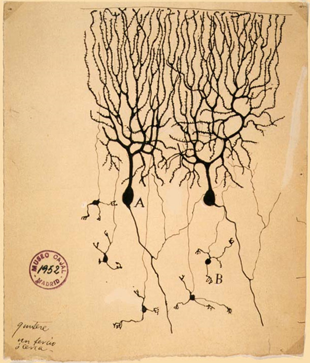
Cerebellar granule cells account for the majority of neurons in the human brain.[1] These granule cells receive excitatory input from mossy fibers originating from pontine nuclei. Cerebellar granule cells project up through the Purkinje layer into the molecular layer where they branch out into parallel fibers that spread through Purkinje cell dendritic arbors. These parallel fibers form thousands of excitatory granule-cell–Purkinje-cell synapses onto the intermediate and distal dendrites of Purkinje cells using glutamate as a neurotransmitter.

Layer 4 granule cells of the cerebral cortex receive inputs from the thalamus and send projections to supragranular layers 2–3, but also to infragranular layers of the cerebral cortex.

Structure

[edit]

Granule cells in different brain regions are both functionally and anatomically diverse: the only thing they have in common is smallness. For instance, olfactory bulb granule cells are GABAergic and axonless, while granule cells in the dentate gyrus have glutamatergic projection axons. These two populations of granule cells are also the only major neuronal populations that undergo adult neurogenesis, while cerebellar and cortical granule cells do not. Granule cells (save for those of the olfactory bulb) have a structure typical of a neuron consisting of dendrites, a soma (cell body) and an axon.

Dendrites: Each granule cell has 3 – 4 stubby dendrites which end in a claw. Each of the dendrites are only about 15 μm in length.

Soma: Granule cells all have a small soma diameter of approximately 10 μm.

Axon: Each granule cell sends a single axon onto the Purkinje cell dendritic tree.[citation needed] The axon has an extremely narrow diameter: ½ micrometre.

Synapse: 100–300,000 granule cell axons synapse onto a single Purkinje cell.[citation needed]

The existence of gap junctions between granule cells allows multiple neurons to be coupled to one another, allowing multiple cells to act in synchrony, and allows signalling functions necessary for granule cell development to occur.[2]

Cerebellar granule cell

[edit]

The granule cells, produced by the rhombic lip, are found in the granule cell layer of the cerebellar cortex. They are small and numerous. They are characterized by a very small soma and several short dendrites which terminate with claw-shaped endings. In the transmission electron microscope, these cells are characterized by a darkly stained nucleus surrounded by a thin rim of cytoplasm. The axon ascends into the molecular layer where it splits to form parallel fibers.[1]

Dentate gyrus granule cell

[edit]

The principal cell type of the dentate gyrus is the granule cell. The dentate gyrus granule cell has an elliptical cell body with a width of approximately 10 μm and a height of 18μm.[3]

The granule cell has a characteristic cone-shaped tree of spiny apical dendrites. The dendrite branches project throughout the entire molecular layer, and the furthest tips of the dendritic tree end at the hippocampal fissure or at the ventricular surface.[4] The granule cells are tightly packed in the granular cell layer of the dentate gyrus.

Dorsal cochlear nucleus granule cell

[edit]

The granule cells in the dorsal cochlear nucleus are small neurons with two or three short dendrites that give rise to a few branches with expansions at the terminals. The dendrites are short with claw-like endings that form glomeruli to receive mossy fibers, similar to cerebellar granule cells.[5] Its axon projects to the molecular layer of the dorsal cochlear nucleus where it forms parallel fibers, also similar to cerebellar granule cells.[6] The dorsal cochlear granule cells are small excitatory interneurons which are developmentally related and thus resemble the cerebellar granule cell.

Olfactory bulb granule cell

[edit]

The main intrinsic granule cell in the vertebrate olfactory bulb lacks an axon (as does the accessory neuron). Each cell gives rise to short central dendrites and a single long apical dendrite that expands into the granule cell layer and enters the mitral cell body layer. The dendrite branches terminate within the outer plexiform layer among the dendrites in the olfactory tract.[7] In the mammalian olfactory bulb, granule cells can process both synaptic input and output due to the presence of large spines.[8]

Function

[edit]

Neural pathways and circuits of the cerebellum

[edit]
Cartoon representation of the neural connections that exist between the different types of neurons in the cerebellar cortex. Including Purkinje cells, granule cells and interneurons.
Neural pathways and circuits in the cerebellum. (+) represent excitatory synapse, while (-) represent inhibitory synapses.

Cerebellar granule cells receive excitatory input from 3 or 4 mossy fibers originating from pontine nuclei. Mossy fibers make an excitatory connection onto granule cells, which causes the granule cells to fire an action potential.

The axon of a cerebellar granule cell splits to form a parallel fiber which innervates Purkinje cells. The vast majority of granule cell axonal synapses are found on the parallel fibers.[9]

The parallel fibers are sent up through the Purkinje layer into the molecular layer where they branch out and spread through Purkinje cell dendritic arbors. These parallel fibers form thousands of excitatory Granule-cell-Purkinje-cell synapses onto the dendrites of Purkinje cells.

This connection is excitatory as glutamate is released.

The parallel fibers and ascending axon synapses from the same granule cell fire in synchrony which results in excitatory signals. In the cerebellar cortex there are a variety of inhibitory neurons (interneurons). The only excitatory neurons present in the cerebellar cortex are granule cells.[10]

Plasticity of the synapse between a parallel fiber and a Purkinje cell is believed to be important for motor learning.[11] The function of cerebellar circuits is entirely dependent on processes carried out by the granular layer. Therefore, the function of granule cells determines the cerebellar function as a whole.[12]

Mossy fiber input on cerebellar granule cells

[edit]

Granule cell dendrites also synapse with distinctive unmyelinated axons which Santiago Ramón y Cajal called mossy fibers[4] Mossy fibers and golgi cells both make synaptic connections with granule cells. Together these cells form the glomeruli.[10]

Granule cells are subject to feed-forward inhibition: granule cells excite Purkinje cells but also excite GABAergic interneurons that inhibit Purkinje cells.

Granule cells are also subject to feedback inhibition: Golgi cells receive excitatory stimuli from granule cells and in turn send back inhibitory signals to the granule cell.[13]

Mossy fiber input codes are conserved during synaptic transmission between granule cells, suggesting that innervation is specific to the input that is received.[14] Granule cells do not just relay signals from mossy fibers, rather they perform various, intricate transformations which are required in the spatiotemporal domain.[10]

Each granule cell is receiving an input from two different mossy fiber inputs. The input is thus coming from two different places as opposed to the granule cell receiving multiple inputs from the same source.

The differences in mossy fibers that are sending signals to the granule cells directly effects the type of information that granule cells translate to Purkinje cells. The reliability of this translation will depend on the reliability of synaptic activity in granule cells and on the nature of the stimulus being received.[15] The signal a granule cell receives from a Mossy fiber depends on the function of the mossy fiber itself. Therefore, granule cells are able to integrate information from the different mossy fibers and generate new patterns of activity.[15]

Climbing fiber input on cerebellar granule cells

[edit]

Different patterns of mossy fiber input will produce unique patterns of activity in granule cells that can be modified by a teaching signal conveyed by the climbing fiber input. David Marr and James Albus suggested that the cerebellum operates as an adaptive filter, altering motor behaviour based on the nature of the sensory input.

Since multiple (~200,000) granule cells synapse onto a single Purkinje cell, the effects of each parallel fiber can be altered in response to a “teacher signal” from the climbing fiber input.

Specific functions of different granule cells

[edit]
Cerebellum granule cells

David Marr suggested that the granule cells encode combinations of mossy fiber inputs. In order for the granule cell to respond, it needs to receive active inputs from multiple mossy fibers. The combination of multiple inputs results in the cerebellum being able to make more precise distinctions between input patterns than a single mossy fiber would allow.[16] The cerebellar granule cells also play a role in orchestrating the tonic conductances which control sleep in conjunction with the ambient levels of GABA which are found in the brain.

Dentate granule cells

Loss of dentate gyrus neurons from the hippocampus results in spatial memory deficits. Therefore, dentate granule cells are thought to function in the formation of spatial memories [17] and of episodic memories.[18] Immature and mature dentate granule cells have distinct roles in memory function. Adult-born granule cells are thought to be involved in pattern separation whereas old granule cells contribute to rapid pattern completion.[19]

Dorsal cochlear granule cells

Pyramidal cells from the primary auditory cortex project directly on to the cochlear nucleus. This is important in the acoustic startle reflex, in which the pyramidal cells modulate the secondary orientation reflex and the granule cell input is responsible for appropriate orientation.[20] This is because the signals received by the granule cells contain information about the head position. Granule cells in the dorsal cochlear nucleus play a role in the perception and response to sounds in our environment.

Olfactory bulb granule cells

Inhibition generated by granule cells, the most common GABAergic cell type in the olfactory bulb, plays a critical role in shaping the output of the olfactory bulb.[21] There are two types of excitatory inputs received by GABAergic granule cells; those activated by an AMPA receptor and those activated by a NMDA receptor. This allows the granule cells to regulate the processing of the sensory input in the olfactory bulb.[21] The olfactory bulb transmits smell information from the nose to the brain, and is thus necessary for a proper sense of smell. Granule cells in the olfactory bulb have also been found to be important in forming memories linked with scents.[22]

Critical factors for function

[edit]
Calcium

Calcium dynamics are essential for several functions of granule cells such as changing membrane potential, synaptic plasticity, apoptosis, and regulation of gene transcription.[10] The nature of the calcium signals that control the presynaptic and postsynaptic function of the olfactory bulb granule cells spines is mostly unknown.[8]

Nitric oxide

Granule neurons have high levels of the neuronal isoform of nitric oxide synthase. This enzyme is dependent on the presence of calcium and is responsible for the production of nitric oxide (NO). This neurotransmitter is a negative regulator of granule cell precursor proliferation which promotes the differentiation of different granule cells. NO regulates interactions between granule cells and glia[10] and is essential for protecting the granule cells from damage. NO is also responsible for neuroplasticity and motor learning.[23]

Role in disease

[edit]

Altered morphology of dentate granule cells

[edit]

TrkB is responsible for the maintenance of normal synaptic connectivity of the dentate granule cells. TrkB also regulates the specific morphology (biology) of the granule cells and is thus said to be important in regulating neuronal development, neuronal plasticity, learning, and the development of epilepsy.[24] The TrkB regulation of granule cells is important in preventing memory deficits and limbic epilepsy. This is due to the fact that dentate granule cells play a critical role in the function of the entorhinal-hippocampal circuitry in health and disease. Dentate granule cells are situated to regulate the flow of information into the hippocampus, a structure required for normal learning and memory.[24]

Decreased granule cell neurogenesis

[edit]

Both epilepsy and depression show a disrupted production of adult-born hippocampal granule cells.[25] Epilepsy is associated with increased production - but aberrant integration - of new cells early in the disease and decreased production late in the disease.[25] Aberrant integration of adult-generated cells during the development of epilepsy may impair the ability of the dentate gyrus to prevent excess excitatory activity from reaching hippocampal pyramidal cells, thereby promoting seizures.[25] Long-lasting epileptic seizure stimulate dentate granule cell neurogenesis. These newly born dentate granule cells may result in aberrant connections that result in the hippocampal network plasticity associated with epileptogenesis.[26]

Shorter granule cell dendrites

[edit]

Patients with Alzheimer's have shorter granule cell dendrites. Furthermore, the dendrites were less branched and had fewer spines than those in patients not with Alzheimer's.[27] However, granule cell dendrites are not an essential component of senile plaques and these plaques have no direct effect on granule cells in the dentate gyrus. The specific neurofibrillary changes of dentate granule cells occur in patients with Alzheimer's, Lewy body variant and progressive supranuclear palsy.[28]

See also

[edit]

References

[edit]
[edit]
Revisions and contributorsEdit on WikipediaRead on Wikipedia
from Grokipedia
Granule cells are a class of small, densely packed neurons distinguished by their bodies (typically 5-18 μm in diameter depending on the region), short claw-like dendrites, and unmyelinated axons, serving as the most abundant neuronal type in the mammalian and comprising over 99% of all cerebellar neurons. Found primarily in the granule cell layer of the , the of the hippocampus, the granule cell layer of the , and the superficial layer of the dorsal cochlear nucleus, these neurons exhibit region-specific morphologies and functions but share a common role in integrating sensory and contextual inputs for higher-order processing. In the , granule cells originate from progenitors in the upper rhombic lip during embryonic development, undergoing massive proliferation in the external granular layer before migrating inward to form the mature granule cell layer by postnatal day 25 in mice, resulting in an estimated 70 million cells per cerebellum. These excitatory neurons receive convergent inputs from mossy fibers via their T-shaped dendritic claws and extend parallel fibers that ascend to the molecular layer, synapsing onto dendrites and inhibitory to relay mossy fiber signals in a excitatory pathway essential for cerebellar circuitry. This organization enables granule cells to support , learning, and error correction by transforming diverse mossy fiber patterns into sparse, combinatorial representations across their parallel fiber outputs. Within the hippocampal , granule cells function as the principal output neurons of this subregion, featuring cone-shaped apical dendrites that extend into the molecular layer to receive perforant path inputs from the , while their mossy fiber axons project to CA3 pyramidal cells. Characterized by ongoing —where new granule cells are generated from subgranular zone progenitors and integrate into existing circuits over weeks—these neurons employ sparse firing to perform pattern separation, orthogonalizing similar input representations to facilitate distinct memory encoding and reduce interference in the trisynaptic hippocampal circuit. Disruptions in dentate granule cell function or have been linked to impairments in spatial memory and contextual learning, underscoring their role in . In the , granule cells represent a diverse inhibitory population, often lacking a prominent (anaxonic) and utilizing GABA as their primary to form reciprocal dendrodendritic synapses with mitral and tufted cells in the external plexiform layer. These periglomerular and deeper granule cells modulate odor-evoked activity by providing , enhancing odor discrimination and sensory contrast through feedback and mechanisms that sharpen glomerular output patterns. Unlike their excitatory counterparts in other regions, olfactory granule cells also undergo from the , contributing to experience-dependent plasticity in olfactory processing.

Overview

Definition and characteristics

Granule cells represent the smallest and most abundant class of neurons in the , characterized by their diminutive size and high packing density that enables extensive neural computation. These neurons typically possess a compact soma with a diameter ranging from 5 to 10 μm, allowing for dense layering in specific brain structures. In humans, the alone contains approximately 69 billion granule cells (as of 2009 estimates), underscoring their numerical dominance and contribution to the 's overall neuronal population, which exceeds half of all neurons. A defining feature of granule cells is their minimalist morphology, including a small soma, limited dendritic arborization with typically 3 to 5 short dendrites, and elongated unmyelinated axons that form either extensive parallel fiber networks or localized connections depending on the region. In the , these cells are excitatory and release glutamate as their primary , facilitating signal integration. In contrast, granule cells in regions such as the are inhibitory, utilizing GABA as their to modulate . This variability in neurochemistry highlights their adaptability while maintaining core structural traits that support roles as or relay cells in sensory-motor pathways. The granule cell was first described in the 1870s by , who utilized his newly developed black reaction staining method to visualize the fine anatomy of the human , revealing these neurons as key components of the granular layer. Within the , granule cells constitute approximately 99% of all neurons, providing the substrate for its vast computational capacity in coordinating movement and learning. Granule cells are present across various brain regions, including the , , and hippocampus, where they contribute to localized circuit functions.

Distribution in the brain

Granule cells are predominantly located in the granular layer of the , where they form the vast majority of neurons in this structure, estimated at approximately 69 billion cells in humans (as of 2009). They are also found in the of the hippocampus, with around 1.2 \times 10^6 granule cells in and approximately 9 \times 10^6 in humans. In the , granule cells populate both the glomerular and granule cell layers, constituting the largest neuronal population in this structure and comprising over 90% of its neurons in many mammals. Additionally, granule cells reside in the superficial layer of the dorsal cochlear nucleus (DCN) in the , where they integrate auditory and nonauditory inputs. The harbors the largest population of granule cells, accounting for over half of all neurons in the and playing a key role in . In contrast, the contains a relatively smaller but functionally significant number, supporting memory formation through pattern separation. The olfactory bulb's granule cells, as the most abundant type, facilitate in olfaction, while those in the DCN contribute to auditory signal integration. Granule cells exhibit evolutionary conservation across all vertebrates, with their presence in cerebellar-like structures dating back to early chordates and notable expansions in mammalian lineages to enhance complex sensory-motor integration. Rare ectopic occurrences of granule-like cells have been identified in other brain regions, such as the , where they appear in healthy individuals and may represent a minor, normally occurring population rather than pathological anomalies, as evidenced by single-nucleus transcriptomic analyses in 2022.

Morphology

General structural features

Granule cells are characterized by a small, round soma with a typically ranging from 5 to 12 μm, making them among the smallest neurons in the in contrast to larger projection neurons such as pyramidal cells, which can exceed 20-50 μm in . The nucleus within the soma is relatively large and oval, occupying a substantial portion of the cell body, while the surrounding is sparse and contains few organelles. Their dendrites are short and claw-like, usually numbering three to four per cell with a total length of 10-50 μm, facilitating multi-input convergence such as up to four mossy rosettes in cerebellar granule cells. The is thin and unmyelinated, often forming parallel fibers that extend considerable distances—up to several millimeters in the —while creating synapses; in other types, such as olfactory granule cells, axons are short and local. At the ultrastructural level, granule cells exhibit a high density of ribosomes in their , supporting robust protein synthesis, and can be identified via using markers like in dentate gyrus granule cells or GAD67 in olfactory granule cells.

Type-specific morphology

Granule cells display region-specific morphological variations that reflect their integration into local neural circuits, with differences in dendritic arborization, axonal projections, and spine distribution. In the cerebellum, granule cells possess 3 to 5 short, claw-like dendrites, each extending 12 to 20 μm from a small soma of 5 to 10 μm diameter, positioned to receive excitatory inputs from mossy rosettes within glomerular structures. Their single axon ascends through the layer before bifurcating in a T-shaped manner to form parallel fibers, which traverse the molecular layer for distances averaging 6 mm and establish numerous en passant synapses onto dendrites. Dentate gyrus granule cells in the hippocampus feature a multipolar architecture, typically with one primary apical branching into spiny processes that span the molecular layer, achieving a total dendritic length of approximately 3,500 μm and individual branches up to 100 μm, thereby sampling inputs from the and other afferents. These cells' mossy fiber axons extend robustly to the CA3 layer, terminating in large, complex giant boutons (5 to 10 μm in diameter) that facilitate strong excitatory connections. Olfactory bulb granule cells, located in the granule cell layer, exhibit short, unbranched or minimally branched dendrites adorned with a high of spines, enabling reciprocal dendrodendritic synapses directly with mitral and tufted cell dendrites in the external plexiform layer; unlike other granule cell types, they lack a distinct , relying instead on dendritic output for signaling. This spine-rich, gemmous morphology supports their role in and is modulated by sensory experience, with spine numbers increasing in enriched environments. Granule cells in the dorsal cochlear nucleus encompass diverse subtypes, including traditional multipolar forms with 4 to 5 short dendrites and ascending axons, as well as specialized unipolar brush cells characterized by a single dendrite terminating in a brush-like tuft of dendrioles (forming large synaptic junctions of 12 to 40 μm²) and short, locally branching axons that create intrinsic mossy fiber circuits within the granular layer. These adaptations are underscored by quantitative disparities, such as cerebellar dendrites limited to about 20 μm versus the more extensive 100 μm branches in cells, and the notably higher spine density in granule cells compared to their sparser counterparts elsewhere.

Development and Neurogenesis

Embryonic development

Granule cell progenitors in the and dorsal originate from the upper rhombic lip, a germinal zone in the dorsal hindbrain expressing the transcription factor Atoh1, which is essential for their specification to an excitatory granule neuron fate. In contrast, progenitors for granule cells arise from the primary dentate neuroepithelium adjacent to the cortical hem, a region of the ventricular zone, beginning around embryonic day (E) 10.5 in mice. Olfactory bulb granule cells similarly derive from embryonic progenitors in the lateral , part of the derived from the ventral telencephalic ventricular zone. In mice, precursors emerge from the rhombic lip around E9.5 and undergo tangential migration to form the external granule layer (EGL) by E13, with the EGL covering the cerebellar surface by E15 and proliferating actively from late embryonic stages through postnatal day (P) 0. These precursors then migrate inward to the internal granule layer between P0 and P21. For granule cells, precursors proliferate in the dentate neuroepithelium from E10 to E12.5, initiate migration around E14 to E15.5, and begin condensing into the granule cell layer by E18.5 to P1, with most embryonic maturation completed by birth. Migration mechanisms differ by region: in the , precursors first migrate tangentially from the rhombic lip to the EGL, followed by radial migration of postmitotic granule cells along Bergmann glial fibers from the inner EGL to the internal granule layer. In the , granule cell precursors undergo radial migration guided by radial glial scaffolds from the ventricular zone toward the forming granule cell layer. Maturation milestones include postnatal axon extension, with cerebellar granule cells forming parallel fiber axons in the molecular layer between P7 and P14. Synapse formation begins prenatally but intensifies postnatally, with parallel fibers establishing connections to dendrites primarily between P10 and P21. Apoptosis affects approximately 5% of granule cell progenitors in the external granular layer (EGL), contributing to population regulation during development, with peaks around P7 to P14 in the cerebellum. In humans, the cerebellar EGL forms between the 9th and 11th gestational weeks and persists with high proliferation until the 34th gestational week, maintaining stable width through the first postnatal month before gradually decreasing and fully disappearing by the 11th postnatal month, longer than the rapid postnatal timeline observed in mice.

Adult neurogenesis

Adult neurogenesis refers to the ongoing generation of new neurons in the mature brain, with granule cells being prominently produced in specific neurogenic niches. This process contributes to neural plasticity, particularly in regions involved in learning and sensory processing. In the hippocampus, new granule cells arise from the subgranular zone (SGZ) of the dentate gyrus, where neural progenitors divide to replenish the granule cell layer. Similarly, in the olfactory system, granule cells are generated in the subventricular zone (SVZ) and migrate through the rostral migratory stream to integrate into the olfactory bulb's granule cell layer. The rate of granule cell production varies by species and region. In adult humans, the dentate gyrus adds approximately 700 new granule cells per day, representing an annual turnover of about 1.75% of the renewing neuronal population. In , the receives a continuous influx of around 50,000 new granule cells per day, supporting ongoing circuit refinement in response to olfactory inputs. These rates underscore the scale of , though only a subset of newborn cells survive and fully integrate into functional networks. The neurogenic process for granule cells involves sequential stages of proliferation, differentiation, migration, and maturation. It begins with the asymmetric division of Nestin-expressing neural stem cells in the SGZ or SVZ, generating intermediate progenitors that differentiate into doublecortin-positive (Dcx+) neuroblasts. These neuroblasts then migrate short distances in the or along the rostral migratory stream to the , where they extend dendrites and axons to form synapses with principal neurons and . Key regulatory mechanisms include (BDNF), which promotes progenitor survival and dendritic growth, and Notch signaling, which maintains the stem cell pool and prevents premature differentiation. Maturation timelines differ between sites but emphasize a period of heightened plasticity. In the , newborn granule cells transition from progenitors to functional, synaptically active neurons over 4-6 weeks, during which they exhibit enhanced excitability and synaptic remodeling critical for circuit incorporation. In the , the process is similarly protracted, with new granule cells achieving maturity in 3-4 weeks post-migration, enabling continuous turnover of inhibitory . This temporal window allows adult-born cells to fine-tune network dynamics before stabilizing. Recent research highlights the functional contributions of adult-born . Studies from 2022 and 2023 demonstrate that these cells in the improve stimulus encoding by enhancing the fidelity of population-level signals and support pattern separation, a computation that distinguishes similar experiences to aid memory formation. Additionally, a 2012 investigation revealed that early-stage adult-born dentate exhibit a transient phenotype, upregulating GABA synthesis enzymes in their soma to modulate local inhibition during initial network integration. These insights, drawn from rodent models, emphasize the role of young neurons in adaptive plasticity. Adult neurogenesis is modulated by environmental and physiological factors that influence proliferation and survival rates. Physical exercise and robustly increase granule cell production in both the and , partly through elevated BDNF levels that enhance progenitor proliferation and neuronal survival. Conversely, and depression suppress by activating pathways that inhibit division and promote newborn cell death, thereby reducing the pool of integrating neurons. These modulations link lifestyle interventions to plasticity outcomes.

Physiology

Electrophysiological properties

Granule cells across regions exhibit characteristic resting potentials typically ranging from -60 to -70 mV, reflecting their compact size and resulting low input resistance, which limits passive signal spread and enhances excitability to synaptic inputs. Action potentials in granule cells are narrow, with durations of 0.5-1 ms, enabling high firing rates that can reach up to 500 Hz during bursts, particularly in cerebellar granule cells responding to mossy fiber inputs. These spikes are followed by mediated by small-conductance calcium-activated (SK) channels, which regulate firing frequency and prevent excessive . Key ionic currents shape these behaviors: a persistent sodium (Na+) current supports pacemaking activity, maintaining subthreshold for rhythmic firing in both cerebellar and hippocampal granule cells. T-type calcium (Ca2+) channels, particularly Cav3.2, contribute to burst firing by generating low-threshold spikes that amplify excitability during transient inputs. In dentate gyrus granule cells, the hyperpolarization-activated cation current (Ih), carried by HCN channels, promotes resonance at frequencies, tuning cells to oscillatory network rhythms. Excitability profiles vary by type and maturity; cerebellar granule cells typically display tonic firing patterns with linear increases in rate against current injection, supporting reliable sensory relay. In contrast, immature dentate gyrus granule cells are hyperexcitable due to depolarizing GABAergic responses, which drive enhanced bursting before the developmental shift to hyperpolarizing inhibition. Patch-clamp recordings have revealed subthreshold oscillations in granule cells, such as theta-frequency resonances in and hippocampal subtypes, underlying their integration into oscillatory circuits. Recent 2025 computational models predict that hippocampal granule cells respond robustly to extracellular fields, with multiscale simulations showing field-induced patterns that modulate excitability without altering intrinsic spike properties.

Synaptic transmission

Granule cells receive primarily excitatory synaptic inputs that activate and NMDA receptors on their dendrites. In the , these inputs arise from glutamatergic mossy fibers, with each granule cell integrating inputs from approximately four mossy fibers within glomerular structures, enabling high convergence that amplifies sensory signals. These mossy fiber-granule cell synapses exhibit rapid transmission mediated by receptors, with NMDA receptors contributing to longer-lasting components during high-frequency activity. In the , excitatory inputs come from the perforant path of the . In the , granule cells receive excitatory inputs from mitral and tufted cells via dendrodendritic synapses. Inhibitory inputs to granule cells are predominantly , originating from local . Cerebellar granule cells receive phasic and tonic inhibition from Golgi cells, which regulate the balance of excitatory drive and prevent overexcitation. In the olfactory bulb, granule cells form reciprocal dendrodendritic synapses with mitral cells, where GABA release provides mutual inhibition to shape processing. Granule cell outputs differ by region: in the and , they are , projecting to downstream principal neurons via specialized axons. In the , parallel fibers from granule cells onto Purkinje cells, conveying recoded mossy information. In the , granule cell axons form mossy s that target CA3 pyramidal cells, often featuring filopodial extensions for additional synaptic contacts. In the , granule cell outputs are , inhibiting mitral cells through reciprocal dendrodendritic connections to modulate sensory output. Synaptic plasticity at granule cell outputs is critical for circuit adaptation, particularly (LTP) and depression (LTD). At cerebellar parallel fiber-Purkinje cell synapses, LTD is induced by coincident activation of parallel and climbing fibers, reducing synaptic strength to refine . Endocannabinoid signaling mediates retrograde suppression at these synapses, with climbing fiber activity enhancing endocannabinoid release to modulate presynaptic glutamate release. LTP can also occur postsynaptically, strengthening transmission during patterned activity. Granule cell synapses feature high densities of receptors to support fast excitatory transmission, with estimates of 20-50 receptors per ensuring reliable signaling. Spillover of glutamate from nearby release sites further activates these receptors, prolonging postsynaptic responses without compromising speed. A 2025 study revealed that the orphan Gpr85, enriched at synapses, modulates excitability by altering synaptic properties; its deficiency leads to heightened neuronal firing and enhanced behavioral responses to stimuli.

Functions in Neural Circuits

Cerebellar granule cells

Cerebellar granule cells (GrCs) integrate diverse sensory and motor inputs from mossy fibers, which originate from precerebellar nuclei and convey information about ongoing movements, sensory stimuli, and contextual cues. These inputs synapse onto the dendrites of GrCs in the granular layer, where each GrC receives excitatory connections from approximately four to five mossy fiber rosettes, enabling the convergence of multimodal signals. GrCs then relay this processed information through their parallel fibers, which extend across the molecular layer to form excitatory s onto Purkinje cells (PCs), the primary output neurons of the cerebellar cortex. This arrangement allows GrCs to expand the input space via combinatorial coding, where the sparse connectivity—each mossy fiber diverging to hundreds of GrCs, while each GrC contacts a single mossy fiber—generates a high-dimensional representation of inputs, facilitating the discrimination of subtle patterns essential for precise . In , GrCs generate sparse and decorrelated activity patterns that support error signaling and predictive computations within the . By producing low-overlap representations across ensembles, GrCs enable the to distinguish between similar inputs, which is crucial for associating sensory contexts with corrective outputs from climbing fibers. A 2024 study demonstrated that during reward-timing tasks, GrCs develop ramping activity that anticipates events over seconds-long intervals, integrating mossy fiber predictions with climbing fiber teaching signals to refine PC responses and facilitate learning of temporal associations between actions and rewards. This ramping emerges as mice acquire predictive behaviors, with GrC ensembles showing graded increases in firing rates that align with expected reward delivery, thereby supporting the 's role in forward modeling of motor outcomes. GrCs receive inhibitory modulation from local , including Golgi cells and Lugaro cells, which fine-tune their output timing and population dynamics. Golgi cells provide direct inhibition onto GrC dendrites, suppressing excessive excitation and promoting burst-pause patterns that enhance signal fidelity. Lugaro cells, which also express glycine transporters, contribute to this network by inhibiting Golgi cells, indirectly regulating GrC activity. Recent 2025 research on GlyT2-positive —encompassing Golgi and Lugaro cells—reveals that their glycinergic and inhibition controls the temporal patterning and variability of GrC population responses, reducing timing in synchronized bursts during sensory-motor tasks and ensuring reliable transmission to PCs. Behaviorally, GrCs are vital for predictive control of movements, allowing the to anticipate sensory consequences and adjust trajectories in real time. Disruption of GrC function impairs this capability, as evidenced by loss-of-function models where selective ablation or genetic knockout in GrC progenitors leads to severe , characterized by uncoordinated , , and due to failed integration of predictive signals. These models underscore the necessity of GrC-mediated expansions for maintaining motor precision, with deficits mirroring cerebellar disorders. While no major subtypes of cerebellar GrCs are widely recognized, ectopic mature GrCs—displaced from the standard granular layer—have been observed to contribute to baseline network activity. These cells, potentially arising from developmental anomalies, maintain tonic firing that sets a permissive state for input processing, influencing overall circuit excitability without dominating task-specific responses. A 2022 study highlighted their role in sustaining low-level activity in otherwise quiescent ensembles, aiding in the maintenance of cerebellar .

Dentate gyrus granule cells

Dentate gyrus granule cells (GCs) are principal neurons in the hippocampal dentate gyrus, receiving inputs from the entorhinal cortex via the perforant path and projecting to CA3 pyramidal cells through mossy fibers, forming a key component of the trisynaptic circuit essential for memory processing. These cells exhibit sparse firing, with low baseline activity rates typically below 0.1 Hz, which contributes to their role in transforming similar inputs into distinct representations. A primary function of GCs is pattern separation, where they convert overlapping entorhinal cortical inputs into orthogonal outputs to CA3, minimizing interference in memory storage. This process relies on sparse firing patterns that decorrelate similar stimuli and mediated by , such as basket cells, which suppress overlapping activity among GCs. Computational and experimental evidence shows that this mechanism enhances the of population activity, with studies demonstrating improved discrimination of similar contexts in when GC activity is intact. Dentate gyrus GCs include morphological subtypes, such as semilunar granule cells (SGCs), which possess hilar basal dendrites and are sparsely distributed near the granule cell layer-hilus border. A 2024 study identified SGCs as preferentially recruited during contextual formation, showing higher activation in fear conditioning tasks compared to typical GCs, potentially due to their unique dendritic integration of hilar inputs. Mossy cells, excitatory hilar neurons, modulate GC activity through feedback projections, providing drive that can enhance or suppress ensembles during memory encoding. Adult-born GCs, generated via hippocampal , integrate into dentate circuits over weeks and enhance pattern separation by improving the fidelity of temporal stimulus encoding in population activity. A 2023 study in mice demonstrated that immature adult-born GCs, despite their higher excitability and variability, boost discrimination of similar time-dependent stimuli, such as sequential odors, by increasing signal reliability across the network. These cells initially express transient GABA-mediated excitation due to delayed transporter expression, which regulates network excitability during integration and supports refined input-output transformations. Behaviorally, dentate gyrus GCs are critical for spatial navigation and contextual , where they enable of similar environments to avoid generalization of fear responses. Optogenetic or silencing of GCs impairs performance in spatial tasks like the Morris water maze and leads to deficits in distinguishing safe from dangerous contexts in fear conditioning paradigms. For instance, post-training of adult-generated GCs degrades previously acquired contextual without affecting overall memory retrieval. Multiscale computational models of GCs simulate their activation in response to extracellular electrical , integrating biophysical details from ion channels to network interactions to predict spike timing and . A 2025 Frontiers study developed such a model, showing that GC responses to extracellular spikes depend on dendritic morphology and synaptic clustering, providing insights into how parameters influence memory-related circuit activity. These models highlight the role of sparse coding in maintaining orthogonal representations under varying input conditions.

Olfactory bulb granule cells

Olfactory bulb granule cells (GCs) are the most abundant interneurons in the mammalian olfactory bulb (OB), comprising up to 90% of all local circuit neurons and primarily located in the granule cell layer. They outnumber the principal output neurons, mitral and tufted cells, at a ratio of approximately 50:1 to 100:1, enabling extensive inhibitory control over olfactory processing. Unlike many other granule cells, OB GCs are axonless and feature spine-covered basal dendrites that extend into the external plexiform layer, where they form reciprocal dendrodendritic synapses with the lateral dendrites of mitral and tufted cells. These GCs also interact reciprocally with periglomerular cells, contributing to contrast enhancement at the glomerular level by modulating excitatory inputs from olfactory sensory neurons. In the OB circuit, GCs provide feedback and to mitral and tufted cells, refining odor representations through transmission at dendrodendritic synapses. This inhibition is often NMDA receptor-dependent, where glutamate release from mitral/tufted cell dendrites excites GC spines via and NMDA receptors, triggering reciprocal GABA release that suppresses mitral/tufted cell activity and controls gain in -evoked responses. Such NMDA-mediated reciprocal inhibition enhances the of principal cell firing, preventing saturation during strong stimuli and promoting sparse coding. mediated by GCs specifically targets coactive glomerular columns, linking related features while suppressing uncorrelated activity to sharpen perceptual boundaries. GCs play essential roles in odor discrimination by implementing lateral inhibition that enhances contrast between similar odorants, as demonstrated in computational models and physiological recordings. Disinhibition of GCs, achieved through optogenetic silencing, accelerates discrimination in mice, reducing response times for both dissimilar and highly similar odor pairs without altering sensitivity. In knockout models lacking key regulators of GC function, such as CPEB4-deficient mice with reduced neonate-born GCs, animals exhibit impaired spontaneous discrimination of novel despite intact and sensitivity. Similarly, of adult-born GCs impairs fine discrimination, underscoring their necessity for behavioral performance in tasks requiring distinction of structurally similar scents. Synaptic plasticity in OB GCs is experience-dependent, supporting learning through strengthening of dendrodendritic connections. enrichment induces long-term potentiation-like changes in GC spines, increasing dendritic complexity and synaptic efficacy in response to repeated exposure, which refines odor tuning in alert animals. Adult-born GCs derived from the (SVZ) integrate into these circuits, with their survival and plasticity modulated by olfactory experience to enhance discrimination of behaviorally relevant odors (see ). Recent studies suggest GC networks contribute to building internal models of olfactory scenes by integrating sensory inputs with contextual feedback, analogous to predictive in motor systems but tuned for dynamic sensory adaptation.

Dorsal cochlear nucleus granule cells

Granule cells in the dorsal cochlear nucleus (DCN) form a critical component of the granule cell layer, integrating auditory and non-auditory inputs to modulate auditory signal processing in the . These cells primarily receive excitatory mossy fiber inputs from unmyelinated type II auditory fibers, collaterals from central auditory nuclei, and somatosensory projections from the dorsal column and spinal trigeminal nuclei. Their axons, known as parallel fibers, extend superficially to onto principal neurons such as pyramidal () cells and like cartwheel cells, facilitating processing and feature extraction of complex sounds. This circuit architecture allows granule cells to convey multimodal information, enhancing the DCN's role in refining auditory representations. Among the subtypes of DCN granule cells, unipolar brush cells (UBCs) stand out for their specialized morphology and function, characterized by a single ending in a brush-like tuft that receives large, calyx-like synapses from mossy fibers. UBCs are divided into ON and OFF subtypes based on their postsynaptic responses: ON UBCs, expressing mGluR1α receptors, generate prolonged excitatory currents via and activation, while OFF UBCs, lacking mGluR1α, produce inhibitory responses through mGluR2-coupled GIRK channels. These subtypes enable delay-line coding, where sustained postsynaptic activity outlasts brief mossy fiber bursts, supporting precise temporal encoding essential for . Unlike typical small granule cells, UBCs project intrinsic mossy fibers to other granule cells, amplifying feedforward excitation with low connection probabilities around 0.12. Functionally, DCN granule cells encode temporal patterns in auditory stimuli, contributing to echo suppression through forward masking mechanisms that inhibit responses to subsequent sounds, as observed in principal cell responses modulated by parallel fiber activity. They play a key role in processing binaural cues by integrating head and pinna position signals with auditory inputs, aiding in spatial localization of sources. Inhibitory modulation arises disynaptically, as parallel fibers excite and glycinergic cartwheel cells, which in turn inhibit cells, sharpening spectral selectivity; disruptions in this pathway, such as reduced glycinergic inhibition following cochlear damage, lead to hyperactivity in cells and tinnitus-like symptoms in animal models. Recent studies highlight the involvement of DCN granule cells in multimodal integration of auditory and somatosensory inputs, potentially modulating auditory processing during head movements, though this area remains less explored compared to granule cells in other nuclei like the cerebellum or olfactory bulb.

Pathological Roles

In neurodegenerative diseases

In Alzheimer's disease (AD), dentate gyrus granule cells exhibit progressive loss, with studies in mouse models such as APP/PS1KI showing age-dependent neuronal depletion in the granule cell layer, contributing to hippocampal atrophy and cognitive decline. Aberrant mossy fiber sprouting from surviving granule cells into the inner molecular layer has been observed in AD transgenic models like hAPP-J20 mice, potentially leading to disrupted hippocampal circuitry and exacerbated memory impairments. Additionally, tau pathology in dentate gyrus mossy cells and granule cells promotes hyperexcitability and impairs pattern separation, a critical function for distinguishing similar experiences, as evidenced by tau accumulation models where soluble tau disrupts dendritic plasticity and synaptic integration. Cerebellar granule cells are implicated in several proteinopathies, with atrophy in the granule layer correlating with motor deficits in spinocerebellar ataxias (SCAs). In SCA models, such as those involving CAPN1 mutations, granule cell loss disrupts cerebellar circuitry and contributes to ataxic gait and coordination impairments, independent of primary Purkinje cell degeneration. A 2025 study in Nature Neuroscience revealed TDP-43 mislocalization in aged cerebellar neurons, leading to splicing dysregulation and heightened vulnerability to neurodegeneration, linking aging-related changes to progressive motor dysfunction in cerebellar pathologies. Mechanistically, amyloid-beta (Aβ) toxicity directly impairs in the , reducing progenitor proliferation and maturation of granule cells in AD models, thereby limiting the brain's regenerative capacity. further exacerbates this by shortening granule cell dendrites and reducing arbor complexity, as demonstrated in superoxide dismutase-deficient models where diminish dendritic length and synaptic connectivity. These processes collectively drive granule cell dysfunction in neurodegenerative contexts. Therapeutic strategies targeting granule cell preservation hold promise for mitigating cognitive and motor symptoms; for instance, PPARγ agonists like normalize dentate granule cell excitability and in AD mouse models, improving memory performance. Boosting hippocampal through pharmacological or interventions has also shown potential to rescue granule cell integration and alleviate AD-related deficits, while similar approaches in SCA models aim to preserve cerebellar granule populations to restore .

In epilepsy and mood disorders

In epilepsy, particularly (TLE), aberrant in the of the hippocampus leads to excessive production of granule cells, which contribute to mossy fiber sprouting and the formation of ectopic excitatory synapses. These sprouted mossy fibers form recurrent excitatory connections onto neighboring granule cells and inhibitory , thereby promoting hyperexcitability and propagation within the dentate network. In TLE animal models, such as those induced by or kainate, this pathological is accompanied by granule cell dispersion, where newly generated cells migrate ectopically into the hilus, further disrupting normal circuit organization and enhancing susceptibility. Granule cell dysfunction also plays a central role in mood disorders, including and anxiety. In depression, chronic stress reduces in the , leading to fewer mature granule cells and impaired hippocampal plasticity, which correlates with depressive symptoms. Selective serotonin reuptake inhibitors (SSRIs), such as , counteract this by promoting granule cell proliferation and survival through upregulation of (BDNF) signaling, which enhances synaptic integration and restores neuroplasticity. In anxiety disorders, diminished granule cell function impairs pattern separation in the , resulting in overgeneralization of fear responses, where similar but distinct stimuli elicit excessive anxiety-like behaviors. Mechanistically, these disorders involve an imbalanced excitation-inhibition ratio in granule cell circuits, where excessive excitatory inputs from sprouted mossy fibers overwhelm inhibitory controls from , fostering hyperexcitability in . In epileptic conditions, morphological changes such as shorter dendrites in dispersed granule cells reduce dendritic arborization and input integration, limiting the cells' to filter noisy signals and exacerbating circuit , as evidenced in recent computational models of TLE. Behaviorally, hypofunction of dentate granule cells impairs extinction, prolonging anxiety by hindering the discrimination of safe versus threatening contexts. Rapid-acting antidepressants like alleviate depression by selectively activating immature adult-born granule cells, enhancing their synaptic integration and boosting dentate output to promote effects within hours. Recent studies highlight the hypersensitivity of semilunar granule cells—a specialized of dentate projection neurons—in epileptic contexts, where they exhibit heightened excitability and preferential during seizures, potentially amplifying aberrant network activity.

Effects of aging

Aging profoundly impacts granule cells across various brain regions, particularly through a marked decline in . In the human dentate gyrus, the production of new granule cells decreases with age, contributing to diminished hippocampal plasticity. Similarly, in the olfactory bulb, neurogenesis of granule cells shows an age-related impairment, though quantification is less precise; rodent models reveal a progressive reduction in the renewal of these , leading to fewer new cells integrating into the circuit and potentially underlying olfactory deficits in older individuals. This decline stems from reduced proliferation of neural stem cells in the subventricular zone and subgranular zone, independent of overt pathology. Morphological alterations in granule cells accompany this neurogenic fade, often manifesting as structural simplifications that impair connectivity. In the , aging neurons exhibit shorter dendrites and reduced spine , particularly in the inner molecular layer, which limits synaptic inputs and arborization . These shifts reflect a broader trend of dendritic retraction and spine loss observed in aging neurons, reducing the surface area available for synaptic contacts. Functionally, these changes translate to diminished and adaptive capacity in granule cells. Aged dentate granule cells display impaired and slower firing rate adaptation, hindering their role in pattern separation and memory encoding. In the , reduced integration of new granule cells leads to weakened inhibitory modulation of mitral cells, contributing to deficits. A 2025 study in Nature Neuroscience highlighted how aging causes mislocalization of splicing proteins like TDP-43 to the in neurons, which disrupts and destabilizes cellular stress responses, further exacerbating plasticity loss. Remaining granule cells may compensate with hyperexcitability, but this often fails to restore efficient circuit function. Underlying these effects are mechanisms such as shortening and chronic low-grade inflammation, which accelerate in granule cells. Telomere attrition limits replicative potential in neural s, while inflammation from microglial activation promotes and progenitor quiescence. These processes can be partially mitigated by factors; for instance, lifelong preserves granule cell integrity by enhancing rates, maintaining dendritic morphology, and supporting memory performance in aging and humans. Such interventions highlight the potential for resilience against normative age-related declines in granule cell function.

References

Add your contribution
Related Hubs
User Avatar
No comments yet.