Hubbry Logo
Commissural fiberCommissural fiberMain
Open search
Commissural fiber
Community hub
Commissural fiber
logo
8 pages, 0 posts
0 subscribers
Be the first to start a discussion here.
Commissural fiber
Commissural fiber
from Wikipedia

Commissural fiber
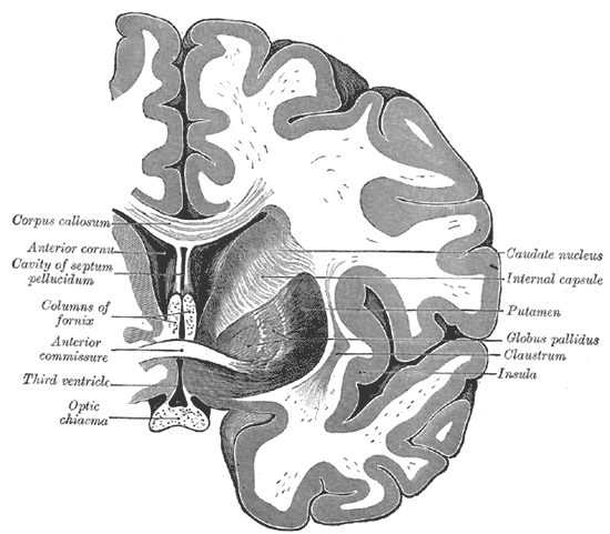
Coronal cross-section of brain showing the corpus callosum at top and the anterior commissure below
Details
Identifiers
Latinfibra commissuralis, fibrae commissurales telencephali
NeuroNames1220
TA98A14.1.00.017
A14.1.09.569
TA25603
FMA75249
Anatomical terms of neuroanatomy

The commissural fibers or transverse fibers are axons that connect the two hemispheres of the brain. Huge numbers of commissural fibers make up the commissural tracts in the brain, the largest of which is the corpus callosum.

In contrast to commissural fibers, association fibers form association tracts that connect regions within the same hemisphere of the brain, and projection fibers connect each region to other parts of the brain or to the spinal cord.[1]

Structure

[edit]

The commissural fibers make up tracts that include the corpus callosum, the anterior commissure, and the posterior commissure, among other pathways.

Corpus callosum

[edit]

The corpus callosum is the largest commissural tract in the human brain. It consists of about 200–300 million axons that connect the two cerebral hemispheres. The corpus callosum is essential to the communication between the two hemispheres.[2]

A recent study of individuals with agenesis of the corpus callosum suggests that the corpus callosum plays a vital role in problem solving strategies, verbal processing speed, and executive performance. Specifically, the absence of a fully developed corpus callosum is shown to have a significant relationship with impaired verbal processing speed and problem solving.[3]

Another study of individuals with multiple sclerosis provides evidence that structural and microstructural abnormalities of the corpus callosum are related to cognitive dysfunction. Particularly, verbal and visual memory, information processing speed, and executive tasks were shown to be impaired when compared to healthy individuals. Physical disabilities in multiple sclerosis patients also seem to be related to abnormalities of the corpus callosum, but not to the same extent of other cognitive functions.[4]

Using diffusion tensor imaging, researchers have been able to produce a visualization of this network of fibers, which shows the corpus callosum has an anteroposterior topographical organization that is uniform with the cerebral cortex.

Anterior commissure

[edit]

The anterior commissure (also known as the precommissure) is a tract that connects the two temporal lobes of the cerebral hemispheres across the midline, and placed in front of the columns of the fornix. The great majority of fibers connecting the two hemispheres travel through the corpus callosum, which is over 10 times larger than the anterior commissure, and other routes of communication pass through the hippocampal commissure or, indirectly, via subcortical connections. Nevertheless, the anterior commissure is a significant pathway that can be clearly distinguished in the brains of all mammals.

Using diffusion tensor imaging, researchers were able to approximate the location of the anterior commissure where it crosses the midline of the brain. This tract can be observed to be in the shape of a bicycle as it branches through various areas of the brain. Through diffusion tensor imaging results, the anterior commissure was categorized into two fiber systems: 1) the olfactory fibers and 2) the non-olfactory fibers.[5]

Posterior commissure

[edit]

The posterior commissure (also known as the epithalamic commissure) is a rounded nerve tract crossing the middle line on the dorsal aspect of the upper end of the cerebral aqueduct. It is important in the bilateral pupillary light reflex.

Evidence suggests the posterior commissure is a tract that plays a role in language processing between the right and left hemispheres of the brain. It connects the pretectal nuclei. A case study described recently in The Irish Medical Journal discussed the role the posterior commissure plays in the connection between the right occipital cortex and the language centers in the left hemisphere. This study explains how visual information from the left side of the visual field is received by the right visual cortex and then transferred to the word form system in the left hemisphere though the posterior commissure and the splenium. Disruption of the posterior commissure can cause alexia without agraphia. It is evident from this case study of alexia without agraphia that the posterior commissure plays a vital role in transferring information from the right occipital cortex to the language centers of the left hemisphere.[6]

Other

[edit]

The lyra or hippocampal commissure, habenular commissure, interthalamic adhesion, commissure of superior colliculus, commissure of inferior colliculus, optic chiasm, supraoptic commissure,[7] and supramammilary commissure.[8]

Aging and function

[edit]

Age-related decline in the commissural fiber tracts that make up the corpus callosum indicate the corpus callosum is involved in memory and executive function. Specifically, the posterior fibers of the corpus callosum are associated with episodic memory. Perceptual processing decline is also related to diminished integrity of occipital fibers of the corpus callosum. Evidence suggests that the genu of the corpus callosum does not contribute significantly to any one cognitive domain in the elderly. As fiber tract connectivity in the corpus callosum declines due to aging, compensatory mechanisms are found in other areas of the corpus callosum and frontal lobe. These compensatory mechanisms, increasing connectivity in other parts of the brain, may explain why elderly individuals still display executive function as a decline of connectivity is seen in regions of the corpus callosum.[9]

Older adults compared to younger adults show poorer performance in balance exercises and tests. A decline in white matter integrity of the corpus callosum in older individuals may explain declines in the ability to balance. Changes in the white matter integrity of the corpus callosum may also be related to cognitive and motor function decline as well. Decreased white matter integrity effects proper transmission and processing of sensorimotor information. White matter degeneration of the genu of the corpus callosum is also associated with gait, balance impairment, and the quality of postural control.[10]

Other animals

[edit]

The corpus callosum allows for communication between the two hemispheres and is found only in placental mammals. The anterior commissure serves as the primary mode of interhemispheric communication in marsupials,[11][12] and which carries all the commissural fibers arising from the neocortex (also known as the neopallium), whereas in placental mammals the anterior commissure carries only some of these fibers).[13]

References

[edit]
[edit]
Revisions and contributorsEdit on WikipediaRead on Wikipedia
from Grokipedia
Commissural fibers are bundles of myelinated axons in the that cross the midline to connect corresponding regions of the left and right cerebral hemispheres, facilitating interhemispheric communication of sensory, motor, and cognitive information. These tracts are essential for integrating bilateral functions, with the serving as the largest and most prominent example, containing over 200 million axons that link homologous cortical areas across the . The primary commissural pathways include the , which is divided into the rostrum, genu, body, and splenium, each connecting specific cortical regions such as the frontal lobes via the forceps minor and the occipital lobes via the forceps major. The , a smaller tract located posterior to the , primarily interconnects the temporal lobes, , and olfactory structures, contributing to functions like olfaction, , and interhemispheric temporal processing. Additional structures, such as the between the and and the hippocampal commissure (or commissure of the fornix) linking the hippocampi, support specialized roles in reflexes and medial temporal lobe integration, respectively. Functionally, commissural fibers enable the synchronization of hemispheric activities, with anterior portions of the transmitting motor signals and posterior regions handling visual, auditory, and somatosensory data. Disruptions, such as or damage from conditions like , can lead to disconnection syndromes characterized by impaired bilateral coordination. These tracts are also implicated in neurodevelopmental disorders and are targeted in surgical interventions like callosotomy for treatment.

Definition and Overview

Anatomical Definition

Commissural fibers are bundles of myelinated axons forming tracts that cross the midline of the through commissures to interconnect homologous regions of the left and right cerebral hemispheres. These tracts facilitate interhemispheric integration by linking corresponding cortical areas, with the serving as the largest example. These fibers typically originate from pyramidal neurons in the of one hemisphere, particularly in layers II/III and V, where they extend axons that decussate through midline structures before terminating in the contralateral cortex or subcortical regions. The pathway ensures bidirectional communication, with axons projecting across the midline to form synaptic connections that mirror the originating cortical architecture. In distinction to other white matter fiber types, commissural fibers specifically enable communication between hemispheres, whereas association fibers connect regions within the same ipsilateral hemisphere, and projection fibers link the cortex to subcortical structures or the . Histologically, these fibers consist of axons with diameters ranging from 0.2 to 20 μm, surrounded by sheaths produced by to support rapid conduction velocities. Their synaptic targets are predominantly in cortical layers II/III and V of the contralateral hemisphere, where they influence excitatory and inhibitory circuits.

Significance in Brain Function

Commissural fibers are essential for integrating sensory, motor, and cognitive processing across the cerebral hemispheres, enabling unified perception and coordinated action in complex tasks. These fibers connect homologous cortical regions, allowing for the synchronization of neural activity that supports bimanual coordination, perceptual synthesis, and higher-order cognitive functions such as memory and problem-solving. The , the largest commissural tract, exemplifies this role by linking nearly all neocortical areas and facilitating the bilateral exchange of information critical for everyday behaviors. In humans, the contains over 200 million axons, highlighting the extensive scale of this interhemispheric connectivity. These fibers also contribute to brain plasticity and recovery after unilateral injury by enabling compensatory rerouting and neural reorganization. Following , callosal neurons in the undamaged hemisphere exhibit selective plasticity, including transient reductions in dendritic spine density followed by increased spine formation and stabilization, which helps re-establish ipsilateral connections and restore neuronal activity over several weeks. This adaptive remodeling supports functional recovery by allowing the intact hemisphere to compensate for deficits in the injured side. From an evolutionary perspective, commissural fibers confer an advantage by enhancing bilateral coordination for sophisticated behaviors, as evidenced by deficits observed in studies. Research by Roger Sperry on patients who underwent callosotomy revealed that severing these fibers results in disconnected hemispheres operating as independent cognitive domains, with impaired integration of sensory inputs, motor outputs, and conscious awareness—such as the right hemisphere's inability to verbally report left-field stimuli despite processing them. These findings underscore the fibers' role in unifying hemispheric functions, a development conserved across vertebrates to support lateralized yet integrated neural processing.

Anatomy

Corpus Callosum

The constitutes the principal commissural pathway in the , forming a broad, arched band of that bridges the to interconnect the neocortices of the left and right cerebral hemispheres. Positioned superior to the , it extends approximately 10 cm in length from anterior to posterior, exhibiting a C-shaped configuration with an upwardly convex arch; its thickness varies regionally from about 1 mm anteriorly to 10 mm posteriorly. This structure facilitates the transfer of sensory, motor, and cognitive information across the midline, underscoring its role as the dominant interhemispheric conduit. Structurally, the corpus callosum is delineated into four primary subdivisions along its anteroposterior axis: the rostrum, genu, body, and splenium. The rostrum represents the slender, beak-like inferior extension at the anterior terminus, merging with the . Immediately posterior lies the genu, a sharply curved segment that gives rise to the forceps minor, linking homologous regions of the prefrontal cortices. The body, comprising the elongated central portion, interconnects premotor, supplementary motor, primary motor, and somatosensory areas of the frontal and parietal lobes via the . The splenium, the thickened posterior bulb, projects the forceps major to unite the parietal, temporal, and occipital cortices, encompassing visual and auditory association areas. These regional divisions reflect a systematic bundling of axons tailored to cortical . Composed of roughly 200 million densely myelinated axons, the demonstrates precise topographic organization, wherein fibers originating from adjacent cortical territories remain grouped during their traversal; for example, somatosensory projections from the course through the midbody. While predominantly homotopic—connecting mirror-symmetric cortical sites—a substantial fraction, estimated at around 75% in core regions, forms heterotopic linkages to non-mirror areas, enabling broader cross-hemispheric influence. diameters vary regionally, averaging 0.8–1.1 μm, with larger fibers in motor-related segments. On (MRI), the manifests as a conspicuous high-signal tract in the midline, readily identifiable in sagittal and coronal planes due to its myelinated composition. Diffusion tensor imaging (DTI) further elucidates its microstructure by mapping fiber orientation and integrity, revealing values typically above 0.7 in healthy tissue. The average volume in adults approximates 10 cm³, with males exhibiting slightly larger measures (around 11 cm³) than females (about 9.6 cm³), independent of overall differences.

Anterior Commissure

The is a compact, oval-shaped tract approximately 4 mm wide, situated in the midline immediately anterior to the anterior columns of the fornix within the of the third ventricle. This structure serves as an evolutionarily conserved commissural bundle that interconnects homologous regions of the bilateral temporal lobes and olfactory bulbs and tracts, facilitating interhemispheric communication in more ventral and limbic-oriented areas of the . The tract is divided into an anterior bundle and a posterior bundle, each carrying distinct fiber pathways. The anterior bundle primarily connects the olfactory bulbs and anterior olfactory nuclei across the midline, supporting bilateral integration of olfactory processing. The posterior bundle, which constitutes the majority of the tract, links limbic and temporal structures, including the , hippocampus, and temporal such as the and inferior temporal cortex. Comprising an estimated 3 million axons, the features a mix of fiber types, with the allocortical (olfactory-related) components largely unmyelinated and the neocortical (temporal-related) components consisting mostly of small myelinated fibers—resulting in overall lower myelination compared to the . These connections emphasize integration, in contrast to the corpus callosum's primary focus on neocortical regions. Clinically, the tract is readily identifiable on coronal MRI slices near the , appearing as a transversely oriented band of high signal intensity in T2-weighted images.

Posterior Commissure

The posterior commissure is a compact bundle of transversely oriented fibers situated in the dorsal aspect of the , forming part of the posterior wall of the third ventricle just caudal to the pineal recess. It extends approximately 4-5 mm in length and 1-2 mm in width in human brains, appearing as a rounded or C-shaped structure that marks the junction between the and mesencephalon. This commissure primarily interconnects contralateral structures in the pretectal and tectal regions, including the superior colliculi, pretectal nuclei, interstitial nucleus of Cajal, and rostral interstitial nucleus of the , as well as thalamic and habenular nuclei. Fibers decussate midline at the level of the pineal recess, facilitating bilateral coordination in these diencephalic and areas. Composed of an estimated 500,000 to 900,000 axons, the contains a mix of heavily myelinated and unmyelinated fibers, with the myelinated components predominating to support rapid signal transmission across the midline. Anatomically, it lies immediately superior to the (aqueduct of Sylvius) and inferior to the habenular commissure, in close proximity to the matter and the . As a key element of the broader commissural fiber system, it contributes to interhemispheric pathways distinct from larger structures like the .

Minor Commissures

The minor commissures of the encompass several smaller tracts that cross the midline to connect homologous structures in the and telencephalon, distinct from the larger , , and . These tracts are notably compact, with the collective number of axons in all minor commissures estimated at less than 1% of the total commissural fiber population in the , which exceeds 200 million primarily within the . Their sizes and trajectories exhibit interindividual variability, often visualized and quantified using diffusion tensor imaging (DTI) in modern studies. The hippocampal commissure, also known as the commissure of the fornix, is a transversely oriented bundle of fibers located immediately below the splenium of the and dorsal to the third ventricle. It interconnects the crura of the fornix, linking the dentate gyri and layers of the left and right hippocampi across the midline. This structure forms a triangular sheet within the posterior aspect of the fornix, facilitating direct interhemispheric communication between these limbic formations. The habenular commissure represents a diminutive tract situated in the , anterior and superior to the . It consists of decussating fibers that connect the habenular nuclei on both sides of the brain, as well as portions of the internal medullary laminae. This commissure lies within the superior wall of the pineal stalk and serves as a key link in the habenular complex, which interfaces with limbic pathways. Additional minor commissures include the supraoptic commissures, located in the near the , which comprise multiple bundles such as the dorsal (Meynert's) supraoptic commissure, ventral supraoptic commissure, and Ganser's commissure. These tracts provide hypothalamic interconnections, crossing the midline to link nuclei involved in autonomic .

Development

Embryonic Origins

Commissural fibers originate during early embryogenesis through the coordinated outgrowth and guidance of axons from neurons in the developing telencephalon. In , pioneering axons begin to extend toward the midline around embryonic day 12 (E12), with initial crossing occurring between E14 and E16, facilitated by interactions with midline and guidance cues such as netrins and . This process corresponds to approximately gestational weeks 6-8 in humans, when the cortical plate forms and initial axonal projections emerge, though definitive midline crossing for the is observed around week 11. The key developmental processes involve axonal outgrowth from the cortical plate, where postmitotic neurons extend growth cones ventrally through the intermediate zone. These axons then fasciculate into bundles, adhering to pioneer fibers via cell adhesion molecules, before approaching the midline. Midline crossing occurs via a glial sling in the ventral telencephalon, a specialized structure of radial that provides a permissive substrate for axons to traverse the midline while avoiding inhibitory cues. Disruptions in glial sling formation, as seen in Nfia mice, lead to defasciculated axons and failure of commissural tracts. Molecular regulators play crucial roles in directing these events, with Slit-2 acting as a repulsive cue via Robo1 receptors to prevent premature midline entry and ensure post-crossing deflection, while DCC receptors mediate attraction to netrin-1 at the midline to promote crossing. Mutations in these pathways, such as in Slit or Robo genes, result in acallosal phenotypes where axons stall or misroute at the midline, highlighting their essential function in commissural assembly.00179-5) Specific commissural tracts form through distinct pioneer populations: the is initiated by axons from early-generated neurons in the , which cross the midline first around E15.5 in mice, providing a scaffold for subsequent neocortical axons. In contrast, the arises from projections originating in the olfactory placode-derived structures, including the anterior olfactory nucleus, with axons bundling and crossing early in ventral development.

Postnatal Maturation

Postnatal maturation of commissural fibers, particularly those in the , involves progressive myelination, structural refinement, and volume expansion that extend from infancy through and into early adulthood. Myelination of these fibers begins in earnest around 4 months of age and accelerates during , with peak rates occurring between 2 and 5 years as precursor cells proliferate and differentiate to form sheaths around axons. This process supports faster interhemispheric and continues at a slower pace thereafter, achieving full structural maturation by approximately 20 to 30 years of age, as evidenced by stabilization in diffusion tensor imaging (DTI) metrics such as (FA). Concurrently, the volume of the increases substantially, expanding 2 to 3 times from infancy to adulthood, driven by axonal growth, myelination, and gliogenesis in tracts. Key cellular processes underpin this maturation, including the proliferation of that wrap axons in sheaths, enhancing conduction efficiency, and that eliminates excess connections to refine interhemispheric pathways.00742-9) proliferation peaks in early postnatal stages, with precursor cells migrating into commissural tracts like the to initiate sheath formation, a process that is tightly coupled to axonal integrity and vascular support in . , mediated by microglial activity, removes superfluous synapses along these fibers during childhood, optimizing connectivity based on activity patterns and contributing to the consolidation of functional circuits. Additionally, experience-dependent plasticity modulates this development, allowing environmental inputs to shape fiber organization and myelination, as seen in enhanced microstructure following enriched sensory or cognitive experiences. Maturation exhibits regional heterogeneity, with posterior segments of the —linking sensory and visual areas—developing earlier than anterior regions connecting prefrontal cortices, reflecting a posterior-to-anterior in myelination and . This aligns with DTI findings, where FA values, indicative of fiber coherence and myelination, rise progressively with age across commissural tracts, increasing from low levels in infancy to peak adulthood values that signify mature axonal alignment. External factors further influence this trajectory; pubertal hormonal surges, particularly in gonadal steroids like and testosterone, accelerate myelination and volume changes in the , with effects more pronounced in females during mid-adolescence. Environmental influences, such as bilingualism, promote enhanced connectivity by increasing volume and integrity, likely through activity-driven plasticity that bolsters interhemispheric communication for language processing.

Function

Interhemispheric Integration

Commissural fibers facilitate interhemispheric integration through bidirectional signaling that enables the phase-locking of neural oscillations across the cerebral hemispheres, particularly for gamma waves in the 30-80 Hz range transmitted via the . This process supports the coordination of distributed neural activity, allowing for the temporal alignment of oscillatory patterns essential for unified brain processing. The contributes similarly to this integration for certain subcortical and temporal connections. The efficiency of signal transfer across commissural fibers is characterized by conduction velocities ranging from approximately 5 to 18 m/s, leading to interhemispheric latencies of 10-20 ms for callosal projections. This transmission involves a balance of excitatory and inhibitory influences, with fibers promoting excitation and fibers mediating inhibition within the . Such neurochemical dynamics ensure precise modulation of interhemispheric communication, preventing excessive synchronization while enabling adaptive neural coupling. Experimental evidence from EEG and MEG studies in acallosal animal models reveals significantly reduced interhemispheric synchrony, underscoring the critical role of commissural fibers in maintaining oscillatory coherence. In humans, procedures result in diminished integration of hemispheric activity, as evidenced by decreased coherence in cross-hemispheric signals, which highlights the fibers' necessity for effective synchronization. Interhemispheric transfer via commissural fibers demonstrates adaptive, frequency-specific properties, with lower frequencies supporting motor-related coordination and higher frequencies aiding attentional mechanisms. This selectivity allows the to optimize communication based on task demands, enhancing overall functional efficiency.

Role in Cognition and Motor Control

Commissural fibers, particularly those in the , play a crucial role in divided attention tasks such as , where simultaneous auditory stimuli are presented to each , facilitating the integration of information across hemispheres for balanced processing. In individuals with intact callosal connections, this enables effective right-ear advantage for verbal material while allowing cross-hemispheric transfer to mitigate left-ear . The contributes to emotional processing by regulating neuronal activity in the amygdalae, thereby influencing behaviors tied to socioemotional responses like social interaction and anxiety-like states in animal models. Split-brain studies, involving surgical sectioning of the to treat intractable , demonstrate impaired cross-hemispheric transfer of information, leading to deficits in unified such as the inability of one hemisphere to access visual or tactile stimuli presented exclusively to the other. These findings highlight how commissural fibers enable the synthesis of hemispheric specializations into coherent perceptual and cognitive experiences, with disruptions revealing independent yet disconnected processing streams. In , the supports bimanual coordination through interhemispheric premotor connections, allowing synchronized movements of both hands by transferring inhibitory and excitatory signals to prevent mirror movements and ensure temporal alignment. The facilitates conjugate gaze, coordinating vertical eye movements via crossing fibers that integrate signals from burst-tonic neurons in the rostral interstitial nucleus of the . Behavioral evidence underscores these roles; for instance, larger size, especially in the anterior region, correlates with enhanced musical ability, particularly in instrumentalists requiring precise bimanual skills, as observed in studies of professional musicians. Commissural fibers exhibit plasticity in recovery processes, such as post- axonal sprouting in the contralesional cortex, where callosal neurons form new spines and connections to compensate for ipsilesional damage, aiding restoration of motor function. This adaptive rewiring, observed in models of ischemic , selectively enhances contralesional callosal projections, supporting behavioral improvements in skilled movements.

Aging and Pathology

As individuals age, commissural fibers, particularly those in the , undergo microstructural decline characterized by demyelination and axonal loss, beginning around the fourth decade of life. Postmortem and studies reveal decreases in density and alterations in sheaths, with histological evidence showing splitting of and accumulation of dense in degenerating fibers. tensor (DTI) further demonstrates these changes through decreased (FA) in white matter tracts, indicating reduced directional coherence of fiber bundles, especially in posterior regions such as the splenium. For instance, in healthy older adults, age is significantly associated with lower FA and higher mean in the splenium, reflecting compromised microstructural integrity. Regional vulnerability is evident in the posterior , where the splenium atrophies more rapidly than anterior segments, potentially due to its role in integrating visual and areas. This accelerated decline in the splenium correlates with cognitive slowing, as reduced interhemispheric connectivity impairs rapid information transfer between hemispheres. Longitudinal MRI data from cohorts like the Birth Cohort 1936 indicate progressive white matter hyperintensities (WMH) in commissural regions, with vascular risk factors accelerating these changes over time and contributing to overall volume reductions in myelinated fibers by late adulthood. No significant neuronal occurs in these tracts during normal aging, but synaptic weakening at axonal terminals and reduced support exacerbate functional disruptions. At the cellular level, these age-related alterations stem from diminished function, which impairs maintenance and repair. Oxidative stress accumulates in , damaging lipid-rich sheaths and promoting via microglial activation, leading to further demyelination without widespread axonal degeneration. Studies highlight that while increases in aging , it primarily affects glial cells rather than causing direct neuronal loss, resulting in synaptic weakening through disrupted signaling efficiency. These mechanisms underscore the progressive, non-pathological nature of commissural fiber changes in healthy aging.

Associated Disorders and Lesions

Agenesis of the corpus callosum (AgCC) is a congenital malformation characterized by the partial or complete absence of the , occurring in approximately 1 in 4,000 births. It is associated with symptoms such as seizures, , speech delays, and visual impairments, with reported in up to 60% of cases and seizures in about 25%. Genetic factors contribute significantly, including mutations in the ARX gene, which are linked to X-linked disorders featuring AgCC alongside and abnormal genitalia. Lesions affecting commissural fibers, particularly through surgical or traumatic means, can lead to disconnection syndromes. Callosotomy, a procedure used to treat severe by severing the , disrupts interhemispheric communication and may result in , where one hand performs involuntary actions perceived as foreign by the patient. In , damage to the anterior fibers often causes , impairing purposeful movements due to impaired coordination between hemispheres. Demyelination in frequently involves commissural fibers, with lesions in the contributing to cognitive dysfunction and motor impairments. In , atrophy of the posterior correlates with memory loss and overall cognitive decline, reflecting disrupted interhemispheric integration in advanced stages. Diagnostic approaches for commissural fiber abnormalities include (MRI), which is the gold standard for identifying or through characteristic features like colpocephaly and the absence of the midline structure. Functional deficits are assessed via neuropsychological testing, revealing impairments in interhemispheric transfer and executive function.

Comparative Anatomy

In Mammals

In mammals, commissural fiber organization exhibits notable variations across species, particularly in the size and connectivity of major tracts like the (CC) and (AC). , especially humans, possess an expanded CC containing approximately 200 million myelinated axons, facilitating extensive interhemispheric communication, whereas rodents such as mice have a substantially smaller CC with around 10 million axons. This disparity is particularly evident in prefrontal regions, where show denser callosal connections supporting advanced cognitive integration, compared to the sparser prefrontal projections in . In contrast, the AC is relatively larger in non-primate mammals, reflecting its prominent role in olfactory processing; for instance, in , the AC conveys a higher proportion of olfactory fibers due to their reliance on olfaction, while in , its olfactory component is diminished alongside reduced size. Among carnivores and ungulates, the of the CC remains broadly similar to that in and , but with scaled-down relative sizes adapted to volume. In dogs, tensor reveals a CC with conserved regional organization—genu for frontal connections, body for sensorimotor, and splenium for occipital—but its overall size is smaller relative to total mass compared to humans, aligning with less elaborate cortical folding. MRI studies in sheep and demonstrate that commissures, such as the , are highly conserved across ruminants, maintaining consistent connectivity for basic visuomotor and auditory integration despite variations in size. These structures show minimal divergence in positioning and fiber density between sheep and , underscoring evolutionary stability in subcortical commissural pathways. Functional scaling of commissural tracts correlates with overall and in mammals, where larger CC volumes support enhanced interhemispheric coordination in socially demanding environments. For example, exhibit a massive CC, with a cross-sectional area comparable to that predicted for their 5 kg brain, enabling synchronized processing across expanded neocortical areas vital for complex social behaviors like matriarchal herd dynamics. In contrast, species with simpler social structures, such as solitary carnivores, show proportionally smaller commissural fibers relative to brain volume. This scaling pattern highlights how commissural expansion facilitates adaptive advantages in group-living mammals. Rodents serve as primary experimental models for studying commissural fiber development due to their rapid axonal growth and genetic tractability, allowing detailed dissection of midline crossing mechanisms during embryogenesis. In these models, manipulations reveal key guidance cues for CC formation, providing insights into congenital dysplasias. Conversely, , particularly macaques, are essential models for investigating commissural roles in , where tract-tracing and demonstrate how callosal fibers integrate sensory and , bridging gaps between findings and human applicability.

In Non-Mammals

In non-mammalian vertebrates, commissural fibers facilitate interhemispheric and midline integration but differ markedly from mammalian structures, lacking a and relying on smaller, more diffuse tracts. In birds and reptiles, pallial commissures such as the anterior and hippocampal commissures serve as primary interhemispheric pathways at the telencephalic level, connecting homologous regions across the midline with myelinated fibers that are notably smaller in scale compared to mammalian counterparts. For instance, in songbirds, these commissures, including anterior and posterior variants, support by integrating bilateral sensory and motor signals essential for production and social communication. Reptiles exhibit similar connectivity patterns, with commissural fibers projecting to heterotopic regions, underscoring a conserved organization that emphasizes functional equivalence over structural elaboration. In and amphibians, commissural systems are adapted for sensory-motor coordination with minimal crossing, focusing instead on and spinal levels. Tectal commissures in these groups enable visual integration, such as interocular transfer of learned responses in , where fibers cross the midline to synchronize binocular inputs for prey detection and spatial localization. In the optic tectum, a non-mammalian homolog of the , these commissures drive spatial summation and behavioral responses to visual stimuli, as seen in larval where intertectal circuits control prey-capture programs. Spinal white commissures, comprising crossing axons in the ventral spinal cord, coordinate locomotion by linking contralateral and motoneurons, facilitating rhythmic movements like in amphibians without extensive supraspinal oversight. This arrangement highlights a simpler suited to aquatic and semi-terrestrial lifestyles, with commissures remaining rudimentary. In , commissural fibers form analogous midline crossings that connect segmental ganglia, though without true cerebral hemispheres, serving to integrate bilateral neural activity across the ventral cord. In , commissures link commissural bundles between ganglia, guided by netrin proteins that act as short-range attractants to promote midline crossing during embryonic development. These netrin-dependent cues ensure precise pathfinding, mirroring mechanisms in vertebrates but adapted to a decentralized for behaviors like locomotion and . Evolutionarily, commissural fibers predate the lineage, with foundational midline-crossing mechanisms evident in and conserved across chordates, though neocortical elaborations like the mammalian represent a derived innovation. The , for example, persists as an ancient structure, co-opting developmental programs to maintain interhemispheric connectivity in non-mammals. This continuity underscores commissures' role in bilateral organization since early metazoan , contrasting with the specialized hemispheric integration unique to mammalian .

References

Add your contribution
Related Hubs
User Avatar
No comments yet.