Hubbry Logo
Lymphatic systemLymphatic systemMain
Open search
Lymphatic system
Community hub
Lymphatic system
logo
8 pages, 0 posts
0 subscribers
Be the first to start a discussion here.
Lymphatic system
Lymphatic system
from Wikipedia
Lymphatic system
Human lymphatic system
Details
Identifiers
Latinsystema lymphoideum
MeSHD008208
TA98A13.0.00.000
TA25149
FMA7162 74594, 7162
Anatomical terminology

The lymphatic system, or lymphoid system, is an organ system in vertebrates that is part of the immune system and complementary to the circulatory system. It consists of a large network of lymphatic vessels, lymph nodes, lymphoid organs, lymphatic tissue and lymph.[1][2] The Latin word for lymph, lympha, refers to the deity of fresh water, "Lympha".[3]

Unlike the circulatory system, which is a closed system, the lymphatic system is open.[4][5] Lymph originates in the interstitial fluid that leaks from blood in the circulatory system into the tissues of the body. This fluid carries nutrients to the cells and collects waste products, bacteria, and damaged cells, before draining into the lymphatic vessels as lymph. The circulatory system processes an average of 20 litres (5.3 US gal) of blood per day through capillary filtration, which removes plasma from the blood. Roughly 17 litres (4.5 US gal) of the filtered blood is reabsorbed directly into the blood vessels, while the remaining 3 litres (0.79 US gal) are left in the interstitial fluid. The lymphatic system provides an accessory return route to the blood for this remainder.[6]

The other main function is that of immune defense. Lymph is very similar to blood plasma, in that it contains waste products and cellular debris, together with bacteria and proteins. The cells of the lymph are mostly lymphocytes. Associated lymphoid organs are composed of lymphoid tissue, and are the sites either of lymphocyte production or of lymphocyte activation. These include the lymph nodes (where the highest lymphocyte concentration is found), the spleen, the thymus, and the tonsils. Lymphocytes are initially generated in the bone marrow. The lymphoid organs also contain other types of cells such as stromal cells for support.[7] Lymphoid tissue is also associated with mucosas such as mucosa-associated lymphoid tissue (MALT).[8]

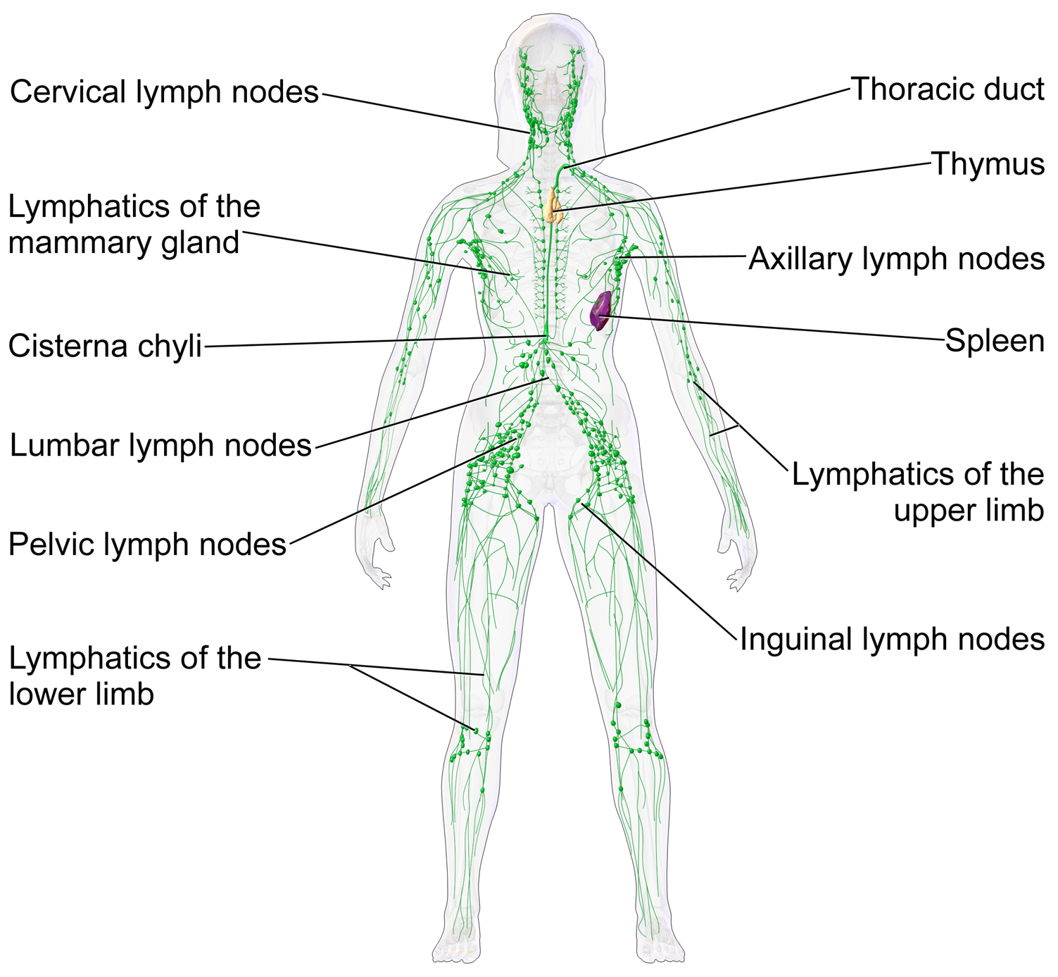
These vessels carry the lymph throughout the body, passing through numerous lymph nodes which filter out unwanted materials such as bacteria and damaged cells. Lymph then passes into much larger lymph vessels known as lymph ducts. The right lymphatic duct drains the right side of the region and the much larger left lymphatic duct, known as the thoracic duct, drains the left side of the body. The ducts empty into the subclavian veins to return to the blood circulation. Lymph is moved through the system by muscle contractions.[9] In some vertebrates, a lymph heart is present that pumps the lymph to the veins.[9][10]

The lymphatic system was first described in the 17th century independently by Olaus Rudbeck and Thomas Bartholin.[11]

Structure

[edit]
Anatomy of the lymphatic system showing primary and secondary lymphoid organs

The lymphatic system consists of a conducting network of lymphatic vessels, lymphoid organs, lymphoid tissues, and the circulating lymph.[1]

Primary lymphoid organs

[edit]

The primary (or central) lymphoid organs, including the thymus, bone marrow, fetal liver and yolk sac, are responsible for generating lymphocytes from immature progenitor cells in the absence of antigens.[12] The thymus and the bone marrow constitute the primary lymphoid organs involved in the production and early clonal selection of lymphocyte tissues.

Bird species' primary lymphoid organs include the bone marrow, thymus, bursa of Fabricius, and yolk sac.[13]

Bone marrow

[edit]

Bone marrow is responsible for both the creation of T cell precursors and the production and maturation of B cells, which are important cell types of the immune system. From the bone marrow, B cells immediately join the circulatory system and travel to secondary lymphoid organs in search of pathogens. T cells, on the other hand, travel from the bone marrow to the thymus, where they develop further and mature. Mature T cells then join B cells in search of pathogens. The other 95% of T cells begin a process of apoptosis, a form of programmed cell death: T cells that cannot interact strongly enough with self-antigens are eliminated during T cell § Positive selection, and T cells that attack the body's own proteins are eliminated during § Negative selection.

Thymus

[edit]

The thymus increases in size from birth in response to postnatal antigen stimulation. It is most active during the neonatal and pre-adolescent periods. The thymus is located between the inferior neck and the superior thorax. At puberty, by the early teens, the thymus begins to atrophy and regress, with adipose tissue mostly replacing the thymic stroma. However, residual T cell lymphopoiesis continues throughout adulthood, providing some immune response. The thymus is where the T lymphocytes mature and become immunocompetent. The loss or lack of the thymus results in severe immunodeficiency and subsequent high susceptibility to infection. In most species, the thymus consists of lobules divided by septa, which are made up of epithelium, which is often considered an epithelial organ. T cells mature from thymocytes, proliferate, and undergo a selection process in the thymic cortex before entering the medulla to interact with epithelial cells.

Research on bony fish showed a buildup of T cells in the thymus and spleen of lymphoid tissues in salmon and showed that there are not many T cells in non-lymphoid tissues.[14]

The thymus provides an inductive environment for developing T cells from hematopoietic progenitor cells. In addition, thymic stromal cells allow for the selection of a functional and self-tolerant T cell repertoire. Therefore, one of the most important roles of the thymus is the induction of central tolerance. However, the thymus is not where the infection is fought, as the T cells have yet to become immunocompetent.

Secondary lymphoid organs

[edit]

The secondary (or peripheral) lymphoid organs, which include lymph nodes and the spleen, maintain mature naive lymphocytes and initiate an adaptive immune response.[15] The secondary lymphoid organs are the sites of lymphocyte activation by antigens.[16] Activation leads to clonal selection and affinity maturation. Mature lymphocytes recirculate between the blood and the secondary lymphoid organs until they encounter their specific antigen.

Spleen

[edit]

The main functions of the spleen are:

  1. to produce immune cells to fight antigens,
  2. to remove particulate matter and aged blood cells, mainly red blood cells, and
  3. to produce blood cells during fetal life.

The spleen synthesizes antibodies in its white pulp and removes antibody-coated bacteria and antibody-coated blood cells by way of blood and lymph node circulation. The white pulp of the spleen provides immune function due to the lymphocytes housed there. The spleen also consists of red pulp, which is responsible for getting rid of aged red blood cells and pathogens. This is carried out by macrophages present in the red pulp. A study published in 2009 using mice found that the spleen contains, in its reserve, half of the body's monocytes within the red pulp.[17] These monocytes, upon moving to injured tissue (e.g., the heart), turn into dendritic cells and macrophages while promoting tissue healing.[17][18][19] The spleen is a center of activity of the mononuclear phagocyte system and can be considered analogous to a large lymph node, as its absence causes a predisposition to certain infections. Notably, the spleen is essential for a multitude of functions. The spleen removes pathogens and old erythrocytes from the blood (red pulp) and produces lymphocytes for immune response (white pulp). The spleen also is responsible for recycling some erythrocyte components and discarding others. For example, hemoglobin is broken down into amino acids that are reused.

Research on bony fish has shown that a high concentration of T cells is found in the spleen's white pulp.[14]

Like the thymus, the spleen has only efferent lymphatic vessels. Both the short gastric arteries and the splenic artery supply it with blood.[20] The germinal centers are supplied by arterioles called penicilliary radicles.[21]

In humans, until the fifth month of prenatal development, the spleen creates red blood cells; after birth, the bone marrow is solely responsible for hematopoiesis. As a major lymphoid organ and a central player in the reticuloendothelial system, the spleen retains the ability to produce lymphocytes. The spleen stores red blood cells and lymphocytes. It can store enough blood cells to help in an emergency. Up to 25% of lymphocytes can be stored at any one time.[22]

Lymph nodes

[edit]
A lymph node showing afferent and efferent lymphatic vessels
Regional lymph nodes

A lymph node is an organized collection of lymphoid tissue through which the lymph passes on its way back to the blood. Lymph nodes are located at intervals along the lymphatic system. Several afferent lymph vessels bring in lymph, which percolates through the substance of the lymph node and is then drained out by an efferent lymph vessel. Of the nearly 800 lymph nodes in the human body, about 300 are located in the head and neck.[23] Many are grouped in clusters in different regions, as in the underarm and abdominal areas. Lymph node clusters are commonly found at the proximal ends of limbs (e.g., groin or armpits) and in the neck, where lymph is collected from body regions likely to sustain pathogen contamination from injuries. Lymph nodes are particularly numerous in the mediastinum in the chest, neck, pelvis, axilla, groin (or inguinal region), and in association with the blood vessels of the intestines.[8]

The substance of a lymph node consists of lymphoid follicles in an outer portion called the cortex. The inner portion of the node is called the medulla, which is surrounded by the cortex on all sides except for a portion known as the hilum. The hilum presents as a depression on the surface of the lymph node, causing the otherwise spherical lymph node to be bean-shaped or ovoid. The efferent lymph vessel directly emerges from the lymph node at the hilum. The arteries and veins supplying the lymph node with blood enter and exit through the hilum. The region of the lymph node called the paracortex immediately surrounds the medulla. Unlike the cortex, which has primarily immature T cells (or thymocytes), the paracortex has a mixture of immature and mature T cells. Lymphocytes enter the lymph nodes through specialised high endothelial venules found in the paracortex.

A lymph follicle is a dense collection of lymphocytes, the number, size, and configuration of which change in accordance with the functional state of the lymph node. For example, the follicles expand significantly when encountering a foreign antigen. The selection of B cells (also known as B lymphocytes) occurs in the germinal centre of the lymph nodes.

Secondary lymphoid tissue provides the environment for the foreign or altered native molecules (antigens) to interact with the lymphocytes. It is exemplified by the lymph nodes, and the lymphoid follicles in tonsils, Peyer's patches, spleen, adenoids, skin, etc. that are associated with the mucosa-associated lymphoid tissue (MALT).

In the gastrointestinal wall, the appendix has mucosa resembling that of the colon, but it is heavily infiltrated with lymphocytes here.

Tertiary lymphoid organs

[edit]

Tertiary lymphoid organs (TLOs) are abnormal lymph node-like structures that form in peripheral tissues at sites of chronic inflammation, such as chronic infection, transplanted organs undergoing graft rejection, some cancers, and autoimmune and autoimmune-related diseases.[24] TLOs are often characterized by CD20+ B cell zone, which is surrounded by CD3+ T cell zone, similar to the lymph follicles in secondary lymphoid organs (SLOs) and are regulated differently from the normal process whereby lymphoid tissues are formed during ontogeny, being dependent on cytokines and hematopoietic cells, but still drain interstitial fluid and transport lymphocytes in response to the same chemical messengers and gradients.[25][26] Mature TLOs often have an active germinal center, surrounded by a network of follicular dendritic cells (FDCs).[27] Although the specific composition of TLOs may vary, within the T cell compartment, the dominant subset of T cells is CD4+ T follicular helper (TFH) cells, but certain number of CD8+ cytotoxic T cells, CD4+ T helper 1 (TH1) cells, and regulatory T cells (Tregs) can also be found within the T cell zone.[25] The B cell zone contains two main areas. The mantle is located at the periphery and composed of naive immunoglobulin D (IgD)+ B cells surrounding the germinal centre. The latter is defined by the presence of proliferating Ki67+CD23+ B cells and a CD21+ FDC network, as observed in SLOs.[28] TLOs typically contain far fewer lymphocytes, and assume an immune role only when challenged with antigens that result in inflammation. They achieve this by importing the lymphocytes from blood and lymph.[29]

According to the composition and activation status of the cells within the lymphoid structures, at least three organizational levels of TLOs have been described. The formation of TLOs starts with the aggregating of lymphoid cells and occasional DCs, but FDCs are lacking at this stage. The next stage is immature TLOs, also known as primary follicle-like TLS, which have an increased number of T cells and B cells with distinct T cell and B cell zones and the formation of FDCs network, but without germinal centres. Finally, fully mature (also known as secondary follicle-like) TLOs often have active germinal centres and high endothelial venules (HEVs), demonstrating a functional capacity by promoting T cell and B cell activation and then leading to expansion of TLS through cell proliferation and recruitment. During TLS formation, T and B cells are separated into two different but adjacent zones, with some cells having the ability to migrate from one to the other, which is a crucial step in developing an effective and coordinated immune response.[28][30]

TLOs may play a key role in the immune response to cancer and serve as a prognostic marker for immunotherapy. TLOs have been reported to present in different cancer types such as melanoma, non-small-cell lung cancer and colorectal cancer (reviewed by Sautès-Fridman and colleagues[31] in 2019), as well as glioma.[32] TLOs are also been seen as a read-out of treatment efficacy. For example, in patients with pancreatic ductal adenocarcinoma (PDAC), vaccination led to the formation of TLOs in responders. Within these patients, lymphocytes in TLOs displayed an activated phenotype, and in vitro experiments showed their capacity to perform effector functions.[28] Patients with the presence of TLOs tend to have a better prognosis,[33][34] even though some certain cancer types showed an opposite effect.[35] Besides, TLOs with an active germinal center seem to show a better prognosis than those with TLOs without a germinal center.[33][34] The reason that these patients tend to live longer is that TLOs can promote an immune response against the tumors. TLOs may also enhance anti-tumor response when patients are treated with immunotherapy such as immune checkpoint blockade treatment.[36]

Other lymphoid tissue

[edit]

Lymphoid tissue associated with the lymphatic system is concerned with immune functions in defending the body against infections and the spread of tumours. It consists of connective tissue formed of reticular fibers, with various types of leukocytes (white blood cells), mostly lymphocytes enmeshed in it, through which the lymph passes.[37] Regions of the lymphoid tissue that are densely packed with lymphocytes are known as lymphoid follicles. Lymphoid tissue can either be structurally well organized as lymph nodes or may consist of loosely organized lymphoid follicles known as the mucosa-associated lymphoid tissue (MALT).

The central nervous system also has lymphatic vessels. The search for T cell gateways into and out of the meninges uncovered functional meningeal lymphatic vessels lining the dural sinuses, anatomically integrated into the membrane surrounding the brain.[38]

Lymphatic vessels

[edit]
Lymph capillaries in the tissue spaces

The lymphatic vessels, also called lymph vessels, are thin-walled vessels that conduct lymph between different parts of the body.[39] They include the tubular vessels of the lymph capillaries, and the larger collecting vessels – the right lymphatic duct and the thoracic duct (the left lymphatic duct). The lymph capillaries are mainly responsible for the absorption of interstitial fluid from the tissues, while lymph vessels propel the absorbed fluid forward into the larger collecting ducts, where it ultimately returns to the bloodstream via one of the subclavian veins.

The tissues of the lymphatic system are responsible for maintaining the balance of the body fluids. Its network of capillaries and collecting lymphatic vessels efficiently drain and transport extravasated fluid, along with proteins and antigens, back to the circulatory system. Numerous intraluminal valves in the vessels ensure a unidirectional flow of lymph without reflux.[40] Two valve systems, a primary and a secondary valve system, are used to achieve this unidirectional flow.[41] The capillaries are blind-ended, and the valves at the ends of capillaries use specialised junctions together with anchoring filaments to allow a unidirectional flow to the primary vessels. When interstitial fluid increases, it causes swelling that stretches collagen fibers anchored to adjacent connective tissue, opening the unidirectional valves at the ends of these capillaries and facilitating the entry and subsequent drainage of excess lymph fluid. The collecting lymphatics, however, propel the lymph by the combined actions of the intraluminal valves and lymphatic muscle cells.[42]

Development

[edit]

The development of the lymphatic vascular system is a highly coordinated process essential for maintaining fluid homeostasis, immune surveillance, and lipid absorption. Lymphangiogenesis, the formation of lymphatic vessels from pre-existing ones, is primarily driven by the migration and differentiation of lymphatic endothelial cells (LECs) under the influence of molecular signals such as vascular endothelial growth factor C (VEGF-C). The maturation and specialization of lymphatic vessels involve complex interactions with surrounding tissues, ensuring the proper function of lymphatic drainage and Immune cell trafficking. Recent advances in vitro and in vivo studies have provided deeper insights into the mechanisms regulating lymphatic vascular development, as well as the processes governing its maintenance and aging. Understanding these pathways is critical, as disruptions in lymphatic development are implicated in congenital disorders, inflammatory diseases,[43] and cancer metastasis.

Lymphatic tissues begin to develop by the end of the fifth week of embryonic development. Lymphatic vessels develop from lymph sacs that arise from developing veins, which are derived from mesoderm. The first lymph sacs to appear are the paired jugular lymph sacs at the junction of the internal jugular and subclavian veins. From the jugular lymph sacs, lymphatic capillary plexuses spread to the thorax, upper limbs, neck, and head. Some of the plexuses enlarge and form lymphatic vessels in their respective regions. Each jugular lymph sac retains at least one connection with its jugular vein, the left one developing into the superior portion of the thoracic duct. The spleen develops from mesenchymal cells between layers of the dorsal mesentery of the stomach. The thymus arises as an outgrowth of the third pharyngeal pouch.[44]

Function

[edit]

The lymphatic system has multiple interrelated functions:[45][46][47][48][49][50][51]

Fat absorption

[edit]
Nutrients in food are absorbed via intestinal vili (greatly enlarged in the picture) to blood and lymph. Long-chain fatty acids (and other lipids with similar fat solubility like some medicines) are absorbed to the lymph and move in it enveloped inside chylomicrons. They move via the thoracic duct of the lymphatic system and finally enter the blood via the left subclavian vein, thus bypassing the liver's first-pass metabolism completely.

Lymph vessels called lacteals are at the beginning of the gastrointestinal tract, predominantly in the small intestine. While most other nutrients absorbed by the small intestine are passed on to the portal venous system to drain via the portal vein into the liver for processing, fats (lipids) are passed on to the lymphatic system to be transported to the blood circulation via the thoracic duct. (There are exceptions, for example medium-chain triglycerides are fatty acid esters of glycerol that passively diffuse from the GI tract to the portal system.) The enriched lymph originating in the lymphatics of the small intestine is called chyle. The nutrients that are released into the circulatory system are processed by the liver, having passed through the systemic circulation.

Immune function

[edit]

The lymphatic system plays a major role in the body's immune system, as the primary site for cells relating to adaptive immune system including T-cells and B-cells.

Cells in the lymphatic system react to antigens presented or found by the cells directly or by other dendritic cells.

When an antigen is recognized, an immunological cascade begins involving the activation and recruitment of more and more cells, the production of antibodies and cytokines and the recruitment of other immunological cells such as macrophages.

Clinical significance

[edit]

The study of lymphatic drainage of various organs is important in the diagnosis, prognosis, and treatment of cancer. The lymphatic system, because of its closeness to many tissues of the body, is responsible for carrying cancerous cells between the various parts of the body in a process called metastasis. The intervening lymph nodes can trap the cancer cells. If they are not successful in destroying the cancer cells the nodes may become sites of secondary tumours.

[52] The lymphatic system (LS) comprises lymphoid organs and a network of vessels responsible for transporting interstitial fluid, antigens, lipids, cholesterol, immune cells, and other materials throughout the body. Dysfunction or abnormal development of the LS has been linked to numerous diseases, making it critical for fluid balance, immune cell trafficking, and inflammation control. Recent advancements, including single-cell technologies, clinical imaging, and biomarker discovery, have improved the ability to study and understand the LS, providing potential pathways for disease prevention and treatment. Studies have shown that the lymphatic system also plays a role in modulating immune responses, with dysfunction linked to chronic inflammatory and autoimmune conditions, as well as cancer progression.

Imaging techniques

[edit]

Recent advancements in imaging modalities have significantly enhanced the visualization and understanding of the lymphatic system. Dynamic contrast-enhanced magnetic resonance lymphangiogram (DCMRL) technology has emerged as a non-invasive technique providing high resolution images of lymphatic vessels and nodes, aiding in the diagnosis of lymphatic disorders and guiding interventional procedures.[53]

Near-infrared fluorescence (NIRF) imaging utilizes indocyanine green as a contrast agent to offer real-time visualization of superficial lymphatic flow, proving valuable in both clinical and research settings. These technologies have revolutionized the assessment of lymphatic function and pathology, facilitating early detection and treatment of related diseases.[54]

Enlarged lymph nodes

[edit]

Lymphadenopathy refers to one or more enlarged lymph nodes. Small groups or individually enlarged lymph nodes are generally reactive in response to infection or inflammation. This is called local lymphadenopathy. When many lymph nodes in different areas of the body are involved, this is called generalised lymphadenopathy. Generalised lymphadenopathy may be caused by infections such as infectious mononucleosis, tuberculosis and HIV, connective tissue diseases such as SLE and rheumatoid arthritis, and cancers, including both cancers of tissue within lymph nodes, discussed below, and metastasis of cancerous cells from other parts of the body, that have arrived via the lymphatic system.[55][56]

Lymphedema

[edit]

Lymphedema is the swelling caused by the accumulation of lymph, which may occur if the lymphatic system is damaged or has malformations. It usually affects limbs, though the face, neck and abdomen may also be affected. In an extreme state, called elephantiasis, the edema progresses to the extent that the skin becomes thick with an appearance similar to the skin on elephant limbs.[57]

Causes are unknown in most cases, but sometimes there is a previous history of severe infection, usually caused by a parasitic disease, such as lymphatic filariasis.

Lymphangiomatosis is a disease involving multiple cysts or lesions formed from lymphatic vessels.[relevant to this paragraph?discuss]

Lymphedema can also occur after surgical removal of lymph nodes in the armpit (causing the arm to swell due to poor lymphatic drainage) or groin (causing swelling of the leg). Conventional treatment is by manual lymphatic drainage and compression garments. Two drugs for the treatment of lymphedema are in clinical trials: Lymfactin[58] and Ubenimex/Bestatin. There is no evidence to suggest that the effects of manual lymphatic drainage are permanent.[59]

Cancer

[edit]
Reed–Sternberg cells

Cancer of the lymphatic system can be primary or secondary. Lymphoma refers to cancer that arises from lymphatic tissue. Lymphoid leukaemias and lymphomas are now considered to be tumours of the same type of cell lineage. They are called "leukaemia" when in the blood or marrow and "lymphoma" when in lymphatic tissue. They are grouped together under the name "lymphoid malignancy".[60]

Lymphoma is generally considered as either Hodgkin lymphoma or non-Hodgkin lymphoma. Hodgkin lymphoma is characterised by a particular type of cell, called a Reed–Sternberg cell, visible under microscope. It is associated with past infection with the Epstein–Barr virus, and generally causes a painless "rubbery" lymphadenopathy. It is staged, using Ann Arbor staging. Chemotherapy generally involves the ABVD and may also involve radiotherapy.[55] Non-Hodgkin lymphoma is a cancer characterised by increased proliferation of B-cells or T-cells, generally occurs in an older age group than Hodgkin lymphoma. It is treated according to whether it is high-grade or low-grade, and carries a poorer prognosis than Hodgkin lymphoma.[55]

Lymphangiosarcoma is a malignant soft tissue tumour, whereas lymphangioma is a benign tumour occurring frequently in association with Turner syndrome. Lymphangioleiomyomatosis is a benign tumour of the smooth muscles of the lymphatics that occurs in the lungs.

Lymphoid leukaemia is another form of cancer where the host is devoid of different lymphatic cells.

Other

[edit]

History

[edit]

Hippocrates, in the 5th century BC, was one of the first people to mention the lymphatic system. In his work On Joints, he briefly mentioned the lymph nodes in one sentence. Rufus of Ephesus, a Roman physician, identified the axillary, inguinal and mesenteric lymph nodes as well as the thymus during the 1st to 2nd century AD.[61] The first mention of lymphatic vessels was in the 3rd century BC by Herophilos, a Greek anatomist living in Alexandria, who incorrectly concluded that the "absorptive veins of the lymphatics," by which he meant the lacteals (lymph vessels of the intestines), drained into the hepatic portal veins, and thus into the liver.[61] The findings of Ruphus and Herophilos were further propagated by the Greek physician Galen, who described the lacteals and mesenteric lymph nodes which he observed in his dissection of apes and pigs in the 2nd century AD.[61]

In the mid 16th century, Gabriele Falloppio (discoverer of the fallopian tubes), described what is now known as the lacteals as "coursing over the intestines full of yellow matter."[61] In about 1563 Bartolomeo Eustachi, a professor of anatomy, described the thoracic duct in horses as vena alba thoracis.[61] The next breakthrough came when in 1622 a physician, Gaspare Aselli, identified lymphatic vessels of the intestines in dogs and termed them venae albae et lacteae, which are now known as simply the lacteals. The lacteals were termed the fourth kind of vessels (the other three being the artery, vein and nerve, which was then believed to be a type of vessel), and disproved Galen's assertion that chyle was carried by the veins. But, he still believed that the lacteals carried the chyle to the liver (as taught by Galen).[62] He also identified the thoracic duct but failed to notice its connection with the lacteals.[61] This connection was established by Jean Pecquet in 1651, who found a white fluid mixing with blood in a dog's heart. He suspected that fluid to be chyle as its flow increased when abdominal pressure was applied. He traced this fluid to the thoracic duct, which he then followed to a chyle-filled sac he called the chyli receptaculum, which is now known as the cisternae chyli; further investigations led him to find that lacteals' contents enter the venous system via the thoracic duct.[61][62] Thus, it was proven convincingly that the lacteals did not terminate in the liver, thus disproving Galen's second idea: that the chyle flowed to the liver.[62] Johann Veslingius drew the earliest sketches of the lacteals in humans in 1641.[63]

The idea that blood recirculates through the body rather than being produced anew by the liver and the heart was first accepted as a result of works of William Harvey—a work he published in 1628. In 1652, Olaus Rudbeck (1630–1702) discovered certain transparent vessels in the liver that contained clear fluid (and not white), and thus named them hepatico-aqueous vessels. He also learned that they emptied into the thoracic duct and that they had valves.[62] He announced his findings in the court of Queen Christina of Sweden, but did not publish his findings for a year,[64] and in the interim similar findings were published by Thomas Bartholin, who additionally published that such vessels are present everywhere in the body, not just in the liver. He is also the one to have named them "lymphatic vessels."[62] This had resulted in a bitter dispute between one of Bartholin's pupils, Martin Bogdan,[65] and Rudbeck, whom he accused of plagiarism.[64]

Galen's ideas prevailed in medicine until the 17th century. It was thought that blood was produced by the liver from chyle contaminated with ailments by the intestine and stomach, to which various spirits were added by other organs, and that this blood was consumed by all the organs of the body. This theory required that the blood be consumed and produced many times over. Even in the 17th century, his ideas were defended by some physicians.[66][67][68]

Alexander Monro, of the University of Edinburgh Medical School, was the first to describe the function of the lymphatic system in detail.[69]

UVA School of Medicine researchers Jonathan Kipnis and Antoine Louveau discovered previously unknown vessels connecting the human brain directly to the lymphatic system. The discovery "redrew the map" of the lymphatic system, rewrote medical textbooks, and struck down long-held beliefs about how the immune system functions in the brain. The discovery may help greatly in combating neurological diseases from multiple sclerosis to Alzheimer's disease.[70]

Etymology

[edit]

Lymph originates in the Classical Latin word lympha "water",[71] which is also the source of the English word limpid. The spelling with y and ph was influenced by folk etymology with Greek νύμϕη (nýmphē) "nymph".[72]

The adjective used for the lymph-transporting system is lymphatic. The adjective used for the tissues where lymphocytes are formed is lymphoid. Lymphatic comes from the Latin word lymphaticus, meaning "connected to water."

See also

[edit]

References

[edit]
[edit]
Revisions and contributorsEdit on WikipediaRead on Wikipedia
from Grokipedia
The lymphatic system is a crucial component of the circulatory and immune systems, comprising a network of lymphatic vessels, lymph nodes, lymphoid organs, and lymph fluid that drains excess interstitial fluid from tissues, returns it to the bloodstream, facilitates immune surveillance, and absorbs dietary fats from the . This system helps maintain fluid by reabsorbing approximately 2 to 3 liters of interstitial fluid daily, preventing while transporting antigens, antibodies, and immune cells like lymphocytes to lymph nodes for adaptive immune responses. Anatomically, the lymphatic system includes blind-ended lymphatic capillaries that collect lymph—a clear, watery fluid containing lymphocytes, proteins, and cellular debris—from interstitial spaces, which then converge into larger collecting vessels equipped with valves to ensure unidirectional flow toward the bloodstream. Key structures encompass over 450 lymph nodes clustered in regions such as the , armpits, , chest, and , which filter and house immune cells including B and T lymphocytes; major lymphoid organs like the , , tonsils, and ; and principal ducts such as the , which drains from most of the body into the venous circulation at the left junction, and the smaller right lymphatic duct serving the upper right quadrant. Physiologically, lymph flow is propelled by intrinsic contractions of lymphatic vessel walls (via smooth muscle and pericytes), external compression from skeletal muscles and respiratory movements, and one-way valves that counteract gravity, ultimately integrating with the cardiovascular system to support overall homeostasis. Beyond fluid balance and immunity—where lymph nodes act as checkpoints for detecting pathogens and initiating responses—the system plays a metabolic role by absorbing chylomicrons (fat particles) through specialized intestinal lacteals, forming chyle that enters the thoracic duct for systemic distribution. Dysfunctions, such as lymphatic obstruction leading to lymphedema or impaired immunity increasing infection risk, underscore its indispensable role in health, while its vascular network also serves as a primary route for cancer metastasis.

Anatomy

Lymph and lymphatic vessels

Lymph is a translucent, yellowish fluid that circulates through the lymphatic system, primarily consisting of , electrolytes, plasma proteins, , and , particularly lymphocytes. It forms when filters out of arterial capillaries into the spaces due to hydrostatic , creating fluid that accumulates and is absorbed by lymphatic capillaries to prevent tissue swelling. In the , known as takes on a milky appearance from emulsified fats absorbed via lacteals. The protein content of is lower than plasma but includes immunoglobulins and other solutes derived from tissue , with its composition varying by region—such as higher levels in intestinal . Lymphatic vessels form a hierarchical network that collects and transports lymph from tissues back to the bloodstream, beginning with blind-ended lymphatic capillaries that originate in nearly all tissues except avascular areas like cartilage, bone marrow, and the central nervous system. These capillaries feature a single layer of endothelial cells with overlapping edges forming one-way flap valves, anchored by filaments to surrounding extracellular matrix, allowing interstitial fluid, proteins, and cells to enter via paracellular and transcellular routes while preventing backflow. Diameters range from 10 to 70 μm, with button-like junctions of and facilitating selective permeability. Capillaries converge into precollecting vessels, which transition to larger collecting lymphatic vessels characterized by a continuous endothelial lining, a , and an outer layer of cells that enable intrinsic phasic contractions. These collecting vessels, forming functional units called lymphangions, contain bicuspid valves spaced 0.5 to 2 mm apart to ensure unidirectional flow, with contractions generating pressures up to 20–30 cm H₂O. coverage increases with vessel size, and adventitial support larger trunks. Lymph flow through these vessels relies on extrinsic factors like contractions, respiratory movements, and arterial pulsations, combined with intrinsic lymphatic pumping, as there is no central heart-like pump. vessels merge into lymphatic trunks that drain specific body regions, ultimately converging into the —which handles about 75% of return from the lower body, left arm, and head via the —or the smaller right lymphatic duct for the upper right quadrant, both emptying into the venous system at the subclavian-jugular junctions. Daily flow averages approximately 2–3 liters in humans, with a 10-fold reserve capacity to accommodate increased fluid loads and prevent .

Primary lymphoid organs

The primary lymphoid organs, the and the , serve as the initial sites for the development and maturation of B and T lymphocytes, respectively, from hematopoietic stem cells. In the , pluripotent hematopoietic stem cells differentiate into lymphoid progenitors that give rise to B cells through a series of stages involving rearrangements and selection processes. The receives T-cell precursors from the and supports their maturation into functional T cells via specialized selection mechanisms. These organs provide unique microenvironments essential for antigen-independent lymphocyte generation. The , located primarily in the red marrow of flat bones such as the , vertebrae, , , and , is the central site for hematopoiesis, including B-cell development. Hematopoietic stem cells reside in this red marrow, supported by stromal cells that provide essential growth factors and adhesion molecules to facilitate B-cell maturation from pro-B to immature B stages. In contrast, the yellow marrow, which is predominantly fatty and less active in hematopoiesis, occupies the medullary cavities of long bones like the and , serving mainly as a reserve. Within the bone marrow, specialized niches maintain the microenvironment for B-cell development, featuring perivascular reticular cells that express cytokines such as interleukin-7 (IL-7) to promote proliferation and differentiation. These niches, including endosteal and vascular regions, interact with lymphoid progenitors through adhesion and signaling molecules, while vascular sinuses enable the egress of mature immature B cells into the bloodstream. The thymus is a bilobed organ situated in the superior mediastinum, divided into an outer cortex and an inner medulla that guide T-cell maturation. The cortex houses immature double-positive (+ +) thymocytes undergoing early differentiation, whereas the medulla contains single-positive mature T cells and specialized structures like , which are aggregates of medullary thymic epithelial cells involved in regulatory T-cell development. Thymic epithelial cells play critical roles in T-cell selection: cortical thymic epithelial cells present self-peptides on molecules to mediate positive selection, ensuring T cells can recognize self-MHC, while medullary thymic epithelial cells facilitate negative selection to eliminate self-reactive clones and promote tolerance. Age-related changes significantly affect these organs, with the undergoing involution after , characterized by progressive loss of lymphoid tissue and reduced output of naive T cells due to decreased epithelial cell function and increased adiposity. In contrast, the maintains its hematopoietic activity throughout life, continuing to produce lymphoid progenitors despite some decline in efficiency.

Secondary lymphoid organs

The secondary lymphoid organs, primarily the spleen and lymph nodes, serve as structured sites where mature lymphocytes from primary organs encounter antigens and initiate adaptive immune responses. These encapsulated organs are strategically positioned to filter lymph or blood, facilitating interactions between immune cells. Unlike primary lymphoid organs, which focus on lymphocyte maturation, secondary organs emphasize compartmentalized architecture to support antigen presentation and lymphocyte activation. The is an encapsulated organ located in the left of the , positioned inferior and medial to the diaphragm and lateral to the . It is surrounded by a dense fibrous capsule of irregular that extends inward as trabeculae, dividing the organ into compartments supported by a framework. The spleen consists of two main regions: the white pulp and the red pulp, separated by the marginal zone. The white pulp surrounds central arterioles and includes periarteriolar lymphoid sheaths () rich in T cells and adjacent B-cell follicles with germinal centers, where provide structural support. The red pulp forms a network of splenic cords (Cords of Billroth) and venous sinusoids populated by macrophages and erythrocytes, enabling and clearance of damaged cells. The marginal zone, a transitional area between white and red pulp, contains specialized macrophages and dendritic cells that trap antigens from circulating . Splenic circulation involves an open system in the red pulp, where blood from trabecular arteries discharges directly into cords before entering sinusoids, contrasting with closed pathways in other vascular beds; this allows efficient scavenging of pathogens and aged cells while lymphocytes enter via the marginal zone. Lymph nodes are bean-shaped, encapsulated structures, typically 1-2 cm in size, distributed throughout the body in clusters along lymphatic vessels, with approximately 600-800 nodes in adults forming chains such as cervical, axillary, inguinal, and mesenteric groups. Each node is enclosed by a capsule that sends trabeculae inward, creating a supportive framework for internal compartments and facilitating lymph flow. The hilum, a medial indentation, serves as the entry point for afferent lymphatic vessels and arteries and the exit for efferent lymphatic vessels and veins. Internally, lymph enters via the subcapsular (afferent) sinus, percolates through trabecular sinuses, and drains via medullary sinuses to efferent vessels at the hilum. The cortex, the outer layer, contains B-cell follicles with germinal centers for B-cell proliferation and deep paracortical regions densely packed with T cells and high endothelial venules for entry. The medulla, the innermost region adjacent to the hilum, consists of medullary cords housing plasma cells, macrophages, and remaining lymphocytes, along with efferent sinuses for lymph egress. This compartmentalized design ensures sequential filtration and cellular interactions within the node.

Tertiary and mucosal lymphoid tissues

Tertiary and mucosal lymphoid tissues encompass diffuse, non-encapsulated aggregates of lymphoid cells primarily located in mucosal surfaces and sites of chronic , serving as inductive sites for localized immune responses. (MALT) represents a key component, comprising organized lymphoid structures adapted for sampling and of mucosal immunity across various epithelial barriers. These tissues facilitate the production of secretory IgA, which neutralizes pathogens at mucosal interfaces without promoting . MALT includes several specialized structures, such as the tonsils forming Waldeyer's ring in the nasopharynx and oropharynx, Peyer's patches in the , and the appendix. Waldeyer's ring consists of the palatine tonsils, adenoids (pharyngeal tonsils), tubal tonsils, and , strategically positioned at the junctions of the respiratory and digestive tracts to sample airborne and ingested antigens via crypts and lymphoepithelium. These tonsils process antigens through dendritic cells and macrophages, activating T and B cells to generate IgA and IgG for mucosal defense and memory responses. Peyer's patches, located in the and of the distal , function as primary inductive sites for IgA-committed B cells, with antigens from the gut lumen initiating T cell-dependent and independent responses. The appendix, part of (GALT) within MALT, contains chains of B cell-rich follicles with germinal centers, , and T cell zones, maturing postnatally to support class-switching to IgA and serving as a reservoir for memory B cells. The structure of Peyer's patches features follicle-associated epithelium (FAE) overlying lymphoid follicles, characterized by reduced villi, a thin mucosa, and a porous to enhance access. Within the FAE, microfold cells (M cells) specialize in and of luminal antigens, delivering them through transcellular pores to underlying antigen-presenting cells like dendritic cells and macrophages for immune activation. This mechanism enables efficient surveillance of and pathogens, promoting IgA production by plasma cells in the adjacent . Bronchus-associated lymphoid tissue (BALT) and nasal-associated lymphoid tissue (NALT) provide analogous structures for respiratory and upper airway immunity. BALT consists of lymphocyte clusters adjacent to major airways, including follicles with germinal centers, surrounding T cell zones, , and high endothelial venules, often forming inducibly (iBALT) in response to or inflammation. It supports local priming of T and B cells, isotype switching to IgA and IgG, and memory maintenance against respiratory pathogens like . NALT, disseminated in the nasal (e.g., middle concha), features lymphoid follicles, lymphoepithelium, and high endothelial venules, acting as an inductive site for IgA responses to inhaled antigens and facilitating nasal efficacy. Diffuse lymphoid tissues complement organized MALT by distributing effector cells throughout mucosal layers, including intraepithelial lymphocytes (IEL) and populations. IEL reside between epithelial cells, with approximately one IEL per 10 epithelial cells in the , comprising mostly TCRαβ+ and TCRγδ+ T cells expressing CD8αα and CD103 for and barrier maintenance against pathogens. populations include IgA-producing plasma cells, mature T cells, dendritic cells, and macrophages, dispersed in to execute effector functions and regulate tolerance to commensals. Tertiary lymphoid structures (TLS) arise de novo in non-lymphoid tissues during chronic , forming ectopic follicles with distinct T/B cell zones, germinal centers, and high endothelial venules to sustain local antigen-specific responses. In , TLS develop in synovial tissues, promoting production via activation and contributing to disease persistence. In tumors, TLS form adjacent to malignant cells, enhancing anti-tumor immunity through T cell priming and correlating with improved prognosis in cancers like colorectal carcinoma.

Development

Embryonic origins

The lymphatic system originates from the venous endothelium during embryonic development. Around the sixth week of gestation, lymphatic endothelial cell (LEC) precursors bud from the anterior cardinal veins, marking the initial specification of the lymphatic lineage. This process is driven by the transcription factors Prox1 and Sox18, which induce LEC fate in a subset of venous endothelial cells, leading to their migration and proliferation to form primitive lymphatic structures. These budding LECs coalesce to form the primary lymph sacs, including the paired jugular lymph sacs near the brachiocephalic veins, the retroperitoneal lymph sac in the , and the in the posterior . Initially, these sacs maintain multiple connections to the venous system for fluid entry, but during further development, most venous communications regress, establishing the lymphatic system's from the bloodstream while retaining key junctions like the outlet. Lymphatic vessel patterning proceeds through sprouting angiogenesis from these lymph sacs and veins, guided by signaling cues from surrounding mesenchymal cells. Vascular endothelial growth factor C (VEGF-C), secreted by mesenchymal tissues, binds to VEGFR3 on LECs, promoting polarized and elongation to form primitive capillaries that extend peripherally in a centrifugal manner. This VEGF-C-dependent mechanism ensures organized network formation, connecting the sacs into a continuous vascular by the eighth week. The primary lymphoid organs also arise from distinct embryonic primordia. The thymus develops from the ventral endoderm of the third pharyngeal pouch around week 6, where epithelial cells interact with neural crest-derived to form the thymic rudiment. Bone marrow hematopoiesis originates from mesodermal progenitors in the during primitive stages (weeks 3-4), transitioning to definitive hematopoietic stem cells generated in the aorta-gonad-mesonephros (AGM) region by week 4-5, which later seed the fetal liver and eventual cavities. Disruptions in these embryonic processes can lead to congenital anomalies, such as Milroy disease, an autosomal dominant form of primary caused by inactivating mutations in the VEGFR3 gene (FLT4). These mutations impair VEGF-C signaling, resulting in hypoplastic or absent lymphatic vessels from early development, manifesting as bilateral lower limb swelling at birth.

Postnatal maturation and lymphangiogenesis

Following birth, the thymus undergoes progressive involution, beginning shortly after infancy and accelerating post-puberty, where thymic epithelial cells decrease and the organ is increasingly replaced by , reducing its overall size by approximately 3% per year through . This process, known as age-related , diminishes the production of naïve T cells but is compensated by enhanced mechanisms, such as activity and homeostatic proliferation, to maintain immune self-tolerance. Despite these adaptations, chronic involution contributes to a narrowed T-cell , increasing susceptibility to infections and in later life. Concurrently, the expands postnatally to assume dominance in hematopoiesis, with the shift from fetal liver dependency occurring rapidly after birth; by 3 to 4 weeks of age, adult hematopoietic stem cells (HSCs) largely replace fetal types, supporting lifelong blood cell production. This transition involves remodeling of the niche, where stromal cells, endothelial components, and dynamically regulate HSC quiescence, self-renewal, and differentiation to sustain steady-state hematopoiesis throughout adulthood. Niche remodeling adapts to physiological demands, such as stress or inflammation, ensuring balanced output of immune cells including lymphocytes critical to the lymphatic system. Postnatal lymphangiogenesis, the formation of new lymphatic vessels, primarily occurs through from existing capillaries, driven by the C (VEGF-C) binding to its receptor VEGFR3 on lymphatic endothelial cells, promoting their proliferation and migration. This pathway is activated in response to stimuli like , where VEGF-C induces transient lymphatic alongside to facilitate tissue repair and immune cell clearance; similarly, in , upregulated VEGF-C/VEGFR3 signaling enhances lymphatic drainage to resolve . In pathological contexts such as tumors, aberrant VEGF-C expression stimulates lymphangiogenesis, enabling metastatic spread, though this process highlights the system's plasticity beyond development. The lymphatic system's adult plasticity allows regeneration after , including surgical resection, where lymphatic vessels regrow via ingrowth from adjacent networks and reconnection of disrupted ends, often supported by growth factors like VEGF-C to restore flow within weeks. In , lymphatic vessels undergo maladaptive remodeling with increased leakiness and reduced pumping efficiency due to adipose accumulation, but exercise training reverses these changes by enhancing vessel contractility and immune cell trafficking. Such adaptations underscore the lymphatic vasculature's responsiveness to factors, maintaining and immune surveillance in dynamic physiological states. With aging, the lymphatic system exhibits functional decline, including reduced lymph flow due to impaired vessel contractility and valve dysfunction, alongside atrophy characterized by loss and disorganized architecture, which hinders immune cell homing and response coordination. Recent studies as of 2025 have shown that rejuvenating meningeal lymphatic vessels in aged mice improves waste clearance and function, highlighting potential therapeutic targets for age-related neurological decline. These changes contribute to immune by limiting and T-cell activation, exacerbating chronic and vulnerability to infections in the elderly. Overall, postnatal maturation balances growth, regeneration, and against progressive deterioration, ensuring the lymphatic system's in immunity and across the lifespan.

Physiology

Fluid balance and circulation

The lymphatic system plays a critical role in maintaining fluid homeostasis by collecting and returning interstitial fluid, derived from capillary filtration, back to the bloodstream, thereby preventing the accumulation of excess fluid that could lead to edema. In a typical adult, capillaries filter approximately 20 liters of plasma per day into the interstitial space, with about 17 liters reabsorbed directly into the venous capillaries, leaving roughly 3 liters as interstitial fluid that enters the lymphatic capillaries as lymph. This lymph is transported through the lymphatic vessels and ultimately returned to the systemic circulation primarily via the thoracic duct, which empties into the left subclavian vein at a rate of about 2-4 liters per day, equivalent to roughly 50-100% of the total plasma volume recycled daily. Lymph flow is driven by a combination of intrinsic and extrinsic mechanisms that generate gradients from peripheral tissues toward the central veins, counteracting the natural opposition to flow due to higher central venous . Lymphatic vessels exhibit intrinsic pumping through rhythmic contractions of cells in their walls, forming segmental units called lymphangions that actively propel lymph forward. These contractions are supplemented by extrinsic pumps, including compression during movement and respiratory movements that create negative intrathoracic to facilitate flow in the . Overall, these dynamics ensure efficient drainage despite low lymphatic , typically ranging from 1-10 mmHg in peripheral vessels to slightly higher in collecting ducts. The regulation of interstitial fluid volume is governed by the Starling principle, which describes the net filtration pressure across capillary walls, with the lymphatic system absorbing the excess fluid not reabsorbed venously to maintain balance. The Starling equation quantifies this as: Jv=Kf[(PcPi)σ(πcπi)]J_v = K_f \left[ (P_c - P_i) - \sigma (\pi_c - \pi_i) \right] where JvJ_v is the fluid movement rate, KfK_f is the filtration coefficient, PcP_c and PiP_i are capillary and interstitial hydrostatic pressures, σ\sigma is the reflection coefficient, and πc\pi_c and πi\pi_i are capillary and interstitial oncotic pressures. Under normal conditions, the slight imbalance favoring filtration at the arterial end of capillaries and reabsorption at the venous end results in a net excess of fluid and proteins in the interstitium, which lymphatics collect to prevent osmotic swelling. Minor physiological lymphaticovenous anastomoses, or direct shunts between lymphatic and venous capillaries, exist in certain tissues such as the skin and mucosa, providing a supplementary pathway for drainage when primary lymphatic routes are overwhelmed, though they account for only a small fraction of total flow. Disruptions in lymphatic function, such as obstruction or impaired pumping, lead to by altering the balance of forces, causing unchecked accumulation of interstitial and proteins that increase tissue and further filtration.

Dietary fat absorption

The lymphatic system plays a crucial role in the absorption and transport of dietary fats, particularly through specialized structures in the known as enteric lacteals. These are blind-ended lymphatic capillaries located within the villi of the intestinal mucosa, designed to uptake lipid-rich particles from enterocytes following the of dietary triglycerides by pancreatic lipases and bile salts in the intestinal lumen. Once digested, the resulting monoglycerides and free fatty acids are absorbed across the apical membrane of enterocytes via passive diffusion and scavenger receptors, where they are re-esterified into triglycerides inside the cell. Chylomicrons, the primary vehicles for dietary transport, are assembled within enterocytes primarily in the smooth , where triglycerides, , phospholipids, and apolipoproteins (notably apoB-48) are packaged into large particles with a hydrophobic core. This process prevents the overload of the with massive lipid loads, which could otherwise disrupt hepatic ; instead, chylomicrons are exocytosed basolaterally into the intercellular space and subsequently enter the lacteals through or paracellular routes facilitated by transient opening of endothelial junctions. The majority of long-chain fatty acids (those with 12 or more carbon atoms) are transported via this lymphatic pathway as chylomicrons, whereas (fewer than 12 carbons) are absorbed directly into the portal bloodstream for rapid hepatic delivery. From the enteric lacteals, chylomicron-laden drains into larger central lacteals at the villus base and then into collecting vessels that converge on mesenteric lymph nodes, where some immune processing may occur before converging into the . The serves as a , propelling the lipid-rich upward through the to empty into the systemic venous circulation at the junction. Postprandially, this process results in lipemia, rendering the milky-white () due to high content, with flow rates increasing 3- to 5-fold within hours of a high-fat to accommodate the surge in absorption. Clinically, disruptions to this pathway, such as from injury during surgery or trauma, can severely impair dietary fat delivery, leading to as long-chain fatty acids accumulate in the pleural space rather than reaching the bloodstream. Management often involves dietary shifts to medium-chain triglycerides, which bypass the lymphatics.

Immune surveillance and response

The lymphatic system plays a crucial role in immune surveillance by facilitating the transport of from peripheral tissues to lymphoid organs, where they can initiate adaptive immune responses. Dendritic cells, as -presenting cells, capture pathogens or in tissues and migrate via afferent lymphatic vessels to draining lymph nodes, carrying processed on their surface to stimulate T cell activation. In mucosal tissues, microfold (M) cells in the overlying Peyer's patches and other lymphoid structures sample luminal and pathogens, transcytosing them to underlying immune cells for sampling and response initiation. This transport mechanism ensures efficient surveillance of potential threats without requiring direct bloodstream exposure, maintaining compartmentalized immunity. Lymphocyte recirculation is essential for continuous immune monitoring, with naive T and B cells constantly trafficking from the blood into lymph nodes through specialized high endothelial venules (HEVs). These venules express molecules and that enable extravasation, guided primarily by the interaction of CCR7 on with CCL19 and CCL21 ligands presented on HEVs. This axis promotes homing to the paracortex for T cells and follicles for B cells, allowing naive to scan for antigens presented by resident dendritic cells. The process supports basal recirculation rates of approximately 10^9 per day in humans, ensuring broad tissue coverage. Within lymph nodes, the adaptive immune response is orchestrated through structured interactions. T cell priming occurs in the paracortex, where antigen-loaded dendritic cells present peptides via to naive CD4+ T cells, leading to their activation, proliferation, and differentiation into effector subsets like helper T cells. Concurrently, B cells in follicles encounter antigens and receive T cell help to form germinal centers, specialized sites for and affinity maturation, resulting in high-affinity production by plasma cells. These compartmentalized reactions amplify specific immunity while minimizing off-target effects. Immune tolerance is maintained through both central and peripheral mechanisms involving the lymphatic system. Central tolerance eliminates self-reactive lymphocytes during development in the for T cells and for B cells, preventing autoreactivity at the source. Peripherally, lymph nodes contribute to tolerance via stromal cell-mediated editing, where lymphatic endothelial cells and fibroblastic reticular cells present peripheral tissue s to induce deletion or anergy of self-reactive T cells entering via HEVs. This process, often involving Aire-dependent antigen expression, broadens tolerance to extrathymic self-s. Activated effector lymphocytes disseminate from lymph nodes to target sites through efferent lymphatic vessels, re-entering the bloodstream at the for systemic distribution. Effector T cells, such as cytotoxic + cells, and antibody-secreting plasma cells exit via the medullary sinuses, guided by gradients that promote egress. This pathway enables rapid deployment to infected or inflamed tissues, completing the cycle while naive cells continue recirculation.

Clinical significance

Diagnostic imaging and assessment

The lymphatic system, being a network of vessels and nodes that is often invisible on standard due to its low-pressure flow and lack of erythrocytes, requires specialized techniques for visualization and functional assessment. Diagnostic plays a crucial role in evaluating lymphatic structure, drainage patterns, and abnormalities such as obstructions or anomalies, aiding in the diagnosis of conditions like and guiding interventions. Common methods include , contrast-enhanced radiological, and optical modalities, each offering unique insights into and . Lymphoscintigraphy is the gold standard for of the lymphatic system, involving the subcutaneous or of a low-dose radiotracer, such as sulfur colloid, followed by detection to map lymph flow and drainage pathways. This technique visualizes lymphatic channels and nodes in real-time, identifying asymmetries, delays, or blockages in drainage, which is particularly valuable for staging primary and secondary by classifying patterns of transport impairment. Dynamic during the procedure allows quantification of transit times and collateral vessel formation, with protocols recommending multiple views (e.g., anterior, posterior, and oblique) over 30-90 minutes post-injection for comprehensive evaluation. Magnetic resonance imaging (MRI) and computed tomography (CT) lymphangiography provide high-resolution anatomical detail of lymphatic vessels and nodes through contrast enhancement, often using -based agents for MRI or for CT. In MRI lymphangiography, non-invasive pedal or intranodal injection of dilute enables T2-weighted and post-contrast T1-weighted sequences to delineate central lymphatic structures like the , detecting leaks, malformations, or obstructions with submillimeter resolution. CT lymphangiography complements this by offering faster acquisition and better bone/soft tissue contrast, useful for identifying peripheral vessel anomalies or postoperative changes, though it involves and is typically reserved for cases where MRI is contraindicated. Both modalities excel in pre-procedural planning for lymphatic interventions, with MRI preferred for its multiplanar capabilities and lack of radiation. Indocyanine green (ICG) fluorescence lymphography utilizes near-infrared imaging to provide real-time visualization of superficial lymphatic vessels during intraoperative or bedside assessments. After intradermal or subcutaneous injection of ICG, a fluorescent dye that binds to albumin and is taken up by lymphatics, excitation with near-infrared light (around 800 nm) allows detection of flow dynamics using specialized cameras, highlighting vessel patency, leaks, or dermal backflow patterns indicative of lymphatic dysfunction. This technique is particularly advantageous for guiding lymphaticovenular anastomosis or node transfers in reconstructive surgery, offering immediate feedback on vessel mapping with depths up to 1-2 cm and minimal invasiveness compared to radiotracer methods. Ultrasound with Doppler enhancement serves as an accessible, non-invasive tool for initial assessment of peripheral lymph nodes and superficial vessels, combining B-mode with color or power Doppler to evaluate size, shape, , and . In B-mode, nodes appear as hypoechoic ovoid structures with a hyperechoic hilum; Doppler assesses hilar versus peripheral blood flow patterns, where avascular or chaotic flow may suggest or . High-frequency transducers (7-15 MHz) enable fine detail for guidance, with sensitivity for detecting enlarged nodes exceeding 90% in accessible regions like the neck or , though it is limited for deep central lymphatics. Applications of -computed (PET-CT) focus on of lymph nodes in , using tracers like 18F-fluorodeoxyglucose (FDG) to highlight metabolic activity in malignant or reactive nodes. This hybrid modality integrates PET's sensitivity for detecting hypermetabolic lesions (e.g., SUVmax thresholds >2.5 for suspicion) with CT's anatomical localization, improving staging accuracy for lymphomas and solid tumors by identifying nodal metastases not visible on conventional . In lymphatic assessment, PET-CT excels in monitoring treatment response through interim scans, showing reduced uptake in responding nodes, and is increasingly used for evaluation in and cases.

Lymphatic disorders and lymphedema

Lymphatic disorders encompass a range of conditions that impair the function of the lymphatic system, leading to inadequate fluid drainage and accumulation of in tissues. Lymphedema, the hallmark manifestation, results from congenital or acquired disruptions in lymphatic transport, causing swelling primarily in the limbs. These disorders can be broadly classified as primary, arising from genetic abnormalities, or secondary, due to external damage or obstruction. Primary stems from inherent developmental defects in the lymphatic vasculature, often involving hypoplastic or absent vessels. It is typically genetic in origin and manifests at predictable life stages. Milroy disease, for instance, is an autosomal dominant condition caused by heterozygous pathogenic variants in the FLT4 gene, which encodes receptor 3 (VEGFR3); these mutations disrupt lymphatic valve formation and lead to congenital-onset lower-limb swelling at birth or shortly thereafter. Meige disease, or praecox, similarly arises from underdeveloped lymph nodes and channels, often linked to genetic variations, and presents between and the mid-20s with gradual swelling in the feet, ankles, and legs. A rarer form, tarda, may emerge later in life due to progressive lymphatic insufficiency from similar hypoplastic changes. Secondary lymphedema develops following damage to otherwise normal lymphatic structures and is far more common in clinical practice. Key causes include surgical interventions such as with axillary dissection, which removes or injures lymphatic channels, and , which induces and scarring of lymph nodes, impairing fluid filtration and increasing proximal pressure. Infections also contribute, particularly in endemic regions, by causing chronic inflammation and lymphatic blockade. The condition progresses through three stages: Stage I features reversible pitting that subsides with elevation; Stage II involves spontaneous irreversibility with and skin thickening, where pitting may persist but elevation offers little relief; and Stage III, known as lymphostatic , presents non-pitting , severe , and dermal changes like papillomas. Lymphatic filariasis represents a major infectious cause of secondary , predominantly in tropical areas. It is transmitted via bites carrying the filarial Wuchereria bancrofti, which invades lymphatic vessels, provoking repeated inflammatory responses. Over years, this chronic inflammation leads to lymphatic dilation, valve incompetence, and eventual —marked by massive limb enlargement from and tissue . Management of lymphatic disorders prioritizes non-invasive strategies to alleviate symptoms and prevent progression. Complete decongestive therapy (CDT) serves as the cornerstone, comprising two phases: an intensive reduction stage with —a gentle technique to redirect fluid—and multilayer compression bandaging to maintain volume reduction, followed by a maintenance phase using custom-fitted garments and education. For advanced cases unresponsive to CDT, surgical interventions like lymphovenous bypass offer targeted relief by anastomosing functional lymphatic vessels to nearby veins, bypassing obstructions and achieving up to 42% limb volume reduction in early-stage patients. Complications of untreated or poorly managed lymphedema significantly impact . The protein-rich nature of accumulated lymph fosters bacterial growth, predisposing affected tissues to recurrent , characterized by acute redness, warmth, and pain requiring prompt antibiotic intervention. Over time, this stagnant fluid triggers progressive tissue remodeling, including , skin hardening, , and fat deposition, culminating in irreversible structural changes that exacerbate functional impairment.

Role in cancer and metastasis

The lymphatic system plays a critical role in cancer progression by facilitating the initial spread of tumor cells from primary sites to regional lymph nodes, a process known as . In approximately 80% of solid tumors, occurs first via the lymphatic system before involving the bloodstream. Tumor cells invade lymphatic vessels through a mechanism involving lymphangiogenesis, where malignant cells or associated stromal elements secrete vascular endothelial growth factor C (VEGF-C), which binds to VEGFR-3 receptors on lymphatic endothelial cells to promote new vessel formation and expansion. This VEGF-C-driven process enhances vessel permeability and density around the tumor, allowing easier intravasation of cancer cells into the lymphatic circulation. The first drainage site, or sentinel lymph node, serves as a primary predictor of further metastatic spread, as tumor cells typically lodge there before disseminating to distant organs. The sentinel lymph node biopsy (SLNB) is a key diagnostic procedure that identifies the first lymph node(s) receiving drainage from a tumor, guiding cancer staging and treatment decisions. Performed using blue dye, radioisotopes, or both to trace lymphatic flow, SLNB is standard for staging early-stage breast cancer and melanoma, reducing the need for more invasive axillary lymph node dissection. In breast cancer, the status of axillary sentinel nodes determines the N-stage in the TNM classification system, where N0 indicates no regional lymph node metastasis, N1 involves 1-3 nodes, and higher stages reflect greater involvement; this nodal status is one of the strongest prognostic factors for recurrence and survival. For micrometastases—small clusters of tumor cells less than 2 mm—immunohistochemistry (IHC) staining enhances detection in sentinel nodes, though its impact on prognosis remains debated, with some studies showing association with poorer outcomes while others find limited influence on survival. Therapeutic strategies increasingly target the lymphatic system's role in to improve outcomes. Anti-VEGF agents, such as , inhibit VEGF-C signaling to suppress tumor-induced lymphangiogenesis, potentially reducing nodal in cancers like colorectal and . In contexts, blocking lymphatic drainage—through agents that modulate function—can enhance anti-tumor immune responses by preventing immunosuppressive signals from draining lymph nodes and promoting T-cell activation. These approaches are particularly relevant for common lymphatic-metastasizing cancers, including (where up to 40% present with nodal involvement at ), (with sentinel node positivity in 15-20% of intermediate-thickness cases), and colorectal (where mesenteric nodes are frequent first sites of spread). The lymphatic system plays a critical role in immune defense, but it is frequently targeted by infectious agents, leading to conditions such as lymphadenitis and that disrupt normal flow and immune surveillance. Bacterial infections often initiate acute in lymphatic vessels and nodes, while viral and parasitic pathogens can cause chronic or systemic involvement, exacerbating immune dysregulation. In autoimmune diseases and immunodeficiencies, lymphatic structures are altered, impairing their function and increasing susceptibility to secondary infections. Bacterial infections commonly affect the lymphatic system by causing lymphadenitis, an of lymph nodes often resulting from direct microbial or spread from adjacent tissues. A representative example is , caused by the bacterium , which typically follows a scratch or bite from an infected cat and leads to regional with fever and . The infection triggers granulomatous within the nodes, and while most cases resolve spontaneously, severe manifestations can involve suppuration or systemic dissemination in immunocompromised individuals. , another bacterial complication, involves acute of lymphatic vessels, most frequently due to or entering through skin breaches, and is characterized by erythematous streaks extending from the infection site toward regional nodes, signaling rapid proximal spread. These streaks, often accompanied by fever and chills, indicate lymphatic obstruction and potential progression to if untreated. Viral infections can profoundly impact lymphatic tissues by inducing hyperplasia or depletion of immune cells within nodes. Infectious mononucleosis, primarily caused by Epstein-Barr virus (EBV), results in prominent lymph node enlargement, particularly in the cervical and posterior chains, due to B-cell proliferation and T-cell response, often with associated fatigue, pharyngitis, and splenomegaly. In chronic cases, EBV can persist in lymphoid tissues, contributing to prolonged nodal reactivity. Human immunodeficiency virus (HIV) infection, conversely, drives progressive CD4+ T-cell depletion in lymph nodes, the primary sites of viral replication, leading to architectural disruption and follicular involution that underlies acquired immunodeficiency. This depletion, occurring through direct cytopathic effects and immune activation-induced apoptosis, correlates with declining peripheral CD4 counts and increased opportunistic infection risk. Parasitic infections extend beyond filariasis to involve lymphatic structures in other ways, with serving as a key example. Caused by the protozoan , acquired via contaminated food or cat feces, toxoplasmosis often presents with painless in immunocompetent hosts, reflecting tachyzoite dissemination and reactive in nodes. The infection elicits a robust CD8+ T-cell response in lymphoid tissues, but in immunocompromised individuals, it can reactivate, causing disseminated disease with nodal . Autoimmune conditions disrupt lymphatic function through chronic inflammation and aberrant lymphoid organization. In Sjögren's syndrome, an autoimmune disorder targeting exocrine glands, patients face a markedly elevated risk of , estimated at 44 times higher than the general population, arising from persistent B-cell stimulation in salivary and lacrimal glands' lymphoid aggregates. This risk stems from chronic antigenic drive and genetic factors like BAFF overexpression, leading to lymphoproliferative transformation. In , tertiary lymphoid structures (TLS) form ectopically in synovial tissues, mimicking architecture with segregated T- and B-cell zones, high endothelial venules, and , which perpetuate production and joint inflammation. These TLS, driven by cytokines like lymphotoxin, correlate with disease severity and erosive progression. Immunodeficiency states highlight the lymphatic system's vulnerability, particularly involving thymic and nodal development. Severe combined immunodeficiency (SCID) features an absent or dysplastic thymus, resulting in profound T-cell deficiency and impaired lymph node maturation, rendering infants susceptible to life-threatening infections from early life. Genetic defects in recombination-activating genes or IL-2 receptor components underlie this, with lymphoid tissues showing depleted cortical thymocytes and absent Hassall's corpuscles on histology. DiGeorge syndrome, caused by 22q11.2 deletion, manifests with thymic hypoplasia of varying degrees, leading to partial T-cell lymphopenia and recurrent infections, as the underdeveloped thymus fails to support adequate T-cell education. The extent of hypoplasia determines immunodeficiency severity, with complete athymia causing SCID-like presentations in severe cases.

History

Key discoveries and anatomists

The earliest references to components of the lymphatic system appear in and Roman medical texts. (c. 460–377 BC) described vessels carrying a milky fluid, which he called "white veins" or "white blood," distinguishing them from the red blood vessels and associating them with , a term derived from the Greek for juice. (c. 129–c. 216 AD), building on Hippocratic ideas, elaborated on the mesenteric lymph nodes and the transport of through these vessels, proposing a nutritional role where from the intestines was conveyed to the liver for processing into blood. These observations, though rudimentary and based on and humoral theory, laid the groundwork for later anatomical studies by identifying lymphatic elements as separate from the bloodstream. The 17th century marked a pivotal era in lymphatic discovery, driven by experimental anatomy during the . In 1622, Italian anatomist Gaspare Aselli (c. 1581–1625) serendipitously observed numerous white, thread-like vessels in the of a living, milk-fed during a , terming them "lacteals" due to their milky appearance when filled with ; his findings were published posthumously in 1627 in De lactibus sive lacteis venis. This challenged Galen's view that chyle reached the liver via the , instead suggesting a direct intestinal pathway. Building on Aselli's work, French anatomist Jean Pecquet (1622–1674) in 1651 demonstrated the continuity of the lacteals with a larger reservoir, the (which he called the receptaculum chyli), and the , which empties chyle into the venous system at the junction. Pecquet's experiments with ligatures and injections in animals refuted earlier misconceptions and established the lymphatic system's role in fat absorption and circulation. In the , advances shifted toward functional and pathological insights. German pathologist (1821–1902) connected lymphatic fluid to immunity through his cellular pathology framework, observing that lymph nodes filter pathogens and that leukocytes in lymph contribute to inflammatory and immune responses, as detailed in his 1858 work Die Cellularpathologie. This integrated lymphatics into broader theories of , emphasizing their role beyond mere fluid transport. Early 20th-century histology further refined understanding of lymphatic-associated organs; in 1905, Swedish anatomist Johan August Hammar provided a comprehensive description of the thymus gland's structure, highlighting its epithelial reticulum, , and thymic vesicles as key to lymphoid development. The mid-20th century introduced diagnostic innovations. British surgeon John B. Kinmonth (1916–2010) pioneered direct lymphangiography in the , developing a technique to inject contrast into lower limb lymphatic vessels during surgery, enabling radiographic visualization of lymphatic and for the first time in living humans; his 1954 paper outlined its clinical application for conditions like . Concurrently, immunological research advanced with the identification of lymphokines in the late —soluble mediators secreted by activated lymphocytes that regulate immune and function; the term was coined by Dumonde et al. in their 1969 study demonstrating migration inhibition factor production by sensitized lymphocytes . Molecular discoveries in the 1990s revolutionized lymphatic research by identifying specific markers and regulators. Banerji et al. (1999) cloned and characterized LYVE-1 (lymphatic vessel endothelial hyaluronan receptor-1), a CD44 homolog expressed selectively on lymphatic endothelial cells, serving as the first reliable molecular marker for distinguishing lymphatics from blood vessels and facilitating studies of hyaluronan transport. Similarly, Joukov et al. (1996) discovered vascular endothelial growth factor C (VEGF-C) as a ligand for the tyrosine kinase receptor VEGFR-3 (Flt4), demonstrating its potent induction of lymphatic endothelial proliferation and vessel sprouting, thus establishing VEGF-C as a central driver of lymphangiogenesis. These findings shifted focus to genetic and signaling mechanisms, enabling targeted research into lymphatic development and disease. In the 2000s and beyond, further milestones included the identification of Prox1 as a master transcription factor for lymphatic endothelial specification (Wigle and Oliver, 1999), with ongoing research as of 2025 exploring lymphatic roles in metabolic disorders, inflammation, and cancer immunotherapy.

Etymology and nomenclature evolution

The term "lymphatic" derives from the Latin adjective lymphaticus, meaning "stricken with nymph-like anger" or "gripped by madness," which itself stems from lympha, referring to clear water and borrowed from the Greek nymphe (nymph), evoking the limpid appearance of the fluid in these vessels. In 1654, Danish anatomist Thomas Bartholin introduced the specific term vasa lymphatica to describe the milky vessels he observed during dissection, marking the first precise nomenclature for the peripheral lymphatic system and distinguishing it from previously known vascular structures. The word "," used to denote the milky formed in the gut from digested fats, originates from the chylos, meaning "juice" or "sap," reflecting its fluid, nutrient-rich nature. This term, first applied in anatomical contexts by early modern scholars to differentiate intestinal from clearer systemic , has persisted in medical usage to highlight the lymphatic system's role in absorption. Early for nodes evolved from the 17th- and 18th-century descriptor "conglobate s," which emphasized their clustered, glandular appearance and presumed secretory function, as noted in anatomical texts of the period. By the , the term shifted to " nodes" or the Greek-derived "lymphaden," combining lympha with adēn (), providing a more standardized and etymologically precise label that aligned with emerging understandings of their role in fluid filtration and immunity. Related lymphoid organs also bear etymological roots tied to ancient observations. The gland's name comes from the Greek thymos, denoting a "warty excrescence" due to its lobulated shape, though it was sometimes linked to thymos as "" or "spirit" in classical , reflecting its central thoracic position. Similarly, the derives from the Greek splēn, simply meaning "spleen" or "milt," and was historically associated with melancholy in humoral theory owing to its proximity to the and perceived influence on temperament via black bile. In the molecular era following the , lymphatic terminology expanded with terms like "lymphangiogenesis," coined to describe the sprouting of new lymphatic vessels analogous to , driven by discoveries such as C (VEGF-C) in 1996. Concurrently, outdated phrases like "lymphatic leukemia" from 19th-century classifications—initially used by in 1856 to describe what he termed "lymphatic" forms of chronic —have been refined to distinguish precise lymphomas, neoplasms of lymphoid tissues, from true leukemias originating in .

References

Add your contribution
Related Hubs
User Avatar
No comments yet.