Hubbry Logo
Genetic recombinationGenetic recombinationMain
Open search
Genetic recombination
Community hub
Genetic recombination
logo
8 pages, 0 posts
0 subscribers
Be the first to start a discussion here.
Genetic recombination
from Wikipedia
A model of meiotic recombination, initiated by a double-strand break or gap, followed by pairing with an homologous chromosome and strand invasion to initiate the recombinational repair process. Repair of the gap can lead to crossover (CO) or non-crossover (NCO) of the flanking regions. CO recombination is thought to occur by the Double Holliday Junction (DHJ) model, illustrated on the right. NCO recombinants are thought to occur primarily by the Synthesis Dependent Strand Annealing (SDSA) model, illustrated on the left. Most recombination events appear to be the SDSA type.

Genetic recombination (also known as genetic reshuffling) is the exchange of genetic material between different organisms which leads to production of offspring with combinations of traits that differ from those found in either parent. In eukaryotes, genetic recombination during meiosis can lead to a novel set of genetic information that can be further passed on from parents to offspring. Most recombination occurs naturally and can be classified into two types: (1) interchromosomal recombination, occurring through independent assortment of alleles whose loci are on different but homologous chromosomes (random orientation of pairs of homologous chromosomes in meiosis I); & (2) intrachromosomal recombination, occurring through crossing over.[1]

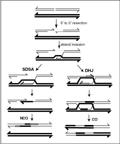
During meiosis in eukaryotes, genetic recombination involves the pairing of homologous chromosomes. This may be followed by information transfer between the chromosomes. The information transfer may occur without physical exchange (a section of genetic material is copied from one chromosome to another, without the donating chromosome being changed) (see SDSA – Synthesis Dependent Strand Annealing pathway in Figure); or by the breaking and rejoining of DNA strands, which forms new molecules of DNA (see DHJ pathway in Figure).

Recombination may also occur during mitosis in eukaryotes where it ordinarily involves the two sister chromatids formed after chromosomal replication. In this case, new combinations of alleles are not produced since the sister chromatids are usually identical. In meiosis and mitosis, recombination occurs between similar molecules of DNA (homologous sequences). In meiosis, non-sister homologous chromosomes pair with each other so that recombination characteristically occurs between non-sister homologues. In both meiotic and mitotic cells, recombination between homologous chromosomes is a common mechanism used in DNA repair.

Gene conversion – the process during which homologous sequences are made identical also falls under genetic recombination.

Genetic recombination and recombinational DNA repair also occurs in bacteria and archaea, which use asexual reproduction.

Recombination can be artificially induced in laboratory (in vitro) settings, producing recombinant DNA for purposes including vaccine development.

V(D)J recombination in organisms with an adaptive immune system is a type of site-specific genetic recombination that helps immune cells rapidly diversify to recognize and adapt to new pathogens.

Synapsis

[edit]

During meiosis, synapsis (the pairing of homologous chromosomes) ordinarily precedes genetic recombination.

Mechanism

[edit]

Genetic recombination is catalyzed by many different enzymes. Recombinases are key enzymes that catalyse the strand transfer step during recombination. RecA, the chief recombinase found in Escherichia coli, is responsible for the repair of DNA double strand breaks (DSBs). In yeast and other eukaryotic organisms there are two recombinases required for repairing DSBs. The RAD51 protein is required for mitotic and meiotic recombination, whereas the DNA repair protein, DMC1, is specific to meiotic recombination. In the archaea, the ortholog of the bacterial RecA protein is RadA.

Bacterial recombination

[edit]

Bacteria regularly undergo genetic recombination in three main ways:

  • Transformation, the uptake of exogenous DNA from the surrounding environment.
  • Transduction, the virus-mediated transfer of DNA between bacteria.
  • Conjugation, the transfer of DNA from one bacterium to another via cell-to-cell contact.[2][3][4][5]

Sometimes a strand of DNA is transferred into the target cell but fails to be copied as the target divides. This is called an abortive transfer.

Chromosomal crossover

[edit]
Thomas Hunt Morgan's illustration of crossing over (1916)

In eukaryotes, recombination during meiosis is facilitated by chromosomal crossover. The crossover process leads to offspring having different combinations of genes from those of their parents, and can occasionally produce new chimeric alleles.[citation needed] The shuffling of genes brought about by genetic recombination produces increased genetic variation. It also allows sexually reproducing organisms to avoid Muller's ratchet, in which the genomes of an asexual population tend to accumulate more deleterious mutations over time than beneficial or reversing mutations.[citation needed]

Chromosomal crossover involves recombination between the paired chromosomes inherited from each of one's parents, generally occurring during meiosis.[citation needed] During prophase I (pachytene stage) the four available chromatids are in tight formation with one another.[citation needed] While in this formation, homologous sites on two chromatids can closely pair with one another, and may exchange genetic information.[6]

Because there is a small probability of recombination at any location along a chromosome, the frequency of recombination between two locations depends on the distance separating them.[citation needed] Therefore, for genes sufficiently distant on the same chromosome, the amount of crossover is high enough to destroy the correlation between alleles.[citation needed]

Tracking the movement of genes resulting from crossovers has proven quite useful to geneticists. Because two genes that are close together are less likely to become separated than genes that are farther apart, geneticists can deduce roughly how far apart two genes are on a chromosome if they know the frequency of the crossovers.[citation needed] Geneticists can also use this method to infer the presence of certain genes. Genes that typically stay together during recombination are said to be linked. One gene in a linked pair can sometimes be used as a marker to deduce the presence of the other gene. This is typically used to detect the presence of a disease-causing gene.[7]

The recombination frequency between two loci observed is the crossing-over value. It is the frequency of crossing over between two linked gene loci (markers), and depends on the distance between the genetic loci observed. For any fixed set of genetic and environmental conditions, recombination in a particular region of a linkage structure (chromosome) tends to be constant, and the same is then true for the crossing-over value which is used in the production of genetic maps.[5][8]

Gene conversion

[edit]

In gene conversion, a section of genetic material is copied from one chromosome to another, without the donating chromosome being changed. Gene conversion occurs at high frequency at the actual site of the recombination event during meiosis. It is a process by which a DNA sequence is copied from one DNA helix (which remains unchanged) to another DNA helix, whose sequence is altered. Gene conversion has often been studied in fungal crosses[9] where the 4 products of individual meioses can be conveniently observed. Gene conversion events can be distinguished as deviations in an individual meiosis from the normal 2:2 segregation pattern (e.g. a 3:1 pattern).

Nonhomologous recombination

[edit]

Recombination can occur between DNA sequences that contain no sequence homology. This can cause chromosomal translocations, sometimes leading to cancer.

In B cells

[edit]

B cells of the immune system perform genetic recombination, called immunoglobulin class switching. It is a biological mechanism that changes an antibody from one class to another, for example, from an isotype called IgM to an isotype called IgG.

Genetic engineering

[edit]

In genetic engineering, recombination can also refer to artificial and deliberate recombination of disparate pieces of DNA, often from different organisms, creating what is called recombinant DNA. A prime example of such a use of genetic recombination is gene targeting, which can be used to add, delete or otherwise change an organism's genes. This technique is important to biomedical researchers as it allows them to study the effects of specific genes. Techniques based on genetic recombination are also applied in protein engineering to develop new proteins of biological interest.

Examples include Restriction enzyme mediated integration, Gibson assembly and Golden Gate Cloning.

Recombinational repair

[edit]

DNA damages caused by a variety of exogenous agents (e.g. UV light, X-rays, chemical cross-linking agents) can be repaired by homologous recombinational repair (HRR).[10][11] These findings suggest that DNA damages arising from natural processes, such as exposure to reactive oxygen species that are byproducts of normal metabolism, are also repaired by HRR. In humans, deficiencies in the gene products necessary for HRR during meiosis likely cause infertility[12] In humans, deficiencies in gene products necessary for HRR, such as BRCA1 and BRCA2, increase the risk of cancer (see DNA repair-deficiency disorder).

In bacteria, transformation is a process of gene transfer that ordinarily occurs between individual cells of the same bacterial species. Transformation involves integration of donor DNA into the recipient chromosome by recombination. This process appears to be an adaptation for repairing DNA damages in the recipient chromosome by HRR.[13] Transformation may provide a benefit to pathogenic bacteria by allowing repair of DNA damage, particularly damages that occur in the inflammatory, oxidizing environment associated with infection of a host.

When two or more viruses, each containing lethal genomic damages, infect the same host cell, the virus genomes can often pair with each other and undergo HRR to produce viable progeny. This process, referred to as multiplicity reactivation, has been studied in lambda and T4 bacteriophages,[14] as well as in several pathogenic viruses. In the case of pathogenic viruses, multiplicity reactivation may be an adaptive benefit to the virus since it allows the repair of DNA damages caused by exposure to the oxidizing environment produced during host infection.[13] See also reassortment.

Meiotic recombination

[edit]

A molecular model for the mechanism of meiotic recombination presented by Anderson and Sekelsky[15] is outlined in the first figure in this article. Two of the four chromatids present early in meiosis (prophase I) are paired with each other and able to interact. Recombination, in this model, is initiated by a double-strand break (or gap) shown in the DNA molecule (chromatid) at the top of the figure. Other types of DNA damage may also initiate recombination. For instance, an inter-strand cross-link (caused by exposure to a cross-linking agent such as mitomycin C) can be repaired by HRR.

Two types of recombinant product are produced. Indicated on the right side is a "crossover" (CO) type, where the flanking regions of the chromosomes are exchanged, and on the left side, a "non-crossover" (NCO) type where the flanking regions are not exchanged. The CO type of recombination involves the intermediate formation of two "Holliday junctions" indicated in the lower right of the figure by two X-shaped structures in each of which there is an exchange of single strands between the two participating chromatids. This pathway is labeled in the figure as the DHJ (double-Holliday junction) pathway.

The NCO recombinants (illustrated on the left in the figure) are produced by a process referred to as "synthesis dependent strand annealing" (SDSA). Recombination events of the NCO/SDSA type appear to be more common than the CO/DHJ type.[16] The NCO/SDSA pathway contributes little to genetic variation, since the arms of the chromosomes flanking the recombination event remain in the parental configuration. Thus, explanations for the adaptive function of meiosis that focus exclusively on crossing-over are inadequate to explain the majority of recombination events.

Achiasmy and heterochiasmy

[edit]

Achiasmy is the phenomenon where autosomal recombination is completely absent in one sex of a species. Achiasmatic chromosomal segregation is well documented in male Drosophila melanogaster. The "Haldane-Huxley rule" states that achiasmy usually occurs in the heterogametic sex.[17]

Heterochiasmy occurs when recombination rates differ between the sexes of a species.[17] In humans, each oocyte has on average 41.6 ± 11.3 recombinations, 1.63-fold higher than sperms. This sexual dimorphic pattern in recombination rate has been observed in many species. In mammals, females most often have higher rates of recombination.[18]

RNA virus recombination

[edit]

Numerous RNA viruses are capable of genetic recombination when at least two viral genomes are present in the same host cell.[19][20] Recombination is largely responsible for RNA virus diversity and immune evasion.[21] RNA recombination appears to be a major driving force in determining genome architecture and the course of viral evolution among picornaviridae ((+)ssRNA) (e.g. poliovirus).[22] In the retroviridae ((+)ssRNA)(e.g. HIV), damage in the RNA genome appears to be avoided during reverse transcription by strand switching, a form of recombination.[23][24]

Recombination also occurs in the reoviridae (dsRNA)(e.g. reovirus), orthomyxoviridae ((-)ssRNA)(e.g. influenza virus)[24] and coronaviridae ((+)ssRNA) (e.g. SARS).[25][26]

Recombination in RNA viruses appears to be an adaptation for coping with genome damage.[19] Switching between template strands during genome replication, referred to as copy-choice recombination, was originally proposed to explain the positive correlation of recombination events over short distances in organisms with a DNA genome (see first Figure, SDSA pathway).[27]

Recombination can occur infrequently between animal viruses of the same species but of divergent lineages. The resulting recombinant viruses may sometimes cause an outbreak of infection in humans.[25]

Especially in coronaviruses, recombination may also occur even among distantly related evolutionary groups (subgenera), due to their characteristic transcription mechanism, that involves subgenomic mRNAs that are formed by template switching.[28][26]

When replicating its (+)ssRNA genome, the poliovirus RNA-dependent RNA polymerase (RdRp) is able to carry out recombination. Recombination appears to occur by a copy choice mechanism in which the RdRp switches (+)ssRNA templates during negative strand synthesis.[29] Recombination by RdRp strand switching also occurs in the (+)ssRNA plant carmoviruses and tombusviruses.[30]

Recombination appears to be a major driving force in determining genetic variability within coronaviruses, as well as the ability of coronavirus species to jump from one host to another and, infrequently, for the emergence of novel species, although the mechanism of recombination in is unclear.[25]

In early 2020, many genomic sequences of Australian SARS‐CoV‐2 isolates have deletions or mutations (29742G>A or 29742G>U; "G19A" or "G19U") in the s2m, suggesting that RNA recombination may have occurred in this RNA element. 29742G("G19"), 29744G("G21"), and 29751G("G28") were predicted as recombination hotspots.[31] During the first months of the COVID-19 pandemic, such a recombination event was suggested to have been a critical step in the evolution of SARS-CoV-2's ability to infect humans.[32] Linkage disequilibrium analysis confirmed that RNA recombination with the 11083G > T mutation also contributed to the increase of mutations among the viral progeny. The findings indicate that the 11083G > T mutation of SARS-CoV-2 spread during Diamond Princess shipboard quarantine and arose through de novo RNA recombination under positive selection pressure. In three patients on the Diamond Princess cruise, two mutations, 29736G > T and 29751G > T (G13 and G28) were located in Coronavirus 3′ stem-loop II-like motif (s2m) of SARS-CoV-2. Although s2m is considered an RNA motif highly conserved in 3' untranslated region among many coronavirus species, this result also suggests that s2m of SARS-CoV-2 is RNA recombination/mutation hotspot.[33]

Schematic representation of the s2m RNA secondary structure, with tertiary structural interactions indicated as long range contacts.

SARS-CoV-2's entire receptor binding motif appeared, based on preliminary observations, to have been introduced through recombination from coronaviruses of pangolins.[34] However, more comprehensive analyses later refuted this suggestion and showed that SARS-CoV-2 likely evolved solely within bats and with little or no recombination.[35][36]

Role of recombination in the origin of life

[edit]

Nowak and Ohtsuki[37] noted that the origin of life (abiogenesis) is also the origin of biological evolution. They pointed out that all known life on earth is based on biopolymers and proposed that any theory for the origin of life must involve biological polymers that act as information carriers and catalysts. Lehman[38] argued that recombination was an evolutionary development as ancient as the origins of life. Smail et al.[39] proposed that in the primordial Earth, recombination played a key role in the expansion of the initially short informational polymers (presumed to be RNA) that were the precursors to life.

See also

[edit]

References

[edit]
[edit]
Revisions and contributorsEdit on WikipediaRead on Wikipedia
from Grokipedia
Genetic recombination is the process by which DNA strands are broken and repaired, producing new combinations of alleles that generate genetic diversity. This fundamental biological mechanism occurs in nearly all organisms, from bacteria to humans, and is essential for sexual reproduction, DNA repair, and evolutionary adaptation. Recombination can be classified into three main types based on the sequence similarity and specificity of the interacting DNA regions: homologous recombination, site-specific recombination, and nonhomologous recombination. Homologous recombination, also known as general recombination, involves the exchange of genetic material between two similar or identical DNA sequences, typically on homologous chromosomes during meiosis or between sister chromatids. It is initiated by double-strand breaks in DNA, which are processed to expose single-stranded ends that then invade a homologous template, forming structures like the Holliday junction that resolve into recombinant molecules. Key proteins such as RecA in bacteria (and its eukaryotic homolog Rad51) facilitate strand invasion and pairing. This type is highly accurate and plays a critical role in repairing DNA damage and ensuring proper chromosome segregation. Site-specific recombination occurs at precise short DNA sequences without requiring extensive homology, enabling the integration, excision, or inversion of mobile genetic elements like transposons and viruses. Mechanisms include transpositional recombination, where elements are cut and pasted or replicated into new sites via enzymes like transposases, and conservative recombination, which reversibly rearranges DNA at specific recognition sites. Examples include the integration of bacteriophage lambda into bacterial genomes or the movement of retrotransposons, which constitute over 45% of the human genome and contribute to genetic variation and disease. Nonhomologous recombination, often exemplified by nonhomologous end joining (NHEJ), joins DNA ends without requiring sequence homology and is a primary pathway for repairing double-strand breaks in mammalian cells. NHEJ is error-prone, as it can result in insertions, deletions, or mutations during the ligation process mediated by proteins like Ku and DNA-PK, but it operates efficiently throughout the cell cycle. This mechanism is vital for immune system development, such as in V(D)J recombination for antibody diversity, though it can also lead to genomic instability if misregulated. Overall, genetic recombination drives evolution by shuffling alleles, maintains genome stability through repair, and underlies biotechnological applications like gene targeting, with rates varying from about 1 in 10^5 cell generations in bacteria to hotspots in eukaryotic meiosis. Dysregulation is linked to diseases including cancer and genetic disorders.

Overview

Definition

Genetic recombination is the process by which a strand of genetic material, typically DNA but also RNA in certain contexts such as viral genomes, is broken and then joined to a different strand, resulting in new combinations of alleles. This exchange occurs between homologous chromosomes or within the same chromosome, producing offspring with genetic variations not present in either parent. The phenomenon was first observed in by and Edward L. Tatum in 1946, through experiments demonstrating the transfer and recombination of genetic markers in . Their work revealed that could exchange genetic information, challenging the prevailing view that such processes were limited to sexually reproducing eukaryotes. A key molecular outcome of genetic recombination is the production of molecules with novel genetic combinations, which enhances across generations. Unlike mutations, which involve changes to the sequence of DNA due to errors in replication or environmental factors, recombination shuffles existing alleles without altering the underlying . In eukaryotes, genetic recombination is particularly important during , where it facilitates the exchange of genetic material between homologous chromosomes to promote diversity in gametes.

Biological significance

Genetic recombination plays a crucial role in generating by shuffling alleles during , which produces gametes with novel combinations of maternal and paternal genetic material, thereby enhancing variability in offspring populations. This random recombination and segregation of chromosomal segments causes each child to inherit varying proportions of parental DNA, resulting in differences in estimated genetic ancestry percentages compared to the parental average. This process is essential for , as it allows populations to respond to environmental pressures through the emergence of beneficial trait combinations, and for , by facilitating the divergence of genetic lineages over time. In evolutionary terms, recombination contributes to adaptive by breaking down , the non-random association of alleles at different loci, which enables the independent assortment and spread of advantageous mutations across the . Without recombination, linked deleterious alleles could hitchhike with beneficial ones, hindering the efficiency of and slowing evolutionary progress. At the cellular level, recombination maintains stability by resolving replication-associated DNA lesions and supports mechanisms, particularly through the accurate restoration of double-strand breaks using homologous sequences. This function is vital for preventing chromosomal aberrations and ensuring faithful transmission of genetic information during . Recombination rates vary across organisms and sexes, influencing patterns of ; for instance, in humans, meiotic recombination typically involves an average of 1 to 2 crossovers per pair, contributing to an overall genetic map length of approximately 42 morgans in females and 26 morgans in males (as of 2025). These rates underscore recombination's quantitative impact on the distribution of and its role in shaping heritable diversity.

Fundamental Mechanisms

Synapsis

Synapsis refers to the precise pairing and alignment of homologous chromosomes during I of , enabling their stable association along their entire length. This process is crucial for ensuring accurate segregation of chromosomes in gametes and is mediated by meiosis-specific protein complexes that facilitate chromosome recognition and stabilization. In most eukaryotes, synapsis begins after the formation of axial elements along individual chromosomes in leptotene and progresses through sequential stages to achieve full alignment. The hallmark of synapsis is the assembly of the (SC), a highly conserved, proteinaceous structure that forms a zipper-like scaffold between paired homologs. The SC exhibits a tripartite organization, consisting of two lateral elements flanking each axis, a central element bridging the homologs, and transverse filaments connecting the lateral elements across the synaptic space, which measures approximately 100 nm in depth. Lateral elements are primarily composed of fibrous proteins that condense and linearize , while transverse filaments tether the axes together, and the central element provides structural integrity. This complex not only stabilizes the physical pairing but also creates a specialized environment that supports subsequent meiotic events. Formation of the SC is initiated at multiple sites along the chromosomes and extends progressively to ensure complete . Key molecular players in mammals include the proteins SYCP1, SYCP2, and SYCP3, which are essential for SC assembly and stability. SYCP1 forms the transverse filaments as a that self-assembles into a lattice, directly linking the lateral elements of homologous and promoting their alignment. SYCP2 and SYCP3 constitute the lateral elements; SYCP3 organizes into paracrystalline fibers that compact the chromosome axes, while SYCP2 interacts with SYCP3 and other components to facilitate axial elongation and pairing initiation. Mutations in these proteins disrupt SC formation, leading to meiotic arrest, as demonstrated in studies in mice. Synapsis unfolds in distinct stages during I. It initiates in zygotene, where double-strand breaks and early pairing signals promote localized SC assembly between homologs, starting from ends or specific interstitial sites. By pachytene, synapsis is complete, with the SC fully extended along the paired chromosomes, stabilizing their alignment and compacting the bivalent structure. In diplotene, the SC begins to disassemble from the ends inward, allowing homologs to separate while chiasmata—points of physical linkage—persist to hold them together until anaphase I. This dynamic process ensures homologous chromosomes are correctly paired as a prerequisite for recombination.

Homologous recombination

Homologous recombination (HR) is a fundamental DNA transaction that enables the exchange of genetic information between homologous DNA molecules, primarily to repair double-strand breaks (DSBs) and promote genetic diversity. This process requires extensive sequence homology between the damaged DNA and its template, distinguishing it from other recombination pathways. HR proceeds through a series of biochemical steps orchestrated by conserved proteins, ensuring accurate repair while minimizing errors such as loss of heterozygosity. The initiation of HR typically begins with the formation of a DSB, which can be induced by specific endonucleases. In meiotic contexts, the topoisomerase-like enzyme SPO11 generates these breaks by forming covalent 5'-phosphotyrosyl intermediates with DNA, requiring dimerization and metal ions like Mg²⁺ for catalysis; this step is essential for subsequent homologous pairing and exchange. In somatic cells or under DNA damage conditions, DSBs arise from other nucleases or exogenous agents, triggering HR as a high-fidelity repair mechanism. Once formed, DSB ends are processed through resection to create single-stranded DNA (ssDNA) overhangs primed for homology search. The MRN complex (comprising Mre11, Rad50, and Nbs1) initiates short-range resection, followed by long-range extension by Exo1 and other exonucleases, yielding 3' ssDNA tails coated with RPA to prevent secondary structures. These tails are then remodeled by the recombinase Rad51 (RecA in prokaryotes), which displaces RPA and assembles an ATP-dependent nucleoprotein filament capable of probing for homologous sequences. Mediators such as BRCA2 facilitate Rad51 loading, enhancing filament stability and search efficiency through an accelerated random sampling mechanism. Strand invasion follows, where the Rad51-ssDNA filament invades a homologous duplex, forming a displacement loop () and initiating base pairing. This can lead to second-end capture in the double Holliday junction (dHJ) model, forming two crossed-strand structures after DNA synthesis fills gaps using the donor template as a primer. In contrast, the single Holliday junction model posits only one junction intermediate, resolved without second-end capture, as evidenced in certain meiotic systems where single junctions predominate over doubles. An alternative non-crossover pathway, synthesis-dependent strand annealing (SDSA), involves extension of the invading 3' end by using the homologous template, followed by dissociation of the D-loop and annealing of the extended strand to the other DSB end, resulting in gene conversion without crossover or HJ formation. SDSA is prevalent in mitotic cells and contributes to error-free DSB repair. Branch migration, driven by helicases like Rad54, then extends the heteroduplex region in HJ-based pathways, allowing misalignment correction and further synthesis. The outcome of HR—crossover or non-crossover—is determined during resolution in HJ-dependent models. Structure-specific endonucleases such as GEN1 and the MUS81-EME1 complex cleave junctions, with symmetric cuts yielding crossovers (reciprocal exchange) and asymmetric cuts producing non-crossover gene conversion. Dissolution pathways, involving BLM and TOP3A, can also process dHJs without cleavage, favoring non-crossover products to suppress genomic rearrangements. Key proteins like and Rad51 drive strand exchange, with their filaments enabling homology recognition over thousands of base pairs. Recombination frequency, a measure of exchange events, is quantified using the RF=(number of recombinantstotal progeny)×100%RF = \left( \frac{\text{number of recombinants}}{\text{total progeny}} \right) \times 100\%, where values approximate genetic distances in centimorgans. This process underpins DSB repair in diverse organisms, briefly intersecting with tolerance mechanisms for replication stress.

Prokaryotic Recombination

Bacterial recombination

Bacterial recombination refers to the processes by which prokaryotes, particularly bacteria, exchange and integrate genetic material, primarily through homologous recombination mechanisms that facilitate DNA repair and genetic diversity. Unlike eukaryotic systems, bacterial recombination often occurs as part of horizontal gene transfer (HGT) and is mediated by three primary natural pathways: transformation, transduction, and conjugation. These pathways enable the uptake, transfer, and incorporation of exogenous DNA into the bacterial genome, often involving RecA protein for strand invasion and homologous sequence alignment. In transformation, competent bacteria actively uptake naked DNA from the environment, a process induced by environmental stresses such as nutrient limitation or DNA damage. The exogenous double-stranded DNA is transported across the cell envelope via specialized machinery, including type IV pili or pseudopili in species like Streptococcus pneumoniae and Bacillus subtilis, and is then converted to single-stranded form for integration. This single-stranded DNA invades the recipient chromosome through RecA-mediated strand exchange, where RecA forms a nucleoprotein filament that searches for and pairs with homologous sequences, leading to recombination via branch migration and resolution. Transduction involves phage-mediated transfer of bacterial DNA, where bacteriophages accidentally package host DNA during lytic cycles and inject it into a new recipient cell. Generalized transduction, as seen with phages like P1 in , transfers random chromosomal fragments, while specialized transduction, exemplified by , transfers specific genes adjacent to the integration site. Upon entry, the transduced DNA undergoes RecA-dependent , with strand invasion facilitating integration into the recipient genome through sequence homology. This process can transfer large chromosomal segments, exceeding the efficiency of other HGT mechanisms in some lineages. Conjugation enables direct cell-to-cell DNA transfer via a pilus structure, primarily involving conjugative plasmids like the F plasmid in E. coli. The donor cell's relaxosome nicks the plasmid DNA, and a single strand is transferred through a type IV secretion system to the recipient, where it circularizes and can integrate via homologous recombination. RecA again plays a central role in strand invasion, promoting pairing with homologous chromosomal regions for stable incorporation. This pathway is highly efficient for disseminating large genetic elements, such as resistance plasmids. A key molecular detail shared across these pathways is the RecA-mediated strand invasion, where RecA polymerizes on single-stranded DNA to form a helical filament that facilitates ATP-dependent homology search and D-loop formation with the recipient duplex. Integration requires sufficient homologous sequences, typically 20-50 base pairs, to ensure stable exchange without disrupting essential genes. In E. coli, double-strand break (DSB) processing prior to recombination often involves the RecBCD pathway: the RecBCD helicase-nuclease complex binds DSB ends, unwinds and degrades DNA asymmetrically until encountering a Chi (crossover hotspot instigator) site, which attenuates nuclease activity and loads RecA onto the resulting 3' single-stranded tail for invasion. This pathway is crucial for initiating recombination in response to DSBs from various sources. Bacterial recombination outcomes include efficient DNA repair, such as recombinational repair of UV-induced lesions like cyclobutane , where HGT pathways provide homologous templates to bypass damage during replication. Additionally, it drives the acquisition of adaptive traits, notably resistance genes; for instance, conjugation-mediated transfer of plasmids carrying genes has been pivotal in the spread of multidrug resistance in pathogens like . These processes enhance bacterial survival and evolution under selective pressures.

Horizontal gene transfer

(HGT) in prokaryotes relies on to integrate exogenous DNA into the recipient , allowing for the stable incorporation of novel genetic material that promotes rapid to environmental pressures such as antibiotics or new hosts. During HGT processes like transformation, the incoming single-stranded DNA is protected by proteins such as DprA and loaded onto filaments, facilitating strand invasion and replacement of homologous chromosomal regions, which can introduce beneficial alleles or excise deleterious mobile elements. This recombination-driven integration enhances and fitness, as evidenced in naturally competent species like Streptococcus pneumoniae, where it purges parasitic prophages and supports the spread of adaptive traits. In bacterial conjugation, mediated by the fertility (F) plasmid in Escherichia coli, DNA transfer occurs through direct cell-to-cell contact via a type IV secretion system, where the relaxase enzyme nicks the plasmid at its origin of transfer (oriT) and directs single-stranded DNA into the recipient. Integration of the F plasmid into the donor chromosome creates high-frequency recombination (Hfr) strains, enabling the transfer of large chromosomal segments alongside plasmid DNA, which then recombines into the recipient's genome via homologous sequences. Similarly, transduction involves bacteriophages as vectors: in generalized transduction, phages like P22 in Salmonella package random bacterial DNA fragments during lytic cycles and inject them into new hosts for recombination; specialized transduction, as seen with lambda phage in E. coli, transfers specific genes adjacent to the prophage integration site through aberrant excision, forming hybrid phage-bacterial DNA that integrates via site-specific or homologous recombination. These mechanisms, building on core bacterial recombination pathways, facilitate the exchange of plasmids or chromosomal loci. The evolutionary implications of recombination-mediated HGT are profound, particularly in the dissemination of virulence factors among pathogens; for instance, in Salmonella enterica, horizontally acquired pathogenicity islands and plasmids encoding type III secretion systems have driven the emergence of new serovars capable of broader host colonization. Such transfers enable pathogens to rapidly acquire traits like toxin production or antibiotic resistance, accelerating adaptation and contributing to outbreaks. HGT frequencies vary by mechanism and environment, typically ranging from 10^{-6} to 10^{-9} per cell per generation for natural transformation and transduction, though conjugation can reach higher rates up to 10^{-3} under optimal conditions in dense populations. However, barriers such as restriction-modification (R-M) systems limit unchecked integration by cleaving unmethylated foreign DNA at specific motifs, with Type II R-M systems particularly shaping plasmid evolution by favoring sequences that evade recognition and reducing interspecies transfer efficiency across bacterial taxa.

Eukaryotic Recombination

Meiotic recombination

Meiotic recombination occurs during prophase I of meiosis, a specialized cell division that produces haploid gametes, where it facilitates the physical linking of homologous chromosomes to ensure their accurate segregation during the first meiotic division. This process begins with the formation of double-strand breaks (DSBs) in DNA, primarily catalyzed by the topoisomerase-like protein SPO11 in complex with accessory factors, which targets hotspots across the genome to initiate strand invasion and repair via homologous recombination pathways.81876-0) These DSBs are essential for establishing chiasmata, the visible manifestations of crossovers that hold homologs together until anaphase I. Crossover interference, a regulatory mechanism, ensures that typically 1-3 crossovers occur per bivalent (pair of homologous ), preventing clustering and promoting even distribution along chromosome arms to maximize segregation fidelity.00616-X) This interference is mediated by proteins that modulate the recombination machinery, including the MutLγ complex (comprising MLH1 and MLH3), which designates and resolves double Holliday junctions into crossovers at designated sites, often visualized as MLH1 foci on synaptonemal complexes. exhibits sex-specific differences, with females generally showing higher overall recombination rates than males in mammals, influenced by factors such as organization and hormonal environments that alter hotspot usage and interference strength. The outcomes of meiotic recombination are critical for through allele shuffling and for preventing ; without sufficient crossovers, homologous chromosomes may fail to segregate properly, leading to gametes with abnormal numbers and increased risk of disorders like in offspring.31000-0) In model organisms, studies in budding yeast () have elucidated the molecular steps, including SPO11-dependent DSB formation and interference via proteins like Zip1, providing a foundation for understanding conserved mechanisms. In mice, electron microscopy and have visualized recombination nodules—early nodules associated with DSB processing and late nodules marking crossover sites—revealing two levels of interference that ensure obligatory crossovers per bivalent. These models highlight how meiotic recombination adapts for , distinct from somatic contexts.

Mitotic recombination

Mitotic recombination is a process of genetic exchange between homologous chromosomes that occurs during in somatic cells, distinct from the more frequent meiotic recombination in germ cells. It is a rare event, occurring at frequencies of approximately 10610^{-6} to 10510^{-5} per in model like and , and is primarily triggered by double-strand breaks (DSBs) arising from sources such as , ultraviolet light, or replication fork stalling during . These DSBs activate machinery to restore genomic integrity, with events favoring repair using the sister but occasionally involving homologous chromosomes. The mechanism of mitotic recombination closely resembles homologous recombination but lacks the extensive synapsis and pairing structures seen in meiosis. It typically begins with DSB resection to generate 3' single-stranded DNA overhangs, followed by strand invasion mediated by Rad51 filaments to form displacement loops or Holliday junctions. Resolution can occur via synthesis-dependent strand annealing (SDSA), producing non-crossover gene conversions, or through double Holliday junction dissolution or cleavage, yielding crossovers. A hallmark outcome is loss of heterozygosity (LOH), where recombination between a heterozygous locus and its homolog results in homozygous regions distal to the breakpoint, often extending to the chromosome end due to segregation patterns. This process is regulated by proteins such as Rad52, Sgs1 helicase, and Mus81 nuclease to suppress excessive crossovers and maintain stability. In biological contexts, mitotic recombination supports DNA repair in proliferating somatic cells, preventing mutations from DSBs and aiding replication restart, though it can contribute to genomic instability if unregulated. It generates somatic variation, creating mosaic tissues that enhance adaptability in plants, such as through sectoral chimeras in Arabidopsis, and in animals, where it drives phenotypic diversity during development. A prominent example is twin spotting in Drosophila melanogaster, where mitotic recombination in heterozygous flies produces adjacent clones of mutant and wild-type tissue—visible as contrasting pigmentation patches—demonstrating how single events propagate into distinct somatic lineages. In mammals, mitotic recombination frequently underlies LOH at tumor suppressor loci, such as RB1 in retinoblastoma, facilitating the second hit in Knudson's two-hit hypothesis and promoting cancer progression.

Types of Recombination

Chromosomal crossover

is the reciprocal exchange of genetic material between non-sister chromatids of homologous chromosomes that occurs during I of . This process involves the breakage and rejoining of DNA strands, resulting in the shuffling of alleles between maternal and paternal chromosomes to generate in gametes. The visible manifestation of this exchange is the formation of chiasmata, which are X-shaped structures that hold the recombined chromatids together. Detection of chromosomal crossovers relies on both genetic and cytological methods. In genetic mapping, linkage analysis measures recombination frequencies between molecular markers or genes; a lower frequency indicates closer linkage and fewer crossovers between loci. Cytological observation during meiotic stages, such as diplotene or I, directly visualizes chiasmata as points of physical exchange between homologs, often using techniques like on pachytene chromosomes. The frequency of chromosomal crossovers varies across the and is concentrated in recombination hotspots, short regions of 1-2 kb where double-strand breaks are preferentially initiated. In mammals, these hotspots are primarily directed by the zinc-finger protein PRDM9, which binds specific motifs and recruits the SPO11 endonuclease to induce breaks, leading to crossover formation. PRDM9 alleles influence hotspot usage, with different variants activating distinct sets of sites, contributing to rapid evolutionary turnover of recombination landscapes. Overall, eukaryotes typically exhibit 1-3 crossovers per chromosome pair to ensure proper segregation. distances between loci, used to quantify crossover frequency, are expressed in centimorgans (cM), where for small intervals, 1 cM ≈ 1% recombination frequency; mapping functions such as Haldane's or Kosambi's adjust for undetected multiple crossovers. A key consequence of chromosomal crossover is the establishment of physical linkages between homologous chromosomes via chiasmata, which persist until anaphase I. These connections promote bipolar attachment to the spindle and ensure balanced segregation of homologs, reducing the risk of aneuploidy in gametes. Without sufficient chiasmata, chromosomes may segregate randomly, leading to genomic instability.

Gene conversion

Gene conversion is a non-reciprocal form of homologous recombination in which genetic information from a donor DNA sequence is unidirectionally transferred to a homologous recipient sequence, resulting in one allele replacing another without reciprocal exchange. This process typically arises during the repair of DNA double-strand breaks (DSBs) when heteroduplex DNA intermediates form between homologous sequences; mismatches in these heteroduplexes are then repaired in a biased manner that favors the donor allele, leading to the conversion of the recipient's sequence. Gene conversion events are associated with approximately half of all homologous recombination (HR) outcomes, particularly as non-crossover products that restore sequence continuity without altering chromosome structure. At the molecular level, the bias in heteroduplex repair is mediated by (MMR) proteins, which recognize and excise mismatched bases to direct synthesis from the donor template. Key enzymes include the MutSα complex (MSH2-MSH6), which detects mismatches, and the MutLα complex (MLH1-PMS2 in mammals or MLH1-PMS1 in ), which coordinates excision and resynthesis to resolve the heteroduplex in favor of the invading donor strand. This directed repair prevents random patch formation and ensures efficient sequence homogenization, often extending over short tracts of 1-2 kilobases (kb). Disruption of these MMR components, such as in MSH2 or MLH1 mutants, reduces conversion efficiency and increases symmetric repair outcomes, highlighting their role in asymmetry. A classic example of gene conversion occurs during mating-type switching in the yeast , where haploid cells change their (from a to α or vice versa) to enable mating. The HO endonuclease induces a DSB at the MAT locus, which is repaired via gene conversion using one of two silent donor cassettes (HMLα or HMRa) as a template, resulting in the replacement of the MAT sequence without altering the donors. This process exemplifies how gene conversion facilitates programmed genetic switches and demonstrates tract lengths typically around 1-2 kb, sufficient to encompass the ~600-base-pair MAT information cassette. Gene conversion tracts are detected through patterns of allele co-conversion, where multiple nearby markers are altered together, revealing the extent and directionality of the event. Tracts can be classified as polar or non-polar: polar tracts exhibit a gradient of conversion frequency, decreasing away from the DSB initiation site due to the directional progression of strand invasion and repair; non-polar tracts show uniform conversion, often from symmetric resection or alternative resolution. This distinction aids in mapping recombination hotspots and understanding initiation biases, as seen in yeast where polar gradients correlate with DSB hotspots. Gene conversion frequently accompanies but can also occur independently of chromosomal crossovers, contributing to sequence diversity without structural rearrangements.

Nonhomologous recombination

Nonhomologous recombination refers to and joining processes that occur without requiring extensive between the participating DNA molecules, distinguishing it from homology-directed mechanisms. This type of recombination is primarily involved in repairing double-strand breaks (DSBs) through error-prone pathways that can lead to genomic alterations. The two main pathways are classical (cNHEJ) and (MMEJ, also known as alternative NHEJ or alt-NHEJ). In cNHEJ, the process begins with the rapid binding of the Ku70/Ku80 heterodimer to the broken DNA ends, which protects them from degradation and recruits the DNA-dependent protein kinase catalytic subunit (DNA-PKcs) to form the DNA-PK holoenzyme. This complex facilitates end tethering and processing, where nucleases like Artemis trim incompatible overhangs, and error-prone polymerases such as pol λ or μ fill gaps, often introducing small insertions or deletions (indels). The final ligation is performed by the DNA ligase IV (Lig4) complex, associated with XRCC4 and XLF, sealing the ends with minimal or no homology required (typically 0 bp). cNHEJ is predominant throughout the cell cycle but operates at higher frequency in the G1 phase, where homologous templates are unavailable.00279-2) MMEJ, in contrast, relies on short stretches of microhomology (generally 5-25 base pairs) at the DSB ends to align and anneal the DNA, followed by resection of non-homologous flaps by exonucleases like MRE11 or EXO1. This pathway is Ku- and Lig4-independent, instead involving poly(ADP-ribose) polymerase 1 (PARP1) for initial end sensing and recruitment of factors like DNA polymerase theta (Polθ) for microhomology-directed synthesis and flap removal. Ligation occurs via Ligase 1 or 3, resulting in larger deletions due to the loss of sequences between microhomologous regions. MMEJ is more active in S and G2 phases and serves as a backup when cNHEJ is impaired. Both pathways are inherently error-prone, frequently producing indels, chromosomal deletions, and translocations that contribute to genomic instability, though cNHEJ tends to cause smaller mutations compared to the more extensive deletions in MMEJ. Unlike homologous recombination, which ensures accurate repair using a sister chromatid template, nonhomologous recombination prioritizes speed over fidelity, making it essential for DSB repair in non-replicative cell phases but a source of mutations in contexts like cancer development.00279-2)

Recombination in DNA Repair

Recombinational repair

Recombinational repair, also known as () repair, serves as a high-fidelity mechanism for repairing double-strand breaks (DSBs) by utilizing a homologous sequence as a template to restore the original genetic information, thereby minimizing errors compared to other pathways. This process is essential for maintaining genomic stability, as unrepaired DSBs can lead to chromosomal aberrations, , or oncogenic transformations. In , the broken ends are processed to generate single-stranded overhangs, which then invade a homologous duplex —typically the sister —to facilitate accurate repair. HR-mediated repair predominantly occurs during the S and G2 phases of the , when the sister chromatid is available as an undamaged template, ensuring the use of identical genetic information for reconstruction. This temporal restriction helps avoid inappropriate recombination with non-allelic sequences that could introduce mutations. In contrast, (NHEJ) serves as an alternative, error-prone pathway active throughout the but less accurate for DSB restoration. A key model for non-crossover HR repair is synthesis-dependent strand annealing (SDSA), in which the resected 3' end of the broken DNA invades the homologous template, extends via to copy the missing sequence, and then anneals back to the other broken end without forming a double , thus preventing crossover products. SDSA is favored in somatic cells to preserve genome integrity by avoiding large-scale rearrangements. The machinery of recombinational repair is highly conserved across species, from bacteria to humans, underscoring its fundamental role in DNA maintenance. In bacteria, the RecA protein initiates strand invasion and filament formation on single-stranded DNA, a process analogous to the eukaryotic RAD51 recombinase. In humans, proteins such as and regulate RAD51 loading and activity, facilitating HR; defects in these genes impair repair and are associated with , a disorder characterized by genomic instability and cancer predisposition. This conservation highlights the evolutionary importance of HR in countering DSB-inducing threats like and replication stress.

Double-strand break repair pathways

Double-strand breaks (DSBs) in DNA represent one of the most severe forms of genomic damage, primarily induced by exogenous factors such as , which directly cleaves both strands of the DNA double helix. Endogenous DSBs also arise frequently from the collapse of replication forks during S-phase, often triggered by encounters with unrepaired single-strand lesions or topological stress.00200-6) These events can lead to chromosomal fragmentation if not promptly repaired, underscoring the cellular need for efficient DSB repair mechanisms. Cells employ distinct DSB repair pathways, with pathway choice tightly regulated by the phase and antagonistic interactions between key proteins. (NHEJ) predominates in , rapidly ligating broken ends with minimal homology but at the risk of small insertions or deletions, whereas (HR) is favored in S and G2 phases to ensure high-fidelity repair using a sister chromatid template.00656-9) This switch is orchestrated by the competition between 53BP1, which shields DSB ends from resection to promote NHEJ, and , which facilitates end resection to enable HR. In BRCA1-deficient cells, unchecked 53BP1 activity biases repair toward error-prone NHEJ, highlighting the balance's role in genomic stability. When both HR and classical NHEJ (c-NHEJ) are compromised, cells resort to alternative end-joining (Alt-EJ) as a mutagenic pathway. Alt-EJ relies on microhomologies (2-15 ) for end ligation, often resulting in larger deletions and chromosomal rearrangements due to its imprecise nature and higher error rate compared to c-NHEJ. This pathway is suppressed in wild-type cells but activated in HR- or NHEJ-deficient contexts, such as during replication stress, contributing to genomic instability. The dysregulation of DSB repair pathways has profound clinical implications, particularly in cancer therapy through . exploit HR deficiencies in BRCA1/2-mutated tumors by trapping PARP on single-strand breaks, leading to replication fork collapse and unrepaired DSBs that cannot be resolved without HR, selectively killing cancer cells while sparing normal ones. This approach, exemplified by drugs like , has revolutionized treatment for BRCA-associated ovarian and breast cancers, with response rates exceeding 40% in HR-deficient patients.

Recombination in the Immune System

V(D)J recombination in B cells

V(D)J recombination in is a site-specific process that assembles the variable regions of immunoglobulin genes, generating a diverse repertoire of B cell receptors (BCRs) essential for adaptive immunity. This recombination occurs during early B cell development in the and involves the ordered joining of variable (V), diversity (D), and joining (J) gene segments flanked by recombination signal sequences (RSSs). The process is initiated by the products and RAG2, which recognize RSSs consisting of conserved heptamer (CACAGTG) and nonamer (ACAAAAACC) motifs separated by 12- or 23-nucleotide spacers, adhering to the 12/23 rule to ensure compatible joining. RAG1/RAG2 form a tetrameric complex that binds RSSs, introduces double-strand breaks, producing hairpin coding ends and blunt signal ends, which are subsequently repaired by (NHEJ) to form coding and signal joints. The recombination proceeds in distinct stages during B cell maturation. In pro-B cells, initial D_H to J_H joining occurs on both immunoglobulin heavy chain (IgH) alleles, followed by V_H to DJ_H rearrangement, which transitions pro-B cells to pre-B cells upon successful expression of a pre-BCR. Light chain recombination, involving V to J joining without D segments, then takes place in pre-B cells, enabling assembly of a complete BCR. This sequential process is confined to the G0/G1 phase of the cell cycle to minimize off-target effects. Errors in V(D)J recombination, such as those caused by mutations in RAG1 or RAG2 (e.g., V779M or C328G), or defects in NHEJ components like Ku or XRCC4, lead to severe combined immunodeficiency (SCID), blocking B cell development and accounting for a significant proportion, approximately 20-25%, of SCID cases. A key outcome of V(D)J recombination is the immense diversity of antibodies, estimated at around 10^{11} unique specificities in humans, arising from combinatorial joining of gene segments—approximately 50 V_H, 27 D_H, and 6 J_H for heavy chains, combined with 40 V_κ and 5 J_κ or 30 V_λ and 4 J_λ for light chains—plus junctional diversity. Junctional diversity is enhanced by exonuclease nibbling at coding ends and the addition of palindromic (P) nucleotides during hairpin opening, but primarily by terminal deoxynucleotidyl transferase (TdT), which adds random non-templated N-nucleotides (typically 2-5, up to 20) to the V-D and D-J junctions, especially in heavy chains during pro-B stages. This TdT-mediated addition, active only during early B cell development, significantly amplifies variability in the complementarity-determining region 3 (CDR3). Regulation of V(D)J recombination includes , which ensures monoallelic expression of a functional BCR to maintain specificity. Successful rearrangement on one allele signals feedback inhibition of /RAG2 expression via pre-BCR signaling, preventing further recombination on the other allele; this is mediated by factors like and histone modifications such as at RSSs, along with RAG2's acidic hinge domain. is strictly enforced at the IgH locus during pro-B stages and extended to light chains in pre-B cells.

Class switch recombination

Class switch recombination (CSR) is a DNA recombination process in activated B cells that enables the production of antibodies with different effector functions by replacing the constant region of the immunoglobulin heavy chain gene while preserving the antigen specificity encoded by the variable region. This process occurs after initial V(D)J recombination has assembled the variable domain, allowing mature B cells to switch from expressing IgM (or IgD) to other isotypes such as IgG, IgA, or IgE. CSR is essential for tailoring humoral immune responses to diverse pathogens and is tightly regulated to prevent genomic instability. The mechanism of CSR begins with the induction of double-strand breaks (DSBs) in repetitive switch (S) regions located upstream of the constant region genes (C_H) by activation-induced cytidine deaminase (AID). AID deaminates cytosines to uracils in the single-stranded DNA exposed during transcription of S regions, leading to mismatch repair or that generates staggered DSBs on both donor (typically Sμ for IgM) and acceptor (e.g., Sγ, Sα, or Sε) regions. These DSBs are then repaired by , primarily the classical pathway involving Ku70/80, , XRCC4, and ligase IV, which joins the upstream VDJ segment to a new downstream C_H gene, resulting in the deletion of intervening DNA as circular extrachromosomal elements. Alternative end-joining pathways can also contribute, particularly in cases of NHEJ deficiency, but classical NHEJ predominates for efficient switching. CSR is triggered in activated B cells within s of secondary lymphoid organs following encounter and T cell help, requiring costimulatory signals such as CD40 ligation and cytokines that direct isotype specificity. For instance, interleukin-4 (IL-4), produced by T follicular helper cells, promotes switching to IgE by inducing transcription of the Sε region through STAT6 activation, while transforming growth factor-β (TGF-β) favors IgA switching. These cytokines enhance chromatin accessibility and AID recruitment to specific S regions, with CSR events occurring infrequently during the germinal center reaction to balance affinity maturation and isotype diversification. Proliferation of B cells, often requiring at least two cell divisions, is also necessary for efficient DSB formation and repair. The primary outcomes of CSR are the production of isotype-switched antibodies with distinct biological roles: IgG subclasses mediate opsonization and complement activation, IgA facilitates mucosal immunity, and IgE drives allergic and anti-parasitic responses. This switching deletes the DNA between the donor and acceptor S regions, irreversibly committing the B cell to the new isotype without altering the variable region assembled during earlier V(D)J recombination. Successful CSR enhances adaptive immunity by providing effector diversity tailored to infection type. Errors in CSR, arising from off-target activity or faulty DSB repair, can lead to chromosomal translocations that juxtapose immunoglobulin loci with proto-oncogenes, contributing to B cell malignancies such as and . For example, translocations like t(8;14) involving c-MYC and IgH often occur at switch regions due to aberrant joining of -induced breaks, with deficiencies in NHEJ components (e.g., 53BP1 or ) increasing translocation frequency by promoting alternative end-joining. Such genomic underscores the double-edged nature of CSR in balancing immune adaptability and cancer risk.

Recombination in Viruses

RNA virus recombination

Genetic recombination in es primarily occurs through a template-switching mechanism during , where the (RdRp) dissociates from one template and reassociates with another, generating chimeric genomes. This copy-choice process is facilitated by the error-prone nature of RdRp, which lacks activity, allowing frequent switches that contribute to . In viruses with segmented genomes, recombination can also manifest as reassortment, involving the exchange of entire genome segments upon co-infection of a host cell. Recombination rates in RNA viruses are notably high, typically ranging from 10410^{-4} to 10310^{-3} per per replication cycle, surpassing rates in many cases and enabling rapid . These elevated frequencies arise from the inherent low fidelity of and the structural features of viral that promote pausing and switching. Factors such as and RNA secondary structures further modulate these rates, with higher incidences observed in viruses like coronaviruses and retroviruses compared to others like picornaviruses. A prominent example is , where reassortment of its eight segments leads to , potentially causing pandemics; the 2009 H1N1 pandemic strain emerged from reassortment among swine, avian, and human influenza viruses, resulting in a novel to which human populations had limited immunity. In human virus (HIV-1), recombination drives within-host by generating diverse quasispecies, aiding immune escape and ; rates can reach up to 20 template switches per replication cycle, shaping intrahost viral populations. Similarly, severe acute respiratory syndrome coronavirus 2 () has produced numerous recombinants post-2020, including Delta-Omicron hybrids like the XD lineage (as of 2022) and more recent subvariant recombinants such as XEC (a KS.1.1 and KP.3.3 hybrid, dominant as of late 2024), which combine variants and potentially alter transmissibility and immune evasion.

DNA virus recombination

Genetic recombination in DNA viruses encompasses both homologous and site-specific mechanisms that facilitate genome replication, integration, and evolution. In herpesviruses, such as herpes simplex virus type 1 (HSV-1), (HR) plays a central role during viral , promoting isomerization and repair of double-strand breaks via the . This process involves host proteins like Rad51 and Rad52, which mediate strand invasion and exchange, with recombination frequencies reaching approximately 0.7% per kilobase pair in wild-type cells. HR in herpesviruses occurs preferentially at recombinogenic junctions between unique and repeated sequences, contributing to and . Site-specific recombination enables precise integration of viral genomes into host DNA, as exemplified by adeno-associated virus (AAV), a single-stranded DNA parvovirus. AAV integrates predominantly at the AAVS1 locus on chromosome 19q13.4, mediated by the viral Rep78/68 proteins that bind to a 34-bp Rep-binding element within AAVS1 and the viral p5 promoter. This integration disrupts the host locus and often involves partial duplication of AAVS1 sequences, achieving efficiencies of 35-40% at high multiplicities of infection. In retroviruses like HIV, which produce a double-stranded DNA intermediate during reverse transcription, recombination occurs via template switching between co-packaged RNA genomes, generating chimeric proviral DNA and facilitating evolutionary leaps. Recombination contributes to in oncogenic DNA viruses, such as human papillomavirus (HPV), where integration into the host genome—often through non-homologous mechanisms—disrupts the E2 repressor, leading to upregulated expression of E6 and E7 oncoproteins that inactivate and Rb tumor suppressors. This event, prevalent in over 80% of HPV-positive cervical cancers, promotes genomic instability and oncogenic progression by forming viral-host fusion transcripts and super-enhancers. In adenoviruses, supports genome replication and can generate diverse progeny, including those with altered packaging signals in the inverted terminal repeats, influencing virion assembly efficiency during infection. Recombination also drives antiviral drug resistance in DNA viruses; for instance, in HSV, intermolecular HR between polymerase mutants produces dual-resistant strains to acyclovir and foscarnet, with fold increases in up to 35-fold. Recent examples include poxviruses, where recombination shaped the 2022 () outbreak, generating at least eight recombinant genomes within the B.1 lineage through variations and single variants, enhancing transmissibility during zoonotic spillover and human-to-human spread.

Applications in Genetic Engineering

Classical recombination techniques

Classical recombination techniques in primarily involve the manipulation of DNA fragments to create recombinant molecules, enabling the study and application of genetic material in controlled settings. These methods emerged in the with the development of restriction enzymes, which act as molecular scissors to cleave DNA at specific recognition sites, allowing precise excision of genes or DNA segments. then joins these fragments, facilitating the construction of hybrid DNA molecules. The foundational demonstration of this approach came from the laboratories of Stanley Cohen and , who in successfully constructed biologically functional bacterial plasmids by ligating restriction endonuclease-generated fragments from different plasmids, marking the birth of technology. This technique revolutionized by permitting the insertion of foreign DNA into host organisms for propagation and expression. Key to these techniques are cloning vectors, which serve as carriers for the inserted DNA. Plasmids, small circular DNA molecules naturally occurring in bacteria, were among the first vectors used due to their ease of manipulation and ability to replicate autonomously in Escherichia coli. For larger DNA inserts, bacterial artificial chromosomes (BACs) were developed in the early 1990s, based on the single-copy F-factor plasmid, enabling stable maintenance of up to 300 kilobase-pair fragments of human DNA in E. coli without rearrangement. Similarly, yeast artificial chromosomes (YACs), introduced in 1987, utilize yeast centromeres, telomeres, and autonomously replicating sequences to clone even larger segments—up to megabase pairs—in Saccharomyces cerevisiae, proving invaluable for genome mapping projects. Applications of classical recombination techniques include and the creation of models. In , recombinant DNA libraries constructed via these methods allowed of genomes by aligning cloned fragments with data derived from recombination frequencies, contributing to early efforts in sequencing large genomes. For functional studies, was harnessed to generate knockout mice, where targeted DNA constructs integrate into embryonic stem cells via sequence homology, disrupting specific . Pioneered by in the 1980s, this approach achieved in mice, enabling the study of function ; for instance, the first targeted disruption of the hypoxanthine phosphoribosyltransferase was reported in 1987. Such models have been essential for understanding and disease mechanisms. Despite their impact, classical recombination techniques suffer from limitations, notably low efficiency and a propensity for random integration. Homologous recombination in mammalian cells occurs at frequencies three to five orders of magnitude lower than non-homologous random integration, often requiring extensive screening of thousands of clones to identify successful targets. Additionally, the reliance on restriction sites can limit flexibility for certain DNA sequences, and large-insert vectors like YACs are prone to instability and chimerism, complicating downstream analyses. These challenges spurred the development of more precise methods in later decades.

CRISPR-Cas and modern advances

The CRISPR-Cas system, particularly the Cas9 variant derived from bacterial adaptive immunity, enables precise genetic recombination by guiding the to a target DNA sequence via a single-guide (sgRNA), where it induces a double-strand break (DSB) at the specified locus. This DSB can be repaired through (HDR), which incorporates a donor template for accurate sequence insertion or modification, or (NHEJ), which often results in small insertions or deletions (indels) that disrupt gene function. The choice between HDR and NHEJ depends on cellular context, with NHEJ predominating in non-dividing cells, limiting precise edits but facilitating applications. Significant advances have refined CRISPR's precision and expanded its recombination capabilities beyond DSB-dependent mechanisms. Base editing, introduced in 2016, fuses a or deaminase to a catalytically inactive Cas9 (dCas9) or nickase (nCas9), enabling single-base conversions (C-to-T or A-to-G) without DSBs, thus avoiding indels and enhancing safety for therapeutic edits. Building on this, , developed in 2019, employs a Cas9 nickase fused to a and a guide RNA (pegRNA) that specifies the edit, allowing versatile insertions, deletions, and all 12 base-to-base transitions directly at the target site with minimal byproducts. In May 2025, the first human trial of was initiated for treating a , marking a milestone in precise applications. Efficiency improvements have also arisen from engineered Cas variants, such as high-fidelity Cas9 (e.g., SpCas9-HF1), which reduce off-target activity by altering DNA-binding dynamics while maintaining on-target cleavage rates up to 90% in human cells. Other variants like xCas9 broaden (PAM) recognition, enabling access to previously intractable genomic regions. In , CRISPR-mediated recombination has achieved clinical breakthroughs, exemplified by Casgevy (exagamglogene autotemcel), approved by the FDA in December 2023 for in patients aged 12 and older with recurrent vaso-occlusive crises. This therapy uses CRISPR-Cas9 to disrupt the BCL11A enhancer in hematopoietic stem cells, reactivating production and alleviating symptoms in 94% of trial participants after one year. Agriculturally, has facilitated non-browning mushrooms by deleting a via NHEJ, exempt from USDA regulation in 2016 due to the absence of foreign DNA, thereby extending and reducing . Despite these advances, challenges persist, particularly off-target effects where unintended DSBs or edits occur at similar sequences, potentially leading to genomic instability; detection methods like GUIDE-seq have identified rates as low as 0.1% with optimized variants, but comprehensive profiling remains essential for clinical translation. As of 2025, efforts to mitigate this include anti-CRISPR proteins that temporally control editing and models predicting off-target sites with over 95% accuracy. Concurrently, Cas12 and Cas13 systems have advanced RNA-level recombination: Cas12a enables multiplexed DNA editing with staggered cuts for efficient HDR templates, while Cas13-based , such as the REPAIR system, allows reversible A-to-I changes in transcripts without genomic alterations, showing up to 40% efficiency for treating RNA-mediated diseases like MECP2 duplication syndrome. These developments underscore CRISPR's evolution toward safer, more versatile recombination tools.

Evolutionary and Pathological Roles

Role in evolution and genetic diversity

Genetic recombination plays a pivotal role in by breaking , enabling alleles on the same to assort more independently than predicted by Mendel's law of independent assortment alone. This process shuffles genetic variants during through random recombination and segregation of chromosomal segments, resulting in offspring whose inherited ancestry proportions deviate from the parental average and generating novel combinations that enhance adaptive potential in populations facing environmental pressures. Without recombination, linked deleterious mutations would hitchhike with beneficial ones, constraining evolutionary flexibility, but crossing over disrupts these associations, promoting the spread of favorable alleles across generations. In asexual populations or non-recombining regions like the , describes the irreversible accumulation of deleterious mutations due to , as the loss of mutation-free lineages cannot be reversed without recombination. Recombination counters this by allowing the combination of mutation-free segments from different individuals, slowing or preventing the ratchet's progression and maintaining genomic integrity over evolutionary time. Similarly, the posits that recombination drives the evolution of sex by generating to evade rapidly coevolving parasites and pathogens; in host-parasite arms races, shuffled genotypes produce rare resistant variants that confer survival advantages, selecting for higher recombination rates. Recombination hotspots, narrow genomic regions with elevated exchange rates, evolve rapidly and vary across species, influencing patterns of . In mammals, the PRDM9 protein directs these hotspots by binding specific DNA motifs and marking for double-strand breaks, leading to high turnover as motifs erode via biased gene conversion. In contrast, birds exhibit generally higher recombination rates—averaging around 2-6 cM/Mb compared to approximately 1-1.6 cM/Mb in humans—and lack PRDM9, resulting in hotspots at default open sites like promoters rather than PRDM9-specified locations. This variation underscores adaptive of recombination landscapes, with rates adjusting to needs for diversity. Quantitatively, recombination rates shape (Ne) estimates via (LD) decay, where the expected LD (r²) between loci separated by recombination fraction r approximates r211+4Nerr^2 \approx \frac{1}{1 + 4 N_e r}, allowing of Ne as Ne1/r214rN_e \approx \frac{1/r^2 - 1}{4 r} for low LD. Higher recombination reduces LD persistence, inflating apparent Ne and facilitating larger effective gene pools that buffer against drift. Evolutionary models suggest recombination rates themselves evolve under selection, increasing in response to mutational load or biotic interactions to optimize diversity without excessive disruption.

Recombination in cancer and disease

Aberrant homologous recombination (HR) resulting from defects in key genes such as BRCA1 and BRCA2 compromises DNA repair fidelity, leading to genomic instability that drives the development of hereditary breast and ovarian cancers. Germline mutations in BRCA1 confer a lifetime breast cancer risk of up to 72% and ovarian cancer risk of 44%, while BRCA2 mutations increase these risks to 69% and 17%, respectively, primarily through impaired HR-mediated repair of double-strand breaks. These defects result in characteristic genomic scars, including large-scale copy number variations and structural rearrangements, which accumulate over time and promote tumorigenesis. Errors in (NHEJ), another double-strand break repair pathway, contribute to leukemogenesis by generating imprecise ligations that introduce insertions, deletions, and chromosomal aberrations. In myeloid leukemias, elevated error-prone NHEJ activity is associated with DNA damage at sites bound by NHEJ core proteins like Ku70/86, fostering genomic instability and clonal expansion of leukemic cells. Alternative NHEJ pathways further exacerbate this by promoting microhomology-mediated deletions, which are prevalent in hematologic malignancies and correlate with poor prognosis. Pathological outcomes of these recombination errors include chromosomal translocations that activate oncogenes, such as the t(9;22) in chronic myeloid leukemia (CML), where erroneous recombination fuses the BCR and ABL1 genes to produce a constitutively active . This fusion occurs preferentially at Alu repeat sequences flanking the breakpoints, highlighting the role of repetitive elements in recombination-mediated oncogenesis. Additionally, (LOH) via inactivates tumor suppressor alleles, accelerating tumor progression; for instance, LOH events spanning megabases often encompass multiple loci and are suppressed during active HR but prevalent in HR-deficient states. In ovarian cancers, copy-neutral LOH is more frequent in advanced stages, correlating with increased genomic instability and metastatic potential. Therapeutic strategies targeting recombination defects have revolutionized cancer treatment, particularly through poly(ADP-ribose) polymerase (PARP) inhibitors that exploit synthetic lethality in HR-deficient cells. Olaparib, the first PARP inhibitor, received FDA approval in December 2014 for maintenance therapy in germline BRCA-mutated advanced ovarian cancer following platinum-based chemotherapy, demonstrating a 65% reduction in disease progression risk. Subsequent approvals expanded to breast, prostate, and pancreatic cancers with BRCA alterations, with PARP trapping on DNA enhancing lethality in HRD tumors. As of 2025, pan-cancer genomic analyses leveraging (TCGA) data have identified distinct HR deficiency (HRD) signatures, including (LOH), telomeric allelic imbalance (TAI), and large-scale transitions (LST), enabling refined subtyping across tumor types. In endometrial cancer cohorts integrated with TCGA, HRD-high subtypes exhibit elevated genomic instability (e.g., score correlation r=0.83) and poorer overall survival (median 30 months), improving prognostic models (c-index 0.903 when combined with TCGA classifications) and guiding precision therapies like PARP inhibition. These signatures also reveal HRD prevalence in 10-20% of non-BRCA tumors, broadening therapeutic opportunities. Regarding immunotherapy, HRD from recombination defects generally enhances tumor via elevated mutational burden and neoantigen load, predicting better responses to inhibitors; however, acquired genomic alterations, including those from adaptive recombination under selective pressure, contribute to resistance by remodeling the tumor immune microenvironment and reducing T-cell infiltration. In metastatic cancers, post-treatment biopsies show recombination-driven changes, such as copy number losses in immune-related genes, correlating with immunotherapy failure in up to 40% of resistant cases. Strategies to overcome this include combining with checkpoint blockade to sustain HRD-induced .

Role in the origin of life

In the hypothesis, genetic recombination is proposed to have played a crucial role in the early stages of by enabling the assembly and diversification of molecules within protocells. Template switching during non-enzymatic RNA replication could facilitate the exchange of genetic segments between RNA strands, allowing for the creation of longer and more complex sequences from shorter oligomers. This process would promote protocell diversity by generating chimeric RNA molecules capable of evolving novel catalytic functions, such as improved or metabolic activities, in prebiotic environments. Laboratory experiments have demonstrated spontaneous RNA-RNA recombination under plausible prebiotic conditions, where short RNA fragments ligate to form extended chains, supporting the feasibility of this mechanism in protocell-like vesicles. Evidence for recombination's ancient role includes widespread (HGT) in the (LUCA), which likely relied on recombination-like processes to assemble modular genetic elements. Analyses of ancient gene families indicate that HGT was prevalent around the time of LUCA, facilitating the modular evolution of genes by shuffling domains and exons to build functional proteins from pre-existing parts. This modularity would have enhanced adaptability in early cellular communities, allowing rapid innovation in metabolic and informational pathways. Such mechanisms are inferred from phylogenetic reconstructions showing shared genetic modules across domains, underscoring recombination's contribution to the genetic toolkit of primordial life. Theoretical models, such as those extending Carl Woese's three-domain framework, suggest that recombination drove the divergence of , , and Eukarya from a common progenitor through extensive HGT and shuffling. Woese's ribosomal RNA-based phylogeny implies that early recombinational events integrated diverse genetic contributions, leading to the distinct domain architectures observed today. Recent prebiotic chemistry simulations, including 2020s lab recreations of RNA ligation in lipid compartments, mimic these processes by showing how recombination could sustain evolutionary dynamics in fluctuating hydrothermal environments. These models highlight recombination as a bridge from chemical networks to Darwinian . Speculatively, recombination may have underpinned the origins of around 1.2 billion years ago, as evidenced by fossil records of exhibiting meiotic-like cycles. In eukaryotic precursors, during would have arisen from primordial HGT mechanisms, enabling genetic exchange to mitigate accumulation and foster multicellularity. This transition likely built on earlier RNA-based recombinational systems, marking a key step in complex life .

References

Add your contribution
Related Hubs
User Avatar
No comments yet.