Hubbry Logo
search
logo
2098273

Atelectasis

logo
Community Hub0 Subscribers
Read side by side
from Wikipedia
Atelectasis
Other namesCollapsed lung[1]
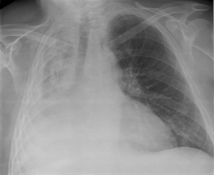
Atelectasis of a person's right lung
Pronunciation
SpecialtyPulmonology

Atelectasis is the partial collapse or closure of a lung resulting in reduced or absence in gas exchange. It is usually unilateral, affecting part or all of one lung.[2] It is a condition where the alveoli are deflated down to little or no volume, as distinct from pulmonary consolidation, in which they are filled with liquid. It is often referred to informally as a collapsed lung, although more accurately it usually involves only a partial collapse, and that ambiguous term is also informally used for a fully collapsed lung caused by a pneumothorax.[1]

It is a very common finding in chest X-rays and other radiological studies, and may be caused by normal exhalation or by various medical conditions. Although frequently described as a collapse of lung tissue, atelectasis is not synonymous with a pneumothorax, which is a more specific condition that can cause atelectasis. Acute atelectasis may occur as a post-operative complication or as a result of surfactant deficiency. In premature babies, this leads to infant respiratory distress syndrome.

The term uses combining forms of atel- + ectasis, from Greek: ἀτελής, "incomplete" + Greek: ἔκτασις, "extension".[3]

Signs and symptoms

[edit]
Atelectasis

May have no signs and symptoms or they may include:[4]

It is a common misconception and pure speculation that atelectasis cause postoperative fever.[5] This claim has been perpetuated in medical textbooks as recently as 2021.[6] Review articles published in 2011 and 2019 summarizing the available evidence on the association between atelectasis and post-operative fever concluded that there is no clinical evidence supporting this speculation.[7][8] A recent article outlined the history of this myth and the true causes of postoperative fever.[9]

Causes

[edit]

The most common cause is post-surgical atelectasis, characterized by splinting, i.e. restricted breathing after abdominal surgery. Atelectasis develops in 75–90% of people undergoing general anesthesia for a surgical procedure.[10]

Another common cause is pulmonary tuberculosis. Smokers and the elderly are also at an increased risk. Outside of this context, atelectasis implies some blockage of a bronchiole or bronchus, which can be within the airway (foreign body, mucus plug), from the wall (tumor, usually squamous cell carcinoma) or compressing from the outside (tumor, lymph node, tubercle). Another cause is poor surfactant spreading during inspiration, causing the surface tension to be at its highest which tends to collapse smaller alveoli. Atelectasis may also occur during suction, as along with sputum, air is withdrawn from the lungs. There are several types of atelectasis according to their underlying mechanisms or the distribution of alveolar collapse; resorption, compression, microatelectasis and contraction atelectasis. Relaxation atelectasis (also called passive atelectasis) is when a pleural effusion or a pneumothorax disrupts the contact between the parietal and visceral pleurae.[11]

Risk factors associated with increased likelihood of the development of atelectasis include: the type of surgery (thoracic and cardiopulmonary surgeries), the use of muscle relaxants, obesity, high oxygen, the lower lung segments.

Factors also associated with the development of atelectasis include: age, presence of chronic obstructive pulmonary disease or asthma, and type of anesthetic.

In the early 1950s, in UK aviation medicine, the condition acceleration atelectasis was given the name "Hunter lung" due to its prevalence in pilots of the transonic fighter jet, the Hawker Hunter, which used a 100% oxygen supply.[12][13]

Diagnosis

[edit]
Atelectasis of the right lower lobe seen on chest X-ray.

Clinically significant atelectasis is generally visible on chest X-ray; findings can include lung opacification and/or loss of lung volume. Post-surgical atelectasis will be bibasal in pattern. Chest CT or bronchoscopy may be necessary if the cause of atelectasis is not clinically apparent. Direct signs of atelectasis include displacement of interlobar fissures and mobile structures within the thorax, overinflation of the unaffected ipsilateral lobe or contralateral lung, and opacification of the collapsed lobe. In addition to clinically significant findings on chest X-rays, patients may present with indirect signs and symptoms such as elevation of the diaphragm, shifting of the trachea, heart and mediastinum; displacement of the hilus and shifting granulomas.[14]

Classification

[edit]
Atelectasis of the middle lobe on a sagittal CT reconstruction.

Atelectasis is broadly categorized into obstructive (resorptive) and non-obstructive types. It may be further classified as an acute or chronic condition. In acute atelectasis, the lung has recently collapsed and is primarily notable only for airlessness. In chronic atelectasis, the affected area is often characterized by a complex mixture of airlessness, infection, widening of the bronchi (bronchiectasis), destruction, and scarring (fibrosis).

Obstructive (absorptive/resorptive) atelectasis

[edit]

This type is defined by blockage of the airway with air trapping and subsequent absorption of air distal to the obstruction.[15] The resulting absorption of air distal to the obstruction results in collapse of the alveoli. It is most commonly due to intrathoracic tumors, aspiration of a foreign body, or mucus plugs.[16] Children are notably more susceptible to atelectasis due to poorly developed collateral airways, which protect against alveolar collapse by maintaining inflation.[15] The Earth's atmosphere is mainly composed of 78% nitrogen and 21% oxygen (+ 1% argon and traces of other gases). Since oxygen is exchanged at the alveoli-capillary membrane, nitrogen is a major component for the alveoli's state of inflation. If a large volume of nitrogen in the lungs is replaced with oxygen, the oxygen may subsequently be absorbed into the blood, reducing the volume of the alveoli, resulting in a form of alveolar collapse known as absorption atelectasis.[17]

Adhesive atelectasis
[edit]

This type of atelectasis is due to lack or dysfunction of surfactant, which normally functions to reduce alveolar surface tension. The increased surface tension in the alveoli then results in alveolar instability and collapse.[18] It is most commonly seen in infant respiratory distress syndrome and acute respiratory distress syndrome (ARDS).[19]

Compressive atelectasis
[edit]

This type occurs when the extra-alveolar pressure overcomes the intra-alveolar pressure, which results in collapse of the lung tissue.[20] While the cause may vary, it is classically associated with the accumulation of blood, fluid, or air within the pleural cavity. These accumulations result in an increase in extra-alveolar pressure which leads to a pressure gradient favoring collapse of the alveoli. This is a frequent occurrence with pleural effusions secondary to congestive heart failure (CHF). Leakage of air into the pleural cavity (pneumothorax) may also lead to compressive atelectasis.[21]

Relaxation atelectasis

This type of atelectasis occurs when there is loss of contact of the lung to the chest wall. It classically occurs as a result of a pleural effusion or pneumothorax. While relaxation and compressive atelectasis share a lot in common, compressive atelectasis tends to be more focal or localized.[18]

Replacement atelectasis

This type of atelectasis occurs when alveoli of an entire lobe of the lung are filled by a tumor, typically bronchioloalveolar carcinoma.[22] The filling of the alveoli results in a loss of lung volume.[20]

Cicatrization (contraction) atelectasis
[edit]

This type occurs when there is contraction of the lung tissue due to the presence of scar tissue. The local or generalized fibrotic changes in the lung or pleura decrease expansion of the lung and increase elastic recoil during expiration.[21][20] Causes include granulomatous disease (e.g., sarcoidosis), necrotizing pneumonia and radiation pneumonitis.[23]

Special cases

[edit]

Right middle lobe syndrome

[edit]

In right middle lobe syndrome, the middle lobe of the right lung contracts due to pressure on the bronchus. This many be secondary to an enlarged lymph node or occasionally a tumor.[24] The blocked, contracted lung may develop pneumonia that fails to resolve completely and leads to chronic inflammation, scarring, and bronchiectasis.[20] Right middle lobe syndrome may occasionally occur in the absence of obvious obstruction. It is hypothesized that the etiology of non-obstructive right middle lobe syndrome is transient hypoventilation secondary to chronic or acute inflammation.[24]

Rounded atelectasis

[edit]

Rounded atelectasis (folded lung or Blesovsky syndrome[25]) is often mistaken for lung cancer on imaging. The most common current theory for rounded atelectasis is that local pleural irritation leads to thickening and shrinkage of the pleura which causes the adjacent lung to shrink with it.[26] The outer portion of the lung slowly collapses as a result of scarring and shrinkage of the membrane layers covering the lungs (pleura), which would show as visceral pleural thickening and entrapment of lung tissue. This produces a rounded appearance on X-ray that doctors may mistake for a tumor. Rounded atelectasis is usually a complication of asbestos-induced disease of the pleura, but it may also result from other types of chronic scarring and thickening of the pleura.[26]

Treatment

[edit]

Treatment is directed at correcting the underlying cause. In atelectasis manifestations that result from the mucus plugging of the airways as seen in patients with cystic fibrosis and pneumonia, mucolytic agents such as acetylcysteine (NAC) is used. This nebulized treatment works by reducing mucous viscosity and elasticity by breaking disulfide bonds in mucoproteins within the mucus complex, thus facilitating mucus clearance.[22] Post-surgical atelectasis is treated by physiotherapy, focusing on deep breathing and encouraging coughing. An incentive spirometer is often used as part of the breathing exercises. Walking is also highly encouraged to improve lung inflation. People with chest deformities or neurologic conditions that cause shallow breathing for long periods may benefit from mechanical devices that assist their breathing.[15]

The primary treatment for acute massive atelectasis is correction of the underlying cause. A blockage that cannot be removed by coughing or by suctioning the airways often can be removed by bronchoscopy. Antibiotics are given for an infection. Chronic atelectasis is often treated with antibiotics because infection is almost inevitable. In certain cases, the affected part of the lung may be surgically removed when recurring or chronic infections become disabling or bleeding is significant. If a tumor is blocking the airway, relieving the obstruction by surgery, radiation therapy, chemotherapy, or laser therapy may prevent atelectasis from progressing and recurrent obstructive pneumonia from developing.[20]

See also

[edit]

References

[edit]
[edit]
Revisions and contributorsEdit on WikipediaRead on Wikipedia
from Grokipedia
Atelectasis is the partial or complete collapse of the lung or a portion of the lung due to deflation of the alveoli, leading to reduced or absent gas exchange and potential respiratory impairment.[1] It represents one of the most common breathing complications following surgery, particularly under general anesthesia, and can occur in both adults and children.[2] The condition arises from several mechanisms, primarily involving airway obstruction (such as from mucus plugs, foreign bodies, or tumors), external compression on the lung (from pleural effusions, pneumothorax, or masses), reduced surfactant production, or hypoventilation due to shallow breathing or prolonged immobility.[3] Obstructive atelectasis, for instance, results specifically from blockage of the bronchi or bronchioles, while compressive forms stem from pressure exerted on lung tissue from adjacent structures.[4] Risk factors include recent surgery, extended bed rest, underlying lung diseases like asthma or chronic obstructive pulmonary disease (COPD), and mechanical ventilation, with postoperative incidence reported in up to 90% of patients under general anesthesia due to impaired diaphragmatic function and mucus clearance.[1] Symptoms of atelectasis are often absent in mild cases but may include shortness of breath, rapid or shallow breathing, cough (which can be productive of sputum), wheezing, decreased breath sounds on auscultation, and in severe instances, cyanosis or respiratory distress.[2] [5] Diagnosis typically involves chest X-rays or computed tomography (CT) scans to visualize areas of collapse, supplemented by pulse oximetry to assess oxygenation and bronchoscopy for identifying obstructions.[3] Treatment focuses on addressing the underlying cause, with interventions such as deep breathing exercises, incentive spirometry, chest physiotherapy (including percussion and postural drainage), bronchodilators or mucolytics to clear secretions, and in refractory cases, bronchoscopy for removal of blockages; mild atelectasis often resolves spontaneously with supportive care.[6] [7] If untreated, atelectasis can lead to complications like pneumonia, hypoxemia, or prolonged ventilation needs, though prognosis is generally favorable with prompt intervention, depending on the extent of collapse and patient comorbidities.[7] Prevention strategies emphasize early mobilization, adequate pain control to encourage deep breathing, and humidified oxygen therapy in at-risk individuals.[1]

Overview and Pathophysiology

Definition

Atelectasis is defined as the partial or complete collapse of lung tissue, leading to reduced or absent gas exchange in the affected alveoli. This condition occurs when the alveoli, the tiny air sacs at the end of the respiratory tree responsible for oxygen and carbon dioxide exchange, deflate and fail to participate in ventilation.[1][3] The term "atelectasis" originates from Greek roots: "atelēs," meaning incomplete or imperfect, and "ektasis," meaning expansion or stretching, thereby describing the incomplete expansion of lung tissue.[1] It was first introduced into medical literature in the early 19th century by the German physician Eduard Jörg in 1832.[8] Maintaining alveolar patency relies on pulmonary surfactant, a lipoprotein complex secreted by type II alveolar cells that lowers surface tension at the air-liquid interface, preventing collapse during exhalation.[9] Atelectasis is distinct from pneumothorax, which involves air accumulation in the pleural space exerting external pressure on the lung, and from consolidation, such as in pneumonia, where alveoli fill with fluid, blood, or pus while preserving overall lung volume.[2][10]

Mechanisms of Lung Collapse

Atelectasis involves several primary mechanisms that lead to lung collapse, categorized as resorptive, compressive, and adhesive processes. Resorptive atelectasis occurs when airway obstruction prevents fresh air from entering the alveoli, resulting in the gradual absorption of trapped alveolar gas into the bloodstream; oxygen is absorbed more rapidly than nitrogen, creating a subatmospheric pressure that causes alveolar collapse.[1] Compressive atelectasis arises from extrinsic forces that reduce lung volume, such as pleural effusions, pneumothorax, or abdominal distension, which mechanically displace lung tissue and promote alveolar emptying without direct airway blockage.[1] Adhesive atelectasis, in contrast, stems from alveolar instability due to insufficient pulmonary surfactant, leading to increased surface tension and spontaneous collapse of alveoli, particularly in conditions impairing surfactant production or function.[11] Biophysically, alveolar stability is governed by Laplace's law, which describes the transmural pressure required to maintain an alveolus open:
P=2Tr P = \frac{2T}{r}

where PP is the pressure difference across the alveolar wall, TT is surface tension, and rr is the alveolar radius. Smaller alveoli (lower rr) experience higher collapsing pressures unless TT is reduced; pulmonary surfactant lowers TT dynamically during expiration, preventing smaller alveoli from emptying into larger ones and averting widespread collapse.[12] In adhesive atelectasis, surfactant deficiency elevates TT, destabilizing alveoli according to this principle and facilitating collapse even without obstruction or compression.[11]
The resulting non-ventilation impairs gas exchange through intrapulmonary shunting, where deoxygenated blood perfuses collapsed regions without participating in oxygenation, leading to systemic hypoxemia that is often refractory to supplemental oxygen.[13] Development of atelectasis progresses in stages: initially, gas resorption or compression causes partial alveolar deflation and volume loss; this advances to complete lobar or segmental collapse over hours to days as hypoxic pulmonary vasoconstriction partially redirects blood flow but fails to fully compensate; re-expansion remains possible if the underlying mechanism is promptly reversed, though prolonged collapse risks fibrosis and permanent impairment.[14]

Etiology and Risk Factors

Primary Causes

Atelectasis arises from several direct etiological factors that disrupt normal lung expansion and aeration, categorized primarily as obstructive, compressive, adhesive, and other postoperative triggers. Obstructive atelectasis occurs when the airways are blocked, preventing air from reaching the alveoli and leading to resorption of trapped gas. Common culprits include mucus plugs formed from tenacious sputum, which are particularly prevalent in patients with impaired mucociliary clearance; foreign bodies aspirated into the bronchi, often seen in children or unconscious individuals; endobronchial tumors that narrow or occlude passages; and conditions like bronchiectasis that promote chronic airway obstruction through dilated, mucus-filled bronchi.[14][1][15] Compressive atelectasis results from external forces that physically collapse lung tissue by increasing intrapleural pressure. This includes pleural effusions, where accumulated fluid in the pleural space compresses adjacent lung segments; pneumothorax, in which air in the pleural cavity displaces the lung; abdominal distension, such as from ascites or post-laparotomy swelling, which elevates the diaphragm and restricts lung base expansion; chronic elevation of the hemidiaphragm due to phrenic nerve injury or diaphragmatic dysfunction, which restricts lung expansion and commonly leads to basilar atelectasis on the affected side (particularly chronic left-sided elevation causing left basilar atelectasis); and space-occupying masses like large tumors or mediastinal shifts that exert pressure on the pulmonary parenchyma.[1][3][2][16] Adhesive atelectasis stems from a deficiency or dysfunction of pulmonary surfactant, which normally reduces alveolar surface tension to prevent collapse. This is frequently observed in premature infants due to immature surfactant production, leading to neonatal respiratory distress syndrome; in adults with acute respiratory distress syndrome (ARDS), where inflammatory damage impairs type II pneumocyte function; and in other surfactant-disrupting states like smoke inhalation or radiation pneumonitis.[1][17][14] Postoperative atelectasis represents a significant direct trigger, often involving retention of secretions due to shallow breathing under anesthesia, endotracheal tube malposition obstructing bronchi, or absorption atelectasis from high inspired oxygen fractions that accelerate nitrogen washout. This form is especially common after abdominal surgery, affecting up to 90% of patients within the first 72 hours, primarily through a combination of reduced inspiratory effort and retained mucus.[1][4][18]

Predisposing Conditions

Several patient-related factors increase susceptibility to atelectasis by impairing lung expansion, clearance mechanisms, or respiratory muscle function. Advanced age is associated with reduced lung compliance and weaker respiratory efforts, elevating the risk particularly in postoperative settings.[19] Obesity contributes as an independent risk factor, primarily through mechanical compression of the lungs and diaphragm in the supine position, which promotes basal atelectasis during anesthesia or bed rest.[20] Neuromuscular diseases, such as muscular dystrophy, weaken respiratory muscles and cough reflex, leading to secretion retention and alveolar collapse.[21] Similarly, chronic obstructive pulmonary disease (COPD) predisposes individuals via airflow obstruction and mucus hypersecretion, though incidence may vary compared to other lung conditions.[22] Iatrogenic factors often arise in medical settings and directly compromise ventilatory dynamics. Prolonged mechanical ventilation can induce atelectasis through alveolar overdistension, reduced surfactant production, and dependent lung collapse due to positive pressure effects.[23] General anesthesia frequently causes rapid-onset atelectasis by suppressing diaphragmatic tone and promoting airway closure, affecting up to 90% of patients within minutes of induction.[24] Opioid use heightens risk by depressing the cough reflex and deep breathing, which impairs secretion clearance and leads to retained mucus.[25] Environmental exposures can exacerbate vulnerabilities in lung function. Tobacco smoking impairs mucociliary clearance, fostering mucus accumulation that predisposes to obstructive atelectasis, with smokers facing up to 4 times higher risk of postoperative complications than non-smokers.[26] In special populations, neonates with respiratory distress syndrome (RDS) are highly prone due to surfactant deficiency, resulting in widespread alveolar instability and collapse.[27] Trauma patients with rib fractures experience splinting from pain, which restricts deep inspiration and promotes atelectasis, often compounded by immobilization.[28] These predisposing conditions can interact with inciting events, such as mucus plugs, to accelerate lung collapse in vulnerable individuals.

Clinical Features

Signs and Symptoms

Atelectasis often presents with a range of respiratory symptoms, the most common of which include shortness of breath (dyspnea), pleuritic chest pain, and cough that may be dry or productive with sputum.[1][7] These manifestations arise due to impaired gas exchange and irritation of the pleural surfaces, though their severity varies widely depending on the underlying cause and extent of lung involvement.[1] Many cases of atelectasis are asymptomatic, particularly when the collapse is limited to a small area or occurs chronically, and this is especially true in elderly patients or those who are sedated or immobile, where subtle changes may go unnoticed.[1][5] In contrast, more extensive involvement, such as lobar atelectasis, can lead to pronounced respiratory distress, while focal atelectasis typically causes only minimal or no symptoms.[1] Systemic effects may include hypoxemia, which in severe cases can manifest as cyanosis, and a low-grade fever if a secondary infection develops in the collapsed lung segments.[29][30] In pediatric patients, particularly infants, atelectasis may present with signs of increased work of breathing, such as grunting respirations and intercostal or subcostal retractions, often alongside tachypnea and nasal flaring.[31] These symptoms reflect the infant's compensatory efforts to maintain adequate oxygenation amid reduced lung volume.[27]

Physical Examination

The physical examination of patients with atelectasis begins with assessment of vital signs, where tachypnea is commonly observed as a compensatory mechanism to maintain oxygenation amid reduced lung volume.[1] Tachycardia may also occur, particularly in cases of significant hypoxemia or increased respiratory effort.[32] These findings are often subtle in mild or basilar atelectasis but become more pronounced with extensive collapse. Inspection reveals asymmetry in chest wall movement, with reduced excursion on the affected side due to diminished lung expansion.[33] In instances of large-volume atelectasis, such as lobar collapse, the trachea may deviate toward the affected side as a result of volume loss pulling mediastinal structures ipsilaterally.[14] Overall, the patient may appear to have increased work of breathing, with visible use of accessory muscles in severe cases.[1] Palpation typically demonstrates decreased tactile fremitus over the region of collapse, reflecting reduced transmission of vibrations through consolidated or airless lung tissue.[34] A palpable mediastinal shift toward the affected side can be noted in significant atelectasis, aligning with the ipsilateral tracheal deviation.30085-9/fulltext) Percussion yields dullness over the involved area, especially in compressive atelectasis where adjacent pleural effusion or mass contributes to the density increase.[35] This finding helps differentiate atelectasis from conditions like pneumothorax, which produce hyperresonance. Auscultation most characteristically shows decreased or absent breath sounds over the affected lung segment, indicating loss of air entry.[33] In cases of partial airway obstruction leading to resorptive atelectasis, localized wheezes may be audible proximal to the blockage.[36] Fine crackles can occasionally be heard upon re-expansion of collapsed alveoli during inspiration.[1]

Diagnosis

Imaging Modalities

Chest X-ray (CXR) serves as the initial imaging modality for evaluating suspected atelectasis, often revealing indirect signs of volume loss such as elevation of the hemidiaphragm, mediastinal or tracheal shift toward the affected side, and crowding of ribs or vessels.[37] Direct signs include opacification of the collapsed lung segment or lobe without visible air bronchograms, which helps differentiate atelectasis from other causes of opacity like pneumonia.[38] CXR typically detects atelectasis when there is significant volume loss, such as greater than 25% of the affected lung, but it frequently misses small peripheral or basilar cases due to its limited sensitivity, particularly in supine or suboptimal views.[39]

Radiographic signs of volume loss

Direct signs of atelectasis/volume loss include displacement of interlobar fissures, crowding of bronchovascular markings (bunching of bronchi and vessels due to reduced lung expansion), and increased density in the affected area. Crowding of bronchovascular markings is particularly common in cases of low lung volumes from poor inspiratory effort or expiratory-phase imaging. This causes the lungs to appear denser with bunched vascular and bronchial structures, especially at the bases, often accompanied by bibasilar subsegmental (platelike) atelectasis. Such findings are frequently technical rather than pathological, mimicking increased markings or infection, but typically resolve on repeat films with full inspiration (assessed by anterior rib count of 6 or posterior 9-10). This is a key pitfall in interpretation, especially in pediatric, elderly, or uncooperative patients. Computed tomography (CT) is considered the gold standard for delineating the precise extent of atelectasis, confirming lobar or segmental collapse through visualization of fissure displacement, bronchial narrowing, and associated opacification.[37] It excels at identifying underlying compressive etiologies, such as tumors or masses obstructing airways or compressing lung tissue, which may not be apparent on CXR.[40] High-resolution CT (HRCT) enhances detection of subtle or early atelectasis, particularly in dependent regions or postoperative settings, by providing detailed cross-sectional views of lung architecture and volume loss.[1] Lung ultrasound is a portable, bedside imaging tool particularly useful for assessing compressive atelectasis secondary to pleural effusions, where it demonstrates hypoechoic consolidation patterns adjacent to anechoic fluid collections.[41] Characteristic findings include static air bronchograms within the consolidated area, absent lung sliding, and a tissue-like echotexture due to volume loss, allowing rapid differentiation from other pathologies like pneumonia.[37] Its high sensitivity, often exceeding that of supine CXR in critically ill patients, makes it ideal for real-time evaluation in settings where radiation exposure should be minimized.[42] Ventilation-perfusion (V/Q) scanning is utilized when atelectasis is suspected to result from pulmonary embolism, assessing for characteristic mismatched defects where perfusion is reduced but ventilation remains relatively preserved.[43] This nuclear medicine technique helps quantify the degree of mismatch in embolism-related cases, guiding further management, though it is less commonly employed as a primary diagnostic tool for atelectasis alone.[44] If imaging findings remain inconclusive, bronchoscopy may be required for definitive confirmation.[37]

Confirmatory Tests

Bronchoscopy serves as a key confirmatory test for atelectasis, enabling direct visualization of the airways to identify and address obstructions such as mucus plugs, tumors, or foreign bodies. Flexible bronchoscopy, utilizing a thin, maneuverable fiberoptic scope inserted through the mouth or nose under sedation, is the preferred technique for most cases due to its ability to reach peripheral bronchi and facilitate therapeutic interventions like suctioning or biopsy. Rigid bronchoscopy, involving a straight metal tube under general anesthesia, is less commonly used but may be employed for central airway obstructions or when larger instruments are needed for removal of substantial foreign material.[1][45] Pulse oximetry is a non-invasive confirmatory test that measures peripheral oxygen saturation (SpO2), often revealing reduced levels in cases of significant atelectasis, providing a quick assessment of oxygenation impairment.[6] Arterial blood gas (ABG) analysis provides functional confirmation of atelectasis by revealing gas exchange impairments, typically showing hypoxemia (reduced PaO2, often below the normal range of 75-100 mmHg depending on the extent of collapse) and normal or low partial pressure of carbon dioxide (PaCO2) due to compensatory hyperventilation. The alveolar-arterial (A-a) oxygen gradient is often elevated, calculated as the difference between alveolar oxygen (estimated from the alveolar gas equation) and arterial oxygen, indicating ventilation-perfusion mismatch or shunt physiology consistent with collapsed lung segments.[1][37] Sputum culture is employed to exclude or confirm infectious etiologies contributing to atelectasis, such as bacterial pneumonia or aspiration-related pathogens, by analyzing expectorated or induced samples for microbial growth and sensitivity. This test is particularly useful when clinical suspicion of infection arises alongside imaging abnormalities.[1][37] Pulmonary function tests (PFTs) quantify the restrictive impact of atelectasis, demonstrating reduced forced vital capacity (FVC) and forced expiratory volume in one second (FEV1) in the affected lung regions, reflecting diminished lung volume and compliance. These tests, including spirometry and lung volume measurements, help assess the extent of collapse and monitor resolution post-intervention.[1][37] While generally safe, bronchoscopy carries risks including bleeding, with an incidence of approximately 1-2% in diagnostic procedures, and pneumothorax, occurring in up to 1% of cases, particularly when biopsies are performed. These complications are more frequent in patients with underlying coagulopathy or severe lung disease.[46][47]

Classification

Obstructive Atelectasis

Obstructive atelectasis, also referred to as resorptive atelectasis, is characterized by the partial or complete collapse of lung tissue due to intrinsic blockage of the bronchial tree, which impedes airflow to the distal alveoli. This obstruction leads to the gradual reabsorption of alveolar gas, primarily oxygen, into the pulmonary circulation, resulting in negative pressure and alveolar deflation; nitrogen, being less soluble, provides temporary stability but cannot prevent eventual collapse as ventilation ceases.[1] The process typically unfolds over hours to days, distinguishing it from more rapid compressive forms, and is driven by the imbalance between gas absorption and lack of replenishment.[4] Common causes of obstructive atelectasis encompass endobronchial tumors, such as primary lung carcinomas or metastatic lesions that narrow or occlude bronchi; aspirated foreign bodies, particularly in children or unconscious adults; and accumulation of thick mucus plugs or secretions, often seen in intubated patients with conditions like chronic obstructive pulmonary disease or post-surgical immobility.[1] Less frequently, extrinsic compression by hilar lymphadenopathy or bronchial stenosis from prior inflammation contributes to the blockage.[14] These etiologies predominantly affect larger airways, leading to lobar or segmental involvement rather than diffuse collapse. Specific clinical features of obstructive atelectasis include persistent unilateral wheezing or diminished breath sounds over the affected area, reflecting the localized airflow limitation, and recurrent episodes of pneumonia or bronchiectasis in the lung segments distal to the obstruction due to stagnant secretions and secondary infection.[1] Patients may also exhibit hypoxemia from ventilation-perfusion mismatch, though systemic symptoms like fever or cough are more attributable to complicating infections than the atelectasis itself.[14] Diagnostic imaging plays a central role, with chest radiography often revealing lobar consolidation or volume loss, such as mediastinal shift toward the affected side; computed tomography (CT) provides greater specificity, demonstrating the "golden S sign" in right upper lobe collapse—where a central obstructing mass forms the apex of an inverted S against the hyperinflated minor fissure—or a "tree-in-bud" pattern indicating endobronchial spread of infection with dilated bronchioles and surrounding centrilobular nodules distal to the blockage.[1] Bronchoscopy confirms the diagnosis by visualizing the obstruction and allowing for biopsy or removal of foreign material.[14] Obstructive atelectasis represents the most common form of atelectasis, accounting for the majority of cases in both adult and pediatric populations, with studies linking many atelectasis episodes to airway obstructions like mucus plugs.[48] Its prevalence is notably higher in patients with underlying malignancy, where endobronchial tumors precipitate collapse in 25–33% of patients at initial presentation.[49][14]

Non-Obstructive Atelectasis

Non-obstructive atelectasis arises from mechanisms that cause lung collapse without airway blockage, primarily through external compression, loss of alveolar stability, or intrinsic lung restriction.[1] This form contrasts with obstructive atelectasis, where gas resorption follows airway occlusion.[1] The main subtypes include passive (also termed compression or relaxation) atelectasis, adhesive atelectasis, and contraction (or cicatrization) atelectasis. Passive atelectasis occurs when external forces, such as pleural effusion, pneumothorax, or abdominal distension, compress lung tissue and reduce alveolar volume without promoting gas resorption.[17] Adhesive atelectasis results from surfactant deficiency or inactivation, which increases alveolar surface tension; according to Laplace's law (P=2TrP = \frac{2T}{r}, where PP is transmural pressure, TT is surface tension, and rr is radius), this elevates the pressure required to maintain small alveoli open, leading to their collapse.[50] Contraction atelectasis stems from fibrotic scarring or parenchymal distortion that restricts lung expansion, often seen in chronic conditions like pulmonary fibrosis.[5] Common examples illustrate these mechanisms in clinical settings. Postoperative shallow breathing, induced by pain or anesthesia, frequently causes basilar plate-like atelectasis through passive compression and reduced ventilation, representing the most prevalent type in this context and affecting up to 90% of patients under general anesthesia.[51] In neonates, surfactant deficiency underlies adhesive atelectasis in conditions like hyaline membrane disease or acute respiratory distress syndrome (ARDS), where immature lungs fail to produce adequate surfactant, promoting widespread alveolar instability.[52] On chest X-ray (CXR), non-obstructive atelectasis typically appears as linear or band-like opacities, often platelike and horizontal, located peripherally or at the lung bases, without air bronchograms due to the absence of distal airway patency issues.[1] These findings reflect localized volume loss without the lobar shift or bronchial crowding seen in other forms.[53]

Special Presentations

Right Middle Lobe Syndrome

Right middle lobe syndrome (RMLS) is a clinical entity characterized by chronic or recurrent atelectasis specifically involving the right middle lobe of the lung, often leading to associated bronchiectasis or recurrent infections. This condition arises primarily from the anatomic vulnerability of the right middle lobe, which features a long and narrow bronchus with an acute take-off angle and an oval "fish mouth" ostium, bordered by the horizontal and oblique fissures that limit collateral ventilation, exacerbating the propensity for collapse and making it particularly susceptible to resorption of air and subsequent atelectasis even without complete obstruction.[54][55][56] The etiologies of RMLS include both intrinsic and extrinsic factors that contribute to bronchial narrowing or compression. Intrinsic causes involve endobronchial lesions such as mucus plugs, infections causing inflammatory stenosis, or neoplasms within the bronchus. Extrinsic factors encompass compression from enlarged hilar lymphadenopathy, peribronchial tumors, or fibrotic tissue, often in the context of chronic infections like tuberculosis or nontuberculous mycobacteria. This distinguishes RMLS as a condition where anatomic predisposition plays a central role, sometimes without an identifiable obstructing lesion.[57][56][54] Patients with RMLS typically present with symptoms related to chronic airway irritation and recurrent lower respiratory tract involvement, including persistent cough, hemoptysis, and episodes of right-sided pneumonia. Other manifestations may include dyspnea or chest pain, particularly in cases with significant volume loss or secondary bronchiectasis, though some individuals remain asymptomatic until advanced disease develops.[57][56][58] Diagnosis relies on imaging and endoscopic evaluation to confirm lobar collapse and identify underlying causes. Chest radiography (CXR) often reveals subtle volume loss in the right middle lobe, such as obscuration of the minor fissure or silhouette sign with the right heart border. Computed tomography (CT) provides more detailed assessment, demonstrating bronchial stenosis, extrinsic compression, or associated bronchiectasis. Bronchoscopy is essential for visualizing intraluminal abnormalities and obtaining biopsies if needed, helping differentiate obstructive from non-obstructive forms. RMLS can present as a special case of non-obstructive atelectasis due to its emphasis on extrinsic and anatomic factors.[57][56][54] The condition was first described in 1948 by Graham et al. in a series of patients with atelectasis and nontuberculous pneumonitis of the right middle lobe. It is classified into two main forms: obstructive, involving intrinsic bronchial blockage, and non-obstructive, driven primarily by extrinsic compression and anatomic vulnerabilities.[57][54][56]

Rounded Atelectasis

Rounded atelectasis represents a distinctive pseudotumoral variant of atelectasis, characterized by focal, rounded collapse of peripheral lung tissue that folds inward against a thickened visceral pleura, often simulating a solitary pulmonary nodule or mass lesion. This infolding occurs due to chronic pleural fibrosis adhering to and contracting the adjacent lung parenchyma, resulting in localized volume loss without complete airway obstruction.[1] The pathogenesis is closely tied to underlying pleural pathology, most frequently chronic pleural thickening from asbestos exposure in asbestosis or scarring following resolved tuberculous pleuritis. In these settings, repeated episodes of pleural effusion or inflammation promote visceral pleural fibrosis, which tethers and retracts the lung, causing the atelectatic segments to curl into a rounded configuration. A hallmark feature, the "comet tail" sign, arises from the curvilinear displacement of bronchi and vessels sweeping into the base of the mass, reflecting the mechanical distortion of normal hilar structures.[59] Patients with rounded atelectasis are typically asymptomatic, with the condition discovered incidentally on chest radiography or computed tomography performed for unrelated indications. Despite its benign nature, it poses a diagnostic challenge due to its mass-like appearance, which can mimic bronchogenic carcinoma or metastatic disease, occasionally prompting unwarranted biopsies or resections.[60] Definitive diagnosis hinges on characteristic computed tomography findings: a subpleural, ovoid opacity measuring 3 to 7 cm, contiguous with focally thickened pleura, accompanied by ipsilateral lobar volume loss, the comet tail sign, and absence of contrast enhancement. Confirmation requires demonstration of lesional stability on follow-up imaging over at least 6 to 12 months, obviating the need for invasive sampling in unequivocal cases. Epidemiologically, rounded atelectasis occurs predominantly in individuals with prior asbestos exposure, comprising 29% to 86% of reported cases and manifesting in a subset of those with benign asbestos-related pleural disease, such as plaques or diffuse thickening.[61]

Management and Prevention

Treatment Approaches

Treatment of atelectasis primarily focuses on re-expanding the collapsed lung tissue and addressing the underlying cause, with approaches varying based on the etiology and severity.[1] Conservative measures form the cornerstone of initial management, particularly for postoperative or non-obstructive atelectasis, and include deep breathing exercises, incentive spirometry, and chest physiotherapy to promote lung expansion and mobilize secretions. Deep breathing exercises encourage voluntary inspiration to counteract shallow breathing patterns that contribute to collapse, while incentive spirometry uses a device to provide visual feedback for sustained maximal inhalation, typically performed 10-15 times per hour when awake. Chest physiotherapy techniques, such as postural drainage, percussion, and vibration, help clear airway secretions and improve ventilation-perfusion matching. These interventions are most effective when initiated early, within the first 24-48 hours of onset. Early physiotherapy is effective in resolving postoperative atelectasis, reducing the need for more invasive interventions and shortening hospital stays. Combining breathing exercises with positive airway pressure can improve oxygenation in high-risk populations.[6][1][3] Pharmacologic therapies target specific reversible components of atelectasis, such as airway obstruction or infection. Bronchodilators, including beta-agonists like albuterol, are administered via nebulizer or metered-dose inhaler to relieve bronchospasm in cases of reversible obstruction, improving airflow and facilitating re-expansion. Mucolytics, such as N-acetylcysteine, reduce the viscosity of mucus plugs, aiding their expulsion, and are particularly useful in patients with thick secretions. If an infectious process, such as pneumonia, is contributing to the atelectasis, antibiotics are prescribed based on identified pathogens, following standard guidelines for respiratory infections. These agents are often combined with conservative measures for synergistic effects.[1][62][63] Interventional procedures are employed when conservative and pharmacologic approaches are insufficient, especially for obstructive atelectasis caused by foreign bodies, tumors, or large effusions. Flexible bronchoscopy allows direct visualization and removal of mucus plugs, endobronchial lesions, or aspirated material using forceps, suction, or laser therapy, often leading to immediate re-expansion. For extrinsic compression from pleural effusions, thoracentesis involves needle aspiration to drain fluid, relieving pressure on the lung. In select cases of central airway stenosis, stent placement during bronchoscopy maintains patency. These procedures carry risks such as bleeding or infection but are guided by imaging and reserved for persistent collapse.[6][64][1] Supportive care is essential in critically ill patients or those on mechanical ventilation, incorporating positive end-expiratory pressure (PEEP) to prevent alveolar collapse by maintaining positive pressure at end-expiration, typically set at 5-10 cm H2O. For irreversible causes like endobronchial tumors, surgical resection via lobectomy or pneumonectomy may be necessary after neoadjuvant therapy to shrink the lesion. Supplemental oxygen is titrated to maintain saturation above 92%, supporting gas exchange during re-expansion. These measures are tailored to the patient's overall condition and integrated into multidisciplinary care.[1][64]

Preventive Measures

Preventive measures for atelectasis focus on mitigating risk in high-incidence settings such as the perioperative period and neonatal care, emphasizing strategies that promote alveolar expansion and reduce compressive or obstructive forces. In perioperative protocols, early mobilization within the first 24 hours post-surgery enhances lung expansion and clearance of secretions, thereby reducing the risk of collapse.[1] Adequate pain control, through multimodal analgesia, facilitates deep breathing and coughing to prevent mucus accumulation and subsequent obstruction.[35] Humidified oxygen delivery during mechanical ventilation maintains airway moisture, minimizing desiccation that could contribute to atelectasis formation.[4] Ventilation strategies during anesthesia play a critical role in prevention by counteracting the compressive effects of supine positioning and high oxygen concentrations. Application of positive end-expiratory pressure (PEEP) at 5-10 cmH₂O during general anesthesia increases functional residual capacity and prevents alveolar collapse, with studies demonstrating reduced atelectasis incidence compared to zero PEEP.[65] Recruitment maneuvers combined with PEEP further optimize lung recruitment, particularly in prolonged procedures.[66] Patient education initiatives target modifiable risk factors to lower atelectasis occurrence preoperatively. Smoking cessation programs, ideally initiated 6-8 weeks before surgery, improve ciliary function and reduce mucus hypersecretion, decreasing postoperative atelectasis risk.[35] Preoperative training in incentive spirometry encourages sustained deep inspirations, with randomized trials showing it reduces postoperative pulmonary complications, including atelectasis, by up to 50% in abdominal surgery patients when used consistently.[18] These measures also address risk factors such as obesity through brief counseling on weight management to alleviate diaphragmatic compression.[67] In neonatal care, particularly for preterm infants, preventive strategies address surfactant deficiency to avert atelectasis in respiratory distress syndrome. Exogenous surfactant replacement therapy, administered prophylactically shortly after birth, stabilizes alveoli and reduces atelectasis by lowering surface tension, with clinical trials indicating improved oxygenation and lower need for mechanical ventilation.[68] Prone positioning minimizes abdominal compression on the diaphragm, promoting even lung expansion and preventing dependent atelectasis in ventilated neonates.[69]

Prognosis and Complications

Expected Outcomes

The prognosis for atelectasis is generally favorable when treated promptly, with most cases demonstrating re-expansion and resolution without lasting impairment. According to a comprehensive review, the majority of atelectasis episodes resolve fully following appropriate intervention, often within days, though the exact timeline depends on the extent and cause.[1] Chronic cases, particularly those involving fibrosis, may persist and resist full re-expansion, leading to potential incomplete recovery if untreated for extended periods.[35] Several factors influence the overall prognosis, including the timeliness of intervention and the presence of underlying conditions. Early detection and treatment significantly enhance recovery rates by preventing progression to more severe collapse.[1] When atelectasis results from malignancy, such as advanced non-small cell lung cancer, outcomes are poorer due to the cancer itself, with 5-year survival rates around 12% for distant stages and about 40% for regional stages; the overall prognosis is primarily determined by the stage of the underlying cancer.[70] Long-term effects are typically minimal, with rare instances of permanent lung function loss except in cicatricial atelectasis where scarring occurs. In postoperative settings, full recovery is achieved in the majority of cases, as atelectasis often self-resolves or responds well to supportive measures without residual deficits.[3] Monitoring involves follow-up chest X-rays approximately 1-2 weeks post-treatment to verify resolution and rule out persistence.[6] Post-2020 studies, including a 2024 meta-analysis, indicate enhanced outcomes through non-invasive ventilation, which reduces ICU length of stay and reintubation risks in postoperative patients (with low to very low certainty evidence). Atelectasis is noted as a frequent reversible postoperative complication.[71]

Associated Complications

Atelectasis impairs gas exchange, leading to hypoxemic respiratory failure characterized by low blood oxygen levels without significant hypercapnia.[35] This occurs primarily through intrapulmonary shunting and ventilation-perfusion (V/Q) mismatch, where collapsed alveoli receive perfusion but no ventilation, exacerbating hypoxemia.[72] In severe or chronic cases, persistent V/Q mismatch can contribute to pulmonary hypertension by altering pulmonary vascular resistance.[35] Infectious complications frequently arise, especially in obstructive atelectasis, where bronchial blockage promotes distal accumulation of secretions and bacterial overgrowth, resulting in post-obstructive pneumonia.[2] Untreated, this can progress to lung abscess formation in approximately 10-15% of cases, particularly those associated with underlying lung malignancies.[73] Among mechanically ventilated patients, atelectasis heightens the risk of sepsis due to secondary bacterial invasion of collapsed lung segments.[33] Additional complications include thromboembolism, as atelectasis often accompanies prolonged immobility in postoperative or critically ill patients, promoting venous stasis and clot formation. In massive atelectasis involving an entire lung, mediastinal shift toward the affected side can induce cardiovascular strain by displacing the heart and great vessels, potentially compromising hemodynamics.[14] Recurrent atelectasis may cause long-term structural changes, such as bronchiectasis, through repeated cycles of inflammation, infection, and bronchial dilation. Unresolved adhesive atelectasis can lead to pulmonary fibrosis, with scarring that diminishes lung compliance and reserve.[1] Prompt treatment, including re-expansion maneuvers and addressing underlying causes, significantly lowers the risk of these complications and enhances recovery.[1]

References

User Avatar
No comments yet.