Hubbry Logo
GlossitisGlossitisMain
Open search
Glossitis
Community hub
Glossitis
logo
8 pages, 0 posts
0 subscribers
Be the first to start a discussion here.
Glossitis
Glossitis
from Wikipedia
Glossitis [1]
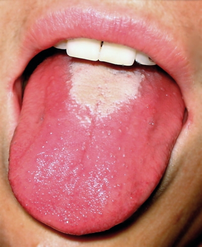
Glossitis in a person with scarlet fever ("red strawberry tongue").
SpecialtyGastroenterology
ComplicationsSome speech impairments

Glossitis can mean soreness of the tongue, or more usually inflammation with depapillation of the dorsal surface of the tongue (loss of the lingual papillae), leaving a smooth and erythematous (reddened) surface,[2][3] (sometimes specifically termed atrophic glossitis). In a wider sense, glossitis can mean inflammation of the tongue generally.[4] Glossitis is often caused by nutritional deficiencies and may be painless or cause discomfort. Glossitis usually responds well to treatment if the cause is identified and corrected. Tongue soreness caused by glossitis is differentiated from burning mouth syndrome, where there is no identifiable change in the appearance of the tongue, and there are no identifiable causes.[5]

Symptoms

[edit]
Patchy depapillation of filiform papillae with prominence of the fungiform papillae.

Depending upon what exact meaning of the word glossitis is implied, signs and symptoms might include:

  • Smooth, shiny appearance of the tongue, caused by loss of lingual papillae.
  • Tongue color changes, usually to a darker red color than the normal white-pink color of a healthy tongue.
  • Tongue swelling.
  • Difficulty with chewing, swallowing, or speaking (either because of tongue soreness or tongue swelling).
  • Burning sensation.[2] Some use the term secondary burning mouth syndrome in cases where a detectable cause, such as glossitis, for an oral burning sensation.[5]

Depending upon the underlying cause, there may be additional signs and symptoms such as pallor, oral ulceration and angular cheilitis.[2]

Causes

[edit]

Anemias

[edit]

Iron-deficiency anemia is mainly caused by blood loss, such as may occur during menses or gastrointestinal hemorrhage. This often results in a depapilled, atrophic glossitis, giving the tongue a bald and shiny appearance, along with pallor (paleness) of the lips and other mucous membranes a tendency towards recurrent oral ulceration,[6] and angular cheilitis (crockings and inflammation at the corners of the lips).[7] The appearance of the tongue in iron-deficiency anemia has been described as diffuse or patchy atrophy with tenderness or burning.[8] One cause of iron-deficiency anemia is sideropenic dysphagia (Plummer–Vinson syndrome, also called Paterson–Brown–Kelly syndrome) which is also characterized by esophageal webbing and dysphagia.[6]

Pernicious anemia is usually caused by autoimmune destruction of gastric parietal cells. Parietal cells secrete intrinsic factor which is required for the absorption of vitamin B12. Vitamin B12 deficiency results in megaloblastic anemia and may present as atrophic glossitis (Hunter's atrophic glossitis). The appearance of the tongue in vitamin B12 deficiency is described as "beefy" or "fiery red and sore".[6] There may be linear or patchy red lesions.[2]

Vitamin B deficiencies

[edit]

Vitamin B1 deficiency (thiamine deficiency) can cause glossitis.[7] Vitamin B2 deficiency (ariboflavinosis) can cause glossitis, along with angular cheilitis, cheilosis, peripheral neuropathy and other signs and symptoms.[7] The glossitis in vitamin B2 deficiency is described as magenta.[9] Vitamin B3 deficiency (pellagra) can cause glossitis.[7] Vitamin B6 deficiency (pyridoxine deficiency) can cause glossitis, along with angular cheilitis, cheilosis, peripheral neuropathy and seborrheic dermatitis.[7] Folate deficiency (vitamin B9 deficiency) can cause glossitis, along with macrocytic anemia, thrombocytopenia, leukopenia, diarrhea, fatigue and possibly neurological signs.[7] Apart from pernicious anemia discussed above, any other cause of vitamin B12 deficiency can cause atrophic glossitis (Hunter's atrophic glossitis), which tends to be painful, smooth and shiny.[7]

Infections

[edit]

Bacterial, viral or fungal infections can cause glossitis. Chronic Candida infections of the tongue can cause an atrophic glossitis known as median rhomboid glossitis.[10]

Syphilis is now relatively rare, but the tertiary stage can cause diffuse glossitis and atrophy of lingual papillae, termed "syphilitic glossitis",[2] "luetic glossitis" or "atrophic glossitis of tertiary syphilis".[8] It is caused by Treponema pallidum and is a sexually transmitted infection.

Other causes

[edit]

Many conditions can cause glossitis via malnutrition or malabsorption,[7] which creates the nutritional deficiencies described above, although other mechanisms may be involved in some of those conditions listed.

A painful tongue may be an indication of an underlying serious medical condition and nearly always merits assessment by a physician or dental surgeon.

Diagnosis

[edit]

Classification

[edit]
Median rhomboid glossitis
Geographic tongue (benign migratory glossitis)

Glossitis could be classified as a group of tongue diseases or gastrointestinal diseases.[4] It may be primary, where there is no underlying cause, or secondary where it is a sign or symptom of another condition.[3] It can be acute or chronic.[4] Generally speaking, there are several clinical patterns of glossitis, some more common than others.

Atrophic glossitis

[edit]

Atrophic glossitis, also known as bald tongue,[3] smooth tongue, Hunter glossitis, Moeller glossitis, or Möller-Hunter glossitis,[14] is a condition characterized by a smooth glossy tongue that is often tender/painful,[15] caused by complete atrophy of the lingual papillae (depapillation).[3] The dorsal tongue surface may be affected totally, or in patches, and may be associated with a burning sensation, pain and/or erythema.[16] Atrophic glossitis is a non-specific finding,[16] and has a great many causes, usually related to iron-deficiency anemia, pernicious anemia, B vitamin complex deficiencies,[16] unrecognized and untreated celiac disease (which often presents without gastrointestinal symptoms),[17][18][19] or other factors such as xerostomia (dry mouth). Although the terms Möller and Hunter glossitis were originally used to refer to specifically the glossitis that occurs in vitamin B12 deficiency secondary to pernicious anemia, they are now used as synonyms for atrophic glossitis generally.[14] In this article, the term glossitis, unless otherwise specified, refers to atrophic glossitis.

Candidiasis may be a concurrent finding or an alternative cause of erythema, burning, and atrophy.

Median rhomboid glossitis

[edit]

This condition is characterized by a persistent erythematous, rhomboidal depapillated lesion in the central area of the dorsum of the tongue, just in front of the circumvallate papillae.[2][15] Median rhomboid glossitis is a type of oral candidiasis, and rarely causes any symptoms. It is treated with antifungal medication. Predisposing factors include use of corticosteroid sprays or inhalers or immunosuppression.

Benign migratory glossitis

[edit]

Geographic tongue, also termed benign migratory glossitis, is a common condition which usually affects the dorsal surface of the tongue. It is characterized by patches of depapillation and erythema bordered by a whitish peripheral zone. These patches give the tongue the appearance of a map, hence the name. Unlike glossitis due to nutritional deficiencies and anemia, the lesions of geographic tongue move around the tongue over time.[20] This is because in geographic tongue, new areas of the tongue become involved with the condition whilst previously affected areas heal, giving the appearance of a moving lesion.[3] The cause is unknown,[21] and there is no curative treatment. Rarely are there any symptoms associated with the lesions, but occasionally a burning sensation may be present, which is exacerbated by eating hot, spicy or acidic foodstuffs. Some consider geographic tongue to be an early stage of fissured tongue, since the two conditions often occur in combination.[22]

Geometric glossitis

[edit]

Geometric glossitis, also termed herpetic geometric glossitis,[4] is a term used by some to refer to a chronic lesion associated with herpes simplex virus (HSV) type I infection,[23] in which there is a deep fissure in the midline of the tongue, which gives off multiple branches.[8] The lesion is usually very painful, and there may be erosions present in the depths of the fissures. Similar fissured lesions which are not associated with HSV, as may occur in fissured tongue, do not tend to be painful.[23] The name comes from the geometric pattern of the fissures which are longitudinal, crossed or branched.[24] It is described as occurring in immunocompromised persons, e.g. who have leukemia. However, the association between herpes simplex and geometric glossitis is disputed by some due to a lack of gold standard techniques for diagnosis of intraoral herpetic lesions, and the high prevalence of asymptomatic viral shedding in immunocompromised individuals.[8] Treatment is with systemic aciclovir.[23]

Strawberry tongue

[edit]
Distinct strawberry tongue with "parched" lips as seen in a young child with Kawasaki disease.

Strawberry tongue, or raspberry tongue,[25] is glossitis which manifests with hyperplastic (enlarged) fungiform papillae, giving the appearance of a strawberry. White strawberry tongue is where there is a white coating on the tongue through which the hyperplastic fungiform papillae protrude. Red strawberry tongue is where the white coating is lost and a dark red, erythematous surface is revealed, interspaced with the hyperplastic fungiform papillae. White strawberry tongue is seen in early scarlet fever (a systemic infection of group A β- hemolytic streptococci),[26] and red strawberry tongue occurs later, after 4–5 days.[8] Strawberry tongue is also seen in Kawasaki disease (a vasculitic disorder primarily occurring in children under 5),[27][28] and toxic shock syndrome.[29] It may mimic other types of glossitis or Vitamin B12 deficiency.[30]

Treatment

[edit]

The goal of treatment is to reduce inflammation. Treatment usually does not require hospitalization unless tongue swelling is severe. Good oral hygiene is necessary, including thorough tooth brushing at least twice a day, and flossing at least daily. Corticosteroids such as prednisone may be given to reduce the inflammation of glossitis. For mild cases, topical applications (such as a prednisone mouth rinse that is not swallowed) may be recommended to avoid the side effects of swallowed or injected corticosteroids. Antibiotics, antifungal medications, or other antimicrobials may be prescribed if the cause of glossitis is an infection. Anemia and nutritional deficiencies (such as a deficiency in niacin, riboflavin, iron, or Vitamin E) must be treated, often by dietary changes or other supplements. Avoiding irritants (such as hot or spicy foods, alcohol, and tobacco) may minimize the discomfort. In some cases, tongue swelling may threaten the airway, a medical emergency that needs immediate attention.

Epidemiology

[edit]

One review reported overall prevalence ranges of 0.1–14.3% for geographic tongue, 1.3–9.0% for "atrophy tongue" (atrophic glossitis), and 0.0–3.35% for median rhomboid glossitis.[31]

References

[edit]
[edit]
Revisions and contributorsEdit on WikipediaRead on Wikipedia
from Grokipedia
Glossitis is an inflammatory condition of the that causes swelling, , and changes in its appearance, often resulting in a smooth surface due to the loss of normal papillae. It can manifest as acute or chronic and is frequently associated with symptoms such as tenderness, redness or , and difficulties with chewing, swallowing, or speaking. While most cases are benign and self-limiting, glossitis may signal underlying systemic issues and requires evaluation to identify and address the root cause. The etiology of glossitis is diverse, encompassing nutritional deficiencies such as those of , iron, , or other , which can lead to atrophic glossitis characterized by a glossy, smooth . Infectious agents, including bacterial, viral (e.g., ), fungal (e.g., Candida species), or parasitic sources, are common triggers, particularly in immunocompromised individuals. Other causes include allergic reactions, mechanical irritation from rough teeth or , chemical irritants like , alcohol, or spicy foods, dry mouth conditions such as Sjögren's syndrome, and certain medications or hormonal factors. Rarely, it may be linked to genetic conditions like or serve as an early sign of more serious disorders, including or malignancies. Diagnosis typically involves a thorough , of the and oral cavity to assess for swelling, color changes, or lesions, and may include blood tests to evaluate for nutritional deficiencies or infections. In persistent or suspicious cases, a may be performed to rule out premalignant or malignant changes. Treatment is tailored to the underlying cause; for instance, supplementation addresses deficiencies, or agents combat infections, and avoidance of irritants provides symptomatic relief. Supportive measures, such as maintaining good and using soothing mouth rinses, are recommended for most patients, with the condition often resolving once the is managed. Prognosis is generally favorable, though chronic forms may require ongoing monitoring by an interprofessional healthcare team.

Overview

Definition

Glossitis is an inflammatory condition affecting the , characterized by soreness, swelling, discoloration, and potential loss of the filiform and fungiform papillae, resulting in a smooth, glossy appearance known as depapillation. This can lead to common symptoms such as pain or a burning sensation, though these are explored further in clinical contexts. The condition manifests in two primary forms: acute glossitis, which has a sudden onset and is often self-limiting, and chronic glossitis, which persists over time with recurrent episodes. Acute cases typically resolve within days to weeks, while chronic forms may require ongoing management. Relevant to glossitis is the , including its dorsal surface covered by filiform papillae—thin, thread-like projections without that provide texture—and fungiform papillae, which are mushroom-shaped, contain , and are distributed on the tip and sides. The mucosa consists of overlying the , with no distinct in most areas, and its vascular supply is primarily from the , a branch of the , facilitating rapid inflammatory responses. The term "glossitis" derives from "glōssa," meaning , combined with the "-itis," denoting , and was first documented in around 1834 during the .

Epidemiology

Precise overall epidemiological data remain limited due to the condition's multifactorial and frequent presentation, leading to potential underreporting. Prevalence varies significantly by subtype. Benign migratory glossitis, also known as , occurs in 0.1-14.3% of individuals, with rates up to 14.3% in children and 1-3% in adults. Atrophic glossitis is reported with prevalence ranging from 1.3-9.0% in various populations, often linked to nutritional issues, while shows a lower range of 0.0-3.35%. Among hospitalized older adults, atrophic glossitis shows higher rates in men (13.2%) compared to women (5.6%). Demographic patterns reveal differences across types and populations. Geographic tongue demonstrates a female predominance, with incidence roughly twice as high in women as in men, and is more common in younger individuals under 30 years. Geographic variations are notable, with elevated prevalence in regions of developing countries where malnutrition is endemic. Key risk factors include malnutrition, particularly vitamin B deficiencies prevalent in low-resource settings; smoking and alcohol use as mucosal irritants; and comorbidities such as diabetes mellitus, with up to 30% of affected individuals developing median rhomboid glossitis due to candidal overgrowth. Underreporting persists for mild or asymptomatic instances.

Clinical Presentation

Symptoms

Patients with glossitis commonly report , which may manifest as soreness or tenderness affecting daily activities. This discomfort often intensifies during eating or speaking, particularly in cases involving from nutritional deficiencies or infections. A burning sensation, known as or glossodynia, is another frequent complaint, especially in atrophic or geographic variants, and can be exacerbated by spicy or acidic foods. Associated symptoms include difficulties with , , or speaking due to the tongue's altered sensitivity and mobility. Patients may also experience altered taste perception, or , which can diminish enjoyment of food and contribute to nutritional challenges. The severity of symptoms varies, ranging from mild, intermittent discomfort in chronic forms to intense, debilitating in acute episodes, such as those triggered by infections. Acute glossitis symptoms typically resolve within days to weeks with appropriate intervention, while chronic cases may persist for months or longer without addressing underlying factors. In severe instances, these complaints can lead to significant impacts on .

Signs

Glossitis manifests through various objective physical findings during clinical examination, distinguishing it from subjective symptoms reported by patients. The tongue commonly exhibits , resulting in swelling that can vary in severity and occasionally progress to , an enlargement that impairs function in advanced cases. This swelling contributes to the altered architecture of the tongue, often accompanied by , a diffuse redness reflecting underlying . A key observable change is the loss of filiform and fungiform papillae, leading to a smooth, shiny, and sometimes dry surface, particularly in atrophic forms of the condition. Color variations provide further diagnostic clues: the tongue may appear pale in anemia-related glossitis, such as that associated with , due to reduced levels affecting mucosal pigmentation. In contrast, nutritional deficiencies like or can produce a bright red, beefy appearance, while infectious etiologies, such as , often feature white, adherent patches on the surface. Additional specific features include linear fissures or cracks, which may form a network-like , superficial ulcers from or trauma, and a coated or pultaceous overlay in certain inflammatory states. To identify these signs, clinicians perform of the 's dorsal and ventral surfaces, noting color, texture, and lesions under adequate lighting. follows to evaluate tenderness, which can correlate with patient-reported burning sensations, and to detect indurated areas or masses. Assessment of mobility is essential, as restricted protrusion or lateral deviation may indicate significant or structural compromise.

Causes

Nutritional and Hematologic Causes

Nutritional deficiencies and hematologic disorders represent significant etiologies of glossitis, particularly atrophic forms characterized by epithelial thinning and loss of . These conditions often arise from inadequate intake, , or increased demand for essential nutrients involved in cellular proliferation and oxygen transport, leading to mucosal and structural changes in the . Iron-deficiency anemia is a prominent hematologic cause, resulting in atrophic glossitis with a smooth, erythematous tongue surface due to impaired oxygen delivery and epithelial maintenance. This deficiency disrupts hemoglobin synthesis, exacerbating tissue hypoxia in rapidly renewing oral epithelia. Vitamin B deficiencies also commonly contribute, each linked to specific mucosal alterations. , often associated with , causes Hunter's glossitis, featuring diffuse and papillae affecting over 50% of the surface, present in approximately 25% of such cases. (B2) deficiency leads to a magenta-colored atrophic glossitis with . (B6) deficiency similarly induces smooth and . Niacin (B3) deficiency, as in , manifests as fiery red glossitis with epithelial . Folate deficiency produces megaloblastic changes, resulting in glossitis alongside anemia, frequently co-occurring with B12 shortfall. The underlying mechanisms involve deficiencies impairing , cell metabolism, and epithelial renewal, as cells exhibit high turnover rates and are thus vulnerable to shortages, culminating in depapillation and . For instance, B12 and deficiencies hinder production essential for , while iron limits oxidative processes in epithelial cells. Nutritional and hematologic causes account for a notable proportion of glossitis cases, with studies indicating in about 21.6% of atrophic glossitis patients and B12 deficiency in 68.2%. Prevalence rises in syndromes such as celiac disease, where atrophic glossitis occurs in 15.3% of affected individuals due to compounded nutrient deficits.

Infectious Causes

Infectious glossitis arises from microbial invasion of the tongue's mucosa, leading to inflammation that manifests as redness, swelling, and pain. These cases are typically acute in onset and often occur in individuals with predisposing factors such as immunosuppression from conditions like or treatments including , as well as poor that facilitates microbial overgrowth. Unlike primary nutritional deficiencies, infectious forms are pathogen-driven and frequently opportunistic, meaning they exploit host vulnerabilities rather than spreading contagiously in most scenarios, though direct contact can transmit certain viruses or bacteria. Fungal infections, predominantly caused by , are the most common infectious etiology of glossitis, particularly in immunocompromised patients where they manifest as oral thrush with white plaques or as featuring a smooth, erythematous patch on the posterior dorsum. This opportunistic fungus thrives in environments of reduced salivary flow or use, leading to pseudomembranous lesions that can be scraped off, revealing underlying . Bacterial causes are rarer in immunocompetent individuals but include streptococci, such as group A Streptococcus pyogenes in scarlet fever, which produces a characteristic strawberry tongue—a swollen, red tongue with enlarged papillae resembling the fruit's surface. Staphylococci, like Staphylococcus aureus, may contribute to polymicrobial infections or ulcerative lesions, often in conjunction with other factors like trauma. Syphilis, induced by Treponema pallidum, notably in its secondary or tertiary stages, can result in ulcerative or atrophic glossitis (luetic glossitis), presenting as mucous patches, gummas, or diffuse tongue atrophy with fissuring. Viral agents also provoke glossitis through vesicular or ulcerative eruptions; herpes simplex virus type 1 (HSV-1) commonly causes herpetic glossitis with painful clusters of vesicles on the that rupture into shallow ulcers, especially in primary infections or reactivations under stress or . , particularly serotype A16 in , leads to multiple small vesicles on the and buccal mucosa, often accompanied by fever and systemic symptoms in children. These viral infections are more contagious via respiratory droplets or direct contact compared to fungal or bacterial forms.

Other Causes

Other causes of glossitis encompass a range of non-nutritional and non-infectious etiologies, including systemic diseases, local irritants, and idiopathic factors, which collectively contribute to chronic of the tongue in a notable proportion of cases. Systemic diseases can precipitate glossitis through mechanisms such as glandular dysfunction or neuropathy. In Sjögren's syndrome, an autoimmune disorder characterized by hypofunction, atrophic glossitis is a frequent oral manifestation due to and mucosal dryness. Diabetes mellitus, particularly in poorly controlled cases, is associated with glossitis variants like and burning sensations from , showing a of such lesions up to 6-10% in diabetic populations compared to 1-2% in controls. leads to tongue involvement via protein deposition, resulting in and secondary glossitis, often as an early sign in systemic forms, with the tongue being the most commonly affected oral site. Local factors often arise from direct injury or exposure to environmental triggers. Trauma, such as accidental bites or thermal burns from hot foods, induces acute glossitis through mechanical disruption of the mucosal , leading to swelling and that typically resolves with avoidance but can recur in habitual cases. Irritants like , alcohol, and spicy foods exacerbate by chemical erosion of the surface, commonly observed in chronic users and contributing to persistent redness. Allergic reactions to , oral care products, or medications manifest as contact glossitis, with causing localized and ulceration, as documented in case series of prosthetic-related sensitivities. Idiopathic forms of glossitis, such as benign migratory glossitis (), lack a clear trigger and may react to stress or unknown factors, presenting as map-like erythematous patches with a prevalence of 1-3% in the general population and often self-limiting. Rare etiologies include malignancies like , which can mimic chronic glossitis with persistent ulcers or plaques, necessitating for differentiation, especially in non-resolving lesions. Radiation therapy for head and neck cancers induces acute glossitis as part of , affecting up to 50-70% of patients due to epithelial damage from , with long-term possible in chronic phases.

Pathophysiology

Mechanisms of Inflammation

The pathophysiology of glossitis varies depending on the underlying cause and is not fully understood in all cases. Inflammation in glossitis involves activation of immune cells and epithelial cells in the tongue mucosa, triggered by insults such as infections, nutritional deficiencies, or irritation. This leads to the release of inflammatory mediators that recruit immune cells, increase blood flow, and cause vascular permeability, resulting in edema and swelling. Nutritional deficiencies, such as or iron, impair epithelial cell turnover and mucosal integrity, leading to and increased susceptibility to secondary infections. In infectious cases, pathogens like Candida species or viruses provoke an inflammatory response through recognition by the . The inflammatory response differs between acute and chronic forms. Acute glossitis typically involves infiltration to combat the insult, while chronic glossitis features lymphocyte-predominant infiltrates that sustain ongoing inflammation.

Tissue and Structural Changes

In glossitis, epithelial changes primarily involve alterations to the tongue's surface mucosa, including , , or ulceration, often accompanied by the loss of filiform papillae. manifests as thinning of the , leading to a smooth, glossy appearance of the dorsum due to partial or complete absence of these papillae, which is particularly evident in atrophic forms. , characterized by excessive production, can occur in response to chronic , resulting in thickened, whitish plaques, while ulceration represents erosive defects in the epithelium from severe . The loss of filiform papillae disrupts the normal textured surface, contributing to the erythematous and depapillated state observed clinically. Vascular effects in glossitis include hyperemia and , which alter the 's color and volume. Hyperemia arises from dilated blood vessels in the subepithelial , causing the characteristic redness by increasing local blood flow to inflamed tissues. results from fluid accumulation in the interstitial spaces, leading to swelling that can enlarge the organ and impair its normal contour. Histopathologically, glossitis features infiltration of the by inflammatory cells, with neutrophils predominating in acute phases and lymphocytes or plasma cells in chronic ones, forming a mixed inflammatory response beneath the . In chronic cases, this progression can lead to , where deposition in the stroma replaces normal tissue architecture, resulting in a fibrotic, less compliant subepithelial layer. These structural changes have notable functional impacts, including reduced numbers of embedded within the papillae, which diminishes gustatory sensation and may lead to . Additionally, and can impair tongue motility, restricting movement and contributing to difficulties in speech, , and oral manipulation of food.

Diagnosis

Clinical Assessment

The clinical assessment of glossitis begins with a thorough taking to identify potential etiologies and guide further evaluation. Clinicians inquire about the onset and duration of symptoms, which may be acute and sudden in cases of allergic reactions or infections, or chronic and gradual in nutritional deficiencies. Associated symptoms such as tongue pain, burning sensation, difficulty chewing or swallowing, and systemic complaints like fatigue—particularly suggestive of underlying —are elicited to contextualize the . Exposures are explored, including dietary habits (e.g., restricted leading to deficiencies), and alcohol use, recent infections, allergens, medications, or irritants like spicy foods. The focuses on direct visualization and of the oral cavity and related structures. Intraoral involves assessing the tongue's color (e.g., redness or ), texture (e.g., due to papillae loss), and surface changes such as swelling or lesions, often using a to depress the and a penlight or head torch for adequate illumination. Extraoral evaluation includes examination of the head and for systemic signs like indicating or lymphadenopathy suggesting or . of the and floor of the mouth checks for tenderness, masses, or induration. Red flags during assessment warrant urgent intervention or referral. Persistent ulcers or non-healing lesions raise concern for and necessitate consideration. Severe tongue swelling that compromises the airway or impairs speech and requires immediate medical attention to prevent respiratory distress.

Laboratory Investigations

Laboratory investigations for glossitis focus on confirming underlying etiologies through targeted blood analyses, microbiological evaluations, , and selective imaging. These tests help differentiate benign inflammatory processes from nutritional deficiencies, infections, or more serious conditions like . Blood tests form the cornerstone of initial evaluation, particularly to identify nutritional and hematologic abnormalities. A (CBC) assesses for , which may indicate deficiencies or chronic inflammation associated with glossitis. Serum , , and iron levels are routinely measured, as their deficiencies—such as those seen in or —can lead to atrophic glossitis with smoothing and . Inflammatory markers like (ESR) and (CRP) are useful in cases suggesting systemic autoimmune involvement, where elevations correlate with disease activity. Microbiological testing targets infectious causes, which are common in immunocompromised patients. Tongue swabs or scrapings are cultured to detect fungal pathogens like Candida species, especially in , where overgrowth leads to characteristic midline lesions. Bacterial cultures may identify secondary invaders, though these are less frequent in healthy individuals. For suspected syphilitic glossitis, serologic tests such as (RPR) and treponemal-specific assays (e.g., TPPA) confirm Treponema pallidum infection, as oral manifestations can mimic other forms of glossitis. Biopsy is reserved for persistent, non-resolving, or atypical lesions to exclude or . Performed under , it reveals subepithelial , vascular dilation, and epithelial atrophy in inflammatory glossitis, while cellular prompts further oncologic evaluation. is infrequently needed but aids in assessing deep extension or masses mimicking glossitis. Ultrasonography delineates submucosal involvement, such as abscesses or infections, offering real-time evaluation of parenchyma. (MRI), particularly dynamic contrast-enhanced sequences, distinguishes inflammatory glossitis from malignancy by highlighting perfusion differences and tissue invasion.

Differential Diagnosis

The differential diagnosis of glossitis encompasses a range of conditions that present with , , or ulceration, requiring careful evaluation to distinguish benign from potentially malignant or systemic processes. Malignancies such as oral must be considered, particularly when lesions appear as indurated, non-healing ulcers on the dorsum that mimic forms of glossitis like . These lesions often persist despite conservative management and are associated with risk factors including use and alcohol consumption; revealing cellular or confirms the diagnosis, while imaging such as CT may assess for regional spread. Dermatological disorders like oral can imitate glossitis through bilateral white lacy reticular lines or erosive patches on the , typically chronic in nature and linked to immunologic factors. Distinction relies on showing lichenoid and lack of resolution with anti-inflammatory therapy alone, often accompanied by extraoral skin involvement. Similarly, presents with painful blisters and erosions on the that may resemble atrophic glossitis, frequently with systemic symptoms like skin bullae and desquamation. involves with demonstrating and intraepithelial autoantibodies, and poor response to topical treatments without systemic . Other mimicking conditions include , characterized by precancerous white patches on the that do not scrape off and may evolve into erythematous areas resembling glossitis. These are distinguished by persistence beyond two weeks, association with , and to exclude . , involving fissured inflammation at the corners potentially extending to tongue margins, differs from isolated glossitis by its perioral location and frequent ties to candidal overgrowth or nutritional deficits. It responds to antifungal or barrier therapies, unlike non-infectious glossitis variants. The diagnostic approach emphasizes lesion duration (chronic suggesting or ), presence of systemic symptoms (e.g., in cancer or fever in infections), and risk factors like for neoplastic processes; remains essential for atypical or non-resolving cases to identify cellular or specific . Infectious etiologies, such as , may overlap but are briefly differentiated by response to antimicrobials as detailed elsewhere.

Types of Glossitis

Atrophic Glossitis

Atrophic glossitis, also known as Hunter's glossitis, is an inflammation causing gradual loss of tongue papillae, manifesting as a smooth, glossy, that is often red or pale due to the partial or complete absence of filiform and fungiform papillae on the dorsal surface. This results in a shiny, often dry appearance with loss of the normal textured surface. The primary causes are nutritional deficiencies, particularly , iron, and folic acid, frequently linked to , anemia, malnutrition, and certain digestive disorders such as malabsorption syndromes (e.g., celiac disease or post-gastrectomy states) where autoantibodies impair production, leading to . Other contributing factors include protein-calorie , though nutritional etiologies predominate in most cases. Clinically, patients often experience burning sensations in the mouth resembling , or altered taste perception, and discomfort while eating or speaking. It is particularly prevalent among the elderly due to age-related absorption issues and in individuals post-gastrectomy, where arises from reduced secretion. Diagnosis relies on clinical assessment of the tongue's appearance combined with laboratory tests, such as serum , , and iron levels, to confirm nutritional deficiencies. In select cases, may reveal epithelial with a thin, non-keratinized and chronic inflammatory infiltrate. The condition's prevalence varies from 1.3% to 9.0% across populations, with higher rates observed in elderly cohorts. With prompt identification and supplementation—such as intramuscular or oral for deficiency-related cases—atrophic glossitis is typically reversible, restoring papillae and alleviating symptoms within months.

Median Rhomboid Glossitis

Median rhomboid glossitis (MRG) is a benign inflammatory condition of the characterized by a well-demarcated, symmetric, depapillated erythematous plaque on the midline of the dorsal surface, located anterior to the circumvallate papillae. The typically assumes a rhomboid or shape, measuring 1 to 2 cm in length, with a smooth, flat, or slightly lobulated surface that appears red due to of the filiform papillae. It may occasionally present as raised or nodular, and in some instances, it is associated with . Clinically, MRG is usually and discovered incidentally during oral examination, though a minority of patients report mild , pruritus, burning, or persistent discomfort, particularly if secondary occurs. The remains fixed in location without migratory patterns, distinguishing it from other disorders. In approximately 10% of cases, a corresponding erythematous "kissing " may develop on the midline of the due to mechanical contact during or speech. The of MRG in the general ranges from 0.01% to 1.0%, with broader estimates up to 3.35% in certain studies; it affects males more frequently, with a male-to-female of about 3:1, and incidence increases with age, peaking in middle-aged adults. Etiologically, MRG is strongly linked to chronic overgrowth of Candida species, especially , detected in up to 90% of cases, often representing a form of erythematous superimposed on a possible developmental anomaly of the . Predisposing factors include diabetes mellitus (with odds ratios up to 12.24), , and denture wearing, though associations with and show variable across studies. Diagnosis of MRG is primarily clinical, relying on the distinctive midline location and rhomboid morphology to differentiate it from mimics like or . Confirmation of candidal involvement can be achieved through cytological scraping, potassium hydroxide preparation, or , revealing yeast forms or hyphae. If the lesion persists despite antifungal therapy, exhibits irregular borders, or raises suspicion for , an incisional is indicated to exclude neoplastic processes, showing histologic features of pseudoepitheliomatous with chronic and possible fungal elements.

Benign Migratory Glossitis

Benign migratory glossitis, commonly known as , is characterized by irregular, map-like erythematous patches on the dorsal and lateral surfaces of the , where the filiform papillae are absent, creating smooth, depapillated areas often bordered by a thin white or yellowish line. These lesions typically measure 2 to 5 cm in diameter and exhibit a distinctive migratory pattern, persisting for several days to weeks before resolving and reappearing in different locations, sometimes extending to extralingual sites such as the lips or buccal mucosa. The of benign migratory glossitis remains idiopathic, with no single causative factor identified, though plays a significant role, evidenced by polygenic patterns and a higher prevalence among first-degree relatives (14.4% compared to 4% in controls). It is frequently associated with conditions like , , and , as well as environmental triggers such as stress, which can exacerbate formation, and potential links to allergies or hormonal fluctuations. Clinically, the condition is often , but affected individuals may experience intermittent mild burning, discomfort, or heightened sensitivity to spicy, acidic, or hot foods and beverages, particularly during activity. It impacts approximately 1% to 2.5% of the general population worldwide, with varying from 0.37% in children to as high as 14.3% in certain pediatric cohorts, showing a slight female predominance and peaking in incidence during the third decade of life. Familial clustering further supports a hereditary component, with studies indicating a tendency within families. Diagnosis relies primarily on recognition of the characteristic clinical presentation and history of migration, requiring no laboratory tests or in typical cases. is generally unnecessary and not recommended unless the presentation is atypical, such as persistent unilateral lesions, to exclude differentials like or premalignant conditions.

Geometric Glossitis

Geometric glossitis, also known as herpetic geometric glossitis, is a distinctive subtype of glossitis characterized by painful linear or fissured lesions that form angular, branching, or crosshatched patterns on the dorsum and lateral borders of the , resembling cracks or geometric designs. These lesions typically involve the mobile portion of the tongue and result from superficial ulceration and of the mucosal surface. The condition disrupts the normal filiform papillae, leading to a rough, irregular texture in the affected areas. The primary cause of geometric glossitis is infection with herpes simplex virus type 1 (HSV-1), representing an atypical and severe presentation of oral herpes that predominantly affects immunocompromised individuals, such as those with , post-transplant patients, or those undergoing . Although rare, it has been documented in immunocompetent patients, suggesting possible predisposing factors like local trauma or stress to the oral mucosa. Unlike more common forms of HSV , this variant does not typically involve vesicles but instead manifests as deep fissures, potentially due to the virus's for in vulnerable hosts. Reactive irritation from mechanical factors may exacerbate the presentation, but viral remains central. Clinically, patients experience significant discomfort or in the tongue, which can range from mild soreness to intense burning, often interfering with eating, speaking, or swallowing due to the fissured lesions' sensitivity. The condition may present with erythematous borders around the fissures and, in advanced cases, vegetative plaques or secondary bacterial . It is less commonly associated with systemic symptoms but can coexist with other oral manifestations of . As a rare subtype of glossitis, geometric glossitis is infrequently encountered in and is often grouped with other inflammatory tongue disorders, with prevalence data limited but estimated to be under 1% of glossitis cases overall. Diagnosis relies on of the characteristic linear fissures during clinical examination, supplemented by of or recent HSV exposure. Confirmation involves laboratory tests such as (PCR) assay, , or Tzanck smear from lesion scrapings to detect multinucleated giant cells indicative of HSV. may be performed in atypical cases to rule out differentials like or , revealing acantholytic cells and viral inclusions. Correlation with nutritional status is not routinely indicated unless broader glossitis features suggest deficiencies, but viral testing is prioritized.

Strawberry Tongue

Strawberry tongue is a distinctive manifestation of glossitis characterized by the tongue's bright red color and enlarged, prominent fungiform papillae that give it a bumpy appearance resembling the surface of a . This condition typically progresses in stages: it begins as a "white strawberry tongue" with a white or yellowish coating over the enlarged papillae, which then peels away over 1 to 2 days to reveal the underlying red, swollen papillae. The of these papillae is due to and is most commonly observed in acute infectious or inflammatory contexts. The primary causes of strawberry tongue are , resulting from toxin-producing strains of group A Streptococcus (), and , a primarily affecting young children. In , the erythrogenic toxin produced by the bacteria leads to the characteristic tongue changes alongside systemic symptoms. involves an autoimmune response causing widespread , including the , which manifests as strawberry tongue in up to 75% of cases. Clinically, strawberry tongue is associated with fever, a sandpaper-like , sore throat, and painful swallowing, particularly in , where the tongue changes appear 1 to 2 days after symptom onset. In , it accompanies prolonged fever, bilateral , cervical , and extremity changes such as redness and swelling. The tongue may feel painful or irritated, contributing to discomfort during eating or speaking. Diagnosis of strawberry tongue is primarily clinical, relying on the characteristic appearance in the context of systemic illness, with confirmation through throat swab culture or rapid antigen testing for group A in suspected scarlet fever cases. For Kawasaki disease, it forms part of the diagnostic criteria, requiring at least four of five principal features (including oral mucosal changes) alongside fever lasting five days or more, often supported by to assess coronary involvement. Parents should seek immediate medical attention if a child presents with a red, swollen, or painful tongue, including bumps or lumps that may resemble strawberry tongue, especially if accompanied by fever or other systemic symptoms such as rash or sore throat. Consultation with a pediatrician is recommended to evaluate for underlying conditions such as scarlet fever or Kawasaki disease. The prognosis for strawberry tongue is generally favorable, as it is an acute, reversible change that resolves with appropriate treatment of the underlying condition, such as antibiotics for or intravenous immunoglobulin and aspirin for . Untreated, the associated illnesses can lead to complications like or cardiac issues, but timely intervention typically results in full recovery without long-term tongue sequelae.

Management and Prognosis

Treatment

Treatment of glossitis primarily focuses on addressing the underlying while providing symptomatic relief to alleviate discomfort. General measures include maintaining excellent through gentle brushing with a soft-bristle and , as well as flossing to prevent secondary infections. Patients are advised to avoid irritants such as spicy, acidic, or hot foods, alcohol, and , which can exacerbate . Adequate hydration and the use of saline or over-the-counter mouth rinses help soothe the and promote healing. For , topical anesthetics like viscous lidocaine or compounded "magic mouthwashes" containing lidocaine and corticosteroids can provide temporary relief during acute episodes. In cases linked to nutritional deficiencies, such as , iron, , or other , supplementation is essential for resolution. For , intramuscular injections are often administered initially, followed by oral maintenance therapy if absorption issues are ruled out. requires oral ferrous sulfate supplements, with monitoring for gastrointestinal side effects. These interventions typically lead to improvement within weeks as the deficiency is corrected. Infectious causes necessitate targeted antimicrobial therapy. Fungal infections, commonly due to Candida species, are treated with topical antifungals such as nystatin suspension for swish-and-swallow application or systemic for more severe cases. Bacterial infections may require oral antibiotics like penicillin or clindamycin, selected based on culture results if available. Viral etiologies, such as those associated with , can be managed with antiviral agents like acyclovir, particularly in immunocompromised patients. Underlying conditions contributing to infections, such as uncontrolled , must be optimized concurrently. For severe inflammatory or allergic glossitis, topical corticosteroids like paste or systemic may be prescribed to reduce swelling and , with short courses to minimize side effects. In cases of persistent or suspicious lesions, a is performed to exclude , guiding further oncologic management if needed. Supportive care includes dietary modifications to soft, non-irritating foods and, in instances of significant impairing airway patency, emergency interventions such as may be required. Most cases of glossitis resolve following correction of the underlying cause and implementation of supportive measures, though chronic forms may require ongoing management to prevent recurrences.

Prognosis

The prognosis of glossitis is generally favorable and depends primarily on the underlying cause, with most cases being benign and reversible upon treatment of the . For treatable conditions such as nutritional deficiencies (e.g., or iron) or infections, glossitis typically resolves completely once the deficiency is corrected or the infection is eradicated. In contrast, if glossitis is a manifestation or mimic of , such as oral cancer, the outlook is poor, with inflammatory conditions like glossitis associated with a significantly elevated (adjusted of 5.8 in elderly individuals) of developing oral cancer, often preceding by several years. Early and intervention are key factors that enhance outcomes by preventing progression, while untreated chronic cases may result in persistent , , or loss of taste sensation. Potential complications include secondary bacterial infections from trauma, exacerbation of underlying nutritional deficits, and, in rare severe instances, airway obstruction due to significant swelling that impairs or . Additionally, the cosmetic changes and discomfort can lead to anxiety, necessitating reassurance or further evaluation. Follow-up care is essential for idiopathic or recurrent forms, such as benign migratory glossitis (), which often follows a chronic, relapsing course with lesions that may reappear and migrate over time, though without long-term health threats. Persistent or non-resolving glossitis warrants ongoing monitoring for , particularly in older adults. Long-term, glossitis itself does not increase mortality risk, but chronic symptoms like pain, burning, or heightened sensitivity to foods can impact .

References

Add your contribution
Related Hubs
User Avatar
No comments yet.