Hubbry Logo
ToeToeMain
Open search
Toe
Community hub
Toe
logo
8 pages, 0 posts
0 subscribers
Be the first to start a discussion here.
Toe
Toe
from Wikipedia

Toes
Toes on the human left foot. The innermost toe (left in image), which is normally called the big toe, is the hallux.
Bones of the foot (the toe bones are the ones in green, blue and orange)
Details
Identifiers
Latindigiti pedis
MeSHD014034
TA98A01.1.00.046
TA2170
FMA25046
Anatomical terminology

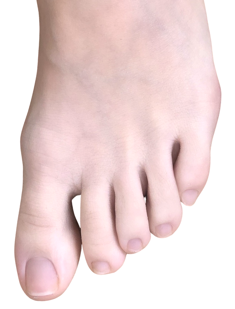
Toes are the digits of the foot of a tetrapod. Animal species such as cats that walk on their toes are described as being digitigrade. Humans, and other animals that walk on the soles of their feet, are described as being plantigrade; unguligrade animals are those that walk on hooves at the tips of their toes.

Structure

[edit]
Bones of the right foot. Plantar surface.
Human toes
A woman's toes decorated with nail polish and henna, and wearing a metti (toe ring) on the second toe, for her wedding

There are normally five toes present on each human foot. Each toe consists of three phalanx bones, the proximal, middle, and distal, with the exception of the big toe (Latin: hallux). For a minority of people, the little toe also is missing a middle bone. The hallux only contains two phalanx bones, the proximal and distal. The joints between each phalanx are the interphalangeal joints. The proximal phalanx bone of each toe articulates with the metatarsal bone of the foot at the metatarsophalangeal joint. Each toe is surrounded by skin, and present on all five toes is a toenail.

The toes are, from medial to lateral:

  • the first toe, also known as the hallux ("big toe", "great toe", "thumb toe"), the innermost toe;
  • the second toe, ("index toe", "pointer toe");
  • the third toe, ("middle toe");
  • the fourth toe, ("fore toe", "ring toe");
  • the fifth toe, ("baby toe", "little toe", "pinky toe", "small toe"), the outermost toe.

Muscles

[edit]

Toe movement is generally flexion and extension (movement toward the sole or the back of the foot, resp.) via muscular tendons that attach to the toes on the anterior and superior surfaces of the phalanx bones.[1]: 573 

With the exception of the hallux, toe movement is generally governed by action of the flexor digitorum brevis and extensor digitorum brevis muscles. These attach to the sides of the bones,[1]: 572–75  making it impossible to move individual toes independently. Muscles between the toes on their top and bottom also help to abduct and adduct the toes.[1]: 579  The hallux and little toe have unique muscles:

Blood supply

[edit]

The toes receive blood from the digital branches of the plantar metatarsal arteries and drain blood into the dorsal venous arch of the foot.[1]: 580–81 

Nerve supply

[edit]

Sensation to the skin of the toes is provided by five nerves. The superficial fibular nerve supplies sensation to the top of the toes, except between the hallux and second toe, which is supplied by the deep fibular nerve, and the outer surface of the fifth toe, supplied by the sural nerve. Sensation to the bottom of the toes is supplied by the medial plantar nerve, which supplies sensation to the great toe and inner three-and-a-half toes, and the lateral plantar nerve, which supplies sensation to the little toe and half of the sensation of the fourth toe.

In humans, the hallux is usually longer than the second toe, but in some individuals, it may not be the longest toe. There is an inherited trait in humans, where the dominant gene causes a longer second toe ("Morton's toe" or "Greek foot") while the homozygous recessive genotype presents with the more common trait: a longer hallux.[2] People with the rare genetic disease fibrodysplasia ossificans progressiva characteristically have a short hallux which appears to turn inward, or medially, in relation to the foot.

Variation

[edit]

Humans usually have five toes on each foot. When more than five toes are present, this is known as polydactyly. Other variants may include syndactyly or arachnodactyly. Forefoot shape, including toe shape, exhibits significant variation among people; these differences can be measured and have been statistically correlated with ethnicity.[3] Such deviations may affect comfort and fit for various shoe types. Research conducted for the U.S. Army indicated that larger feet may still have smaller arches, toe length, and toe-breadth.[4]

Function

[edit]

The human foot consists of multiple bones and soft tissues which support the weight of the upright human. Specifically, the toes assist the human while walking,[5] providing balance, weight-bearing, and thrust during gait.

Clinical significance

[edit]

A sprain or strain to the small interphalangeal joints of the toe is commonly called a stubbed toe. A sprain or strain where the toe joins to the foot is called turf toe.

Long-term use of improperly sized shoes can cause misalignment of toes, as well as other orthopedic problems.

Morton's neuroma commonly results in pain and numbness between the third and fourth toes of the sufferer, due to it affecting the nerve between the third and fourth metatarsal bones.[6]

The big toe is also the most common locus of ingrown nails, and its proximal phalanx joint is the most common locus for gout attacks.

Deformity

[edit]

Deformities of the foot include hammer toe, trigger toe, and claw toe. Hammer toe can be described as an abnormal contraction or “buckling” of a toe. This is done by a partial or complete dislocation of one of the joints, which form the toes. Since the toes are deformed further, these may press against a shoe and cause pain. Deformities of the foot can also be caused by rheumatoid arthritis and diabetes mellitus. Deformities may predispose to ulcers and pain, especially when shoe-wearing.

A common problem involving the big toe is the formation of bunions. These are structural deformities of the bones and the joint between the foot and big toe, and may be painful.[7] Similar deformity involving the fifth toe is described as tailor's bunion or bunionette.

Right-sided duplication of the right little toe in an 8.5 months old male, with two toes (fifth and sixth) apparently forming joints with the fifth metatarsal bone, which is mildly broadened distally. The duplicated toes have almost normal growth. The fifth toe has mild varus angulation, and the sixth toe has substantial valgus angulation.

In polydactyly (which can also affect the fingers) one or more extra toes are present.

In reconstruction

[edit]

A favourable option for the reconstruction of missing adjacent fingers[8]/multiple digit amputations, i.e. such as a metacarpal hand reconstruction, is to have a combined second and third toe transplantation.[9] Third and fourth toe transplantation to the hand in replacing lost fingers is also a viable option.[10]

History

[edit]

Etymology

[edit]

[edit]

The Old English term for toe is (plural tān). This is a contraction of tāhe, and derives from Proto-Germanic *taihwǭ (cognates: Old Norse , Old Frisian tāne, Middle Dutch tee, Dutch teen (perhaps originally a plural), Old High German zēha, German Zehe), perhaps originally meaning 'fingers' as well (many Indo-European languages use one word to mean both 'fingers' and 'toes', e.g. digit), and thus from PIE root *deyḱ- — 'to show'.[11]

Hallux

[edit]
The big toe of a human

In classical Latin, hallex,[12][13] allex,[12][14] hallus[12] and allus,[12] with genitive (h)allicis and (h)alli, are used to refer to the big toe. The form hallux (genitive, hallucis) currently in use is however a blend word of the aforementioned forms.[12][15] Compare pollex, the equivalent term for the thumb.

Evolution

[edit]

Haeckel traces the standard vertebrate five-toed schema from fish fins via amphibian ancestors.[16]

Other animals

[edit]
The skeleton of Pakicetus, an extinct digitigrade mammal.

In birds with anisodactyl or heterodactyl feet, the hallux is opposed or directed backwards and allows for grasping and perching.

While the thumb is often mentioned[by whom?] as one of the signature characteristics in humans, this manual digit remains partially primitive and is actually present in all primates. In humans, the most derived digital feature is the hallux.[17]

See also

[edit]

References

[edit]
Revisions and contributorsEdit on WikipediaRead on Wikipedia
from Grokipedia
A toe is one of the five digits located at the distal end of the human foot, serving as a critical component of the lower limb's anatomy for and stability. Each foot contains five toes, collectively accounting for 14 of the 26 bones in the foot through their phalangeal structure. The great toe, also known as the hallux, consists of two phalanges (proximal and distal), while the lesser toes (second through fifth) each have three phalanges (proximal, middle, and distal). These phalanges articulate via interphalangeal joints, and the proximal phalanges connect to the of the forefoot through metatarsophalangeal (MTP) joints, which are hinge-like structures essential for flexion and extension. The toes are supported by a network of intrinsic and extrinsic muscles, tendons, and ligaments that enable precise movements. Key intrinsic muscles include the flexor digitorum brevis, which flexes the second through fifth toes; the lumbricals, which extend the interphalangeal while flexing the MTP joints; and the interossei muscles, responsible for abducting and adducting the toes. For the great toe, specialized muscles such as the abductor hallucis, flexor hallucis brevis, and adductor hallucis provide additional control for abduction, flexion, and adduction. The fifth toe is supported by the abductor digiti minimi and flexor digiti minimi brevis. These muscular attachments, combined with sesamoid bones in the great toe's MTP , enhance leverage and reduce during activities. Functionally, the toes play a pivotal role in bipedal , balance, and force absorption. The great toe, in particular, contributes significantly to propulsion during the toe-off phase of walking, bearing a significant portion of the body's weight at push-off, while the lesser toes aid in stability and shock absorption upon strike. This structure allows the foot to adapt to varied terrains, distribute pressure across the forefoot, and maintain postural equilibrium. Anatomical variations, such as (extra toes) or (fused toes), can occur but are less common, with the typical configuration optimized for human upright posture.

Anatomy

Bones and Joints

The human foot contains 14 phalanges, which form the skeletal structure of the toes. The hallux, or big toe, consists of two phalanges: a proximal phalanx and a distal phalanx. Each of the lesser toes (second through fifth) has three phalanges: proximal, middle, and distal. These phalanges are elongated bones with a base, shaft, and head, adapted for articulation and weight transmission during locomotion. The toes articulate via synovial joints that enable flexion and extension. The metatarsophalangeal (MTP) joints connect the proximal phalanges to the at the base of each toe, while interphalangeal (IP) joints link the phalanges within each toe: a proximal IP joint between the proximal and middle phalanges (in lesser toes), and a distal IP joint between the middle and distal phalanges (or proximal and distal in the hallux). These joints are stabilized by collateral ligaments on the medial and lateral sides, which prevent excessive lateral deviation, and plantar plates—a fibrocartilaginous structure on the volar aspect—that reinforce the and maintain alignment under load. The hallux differs from the lesser toes in its structure and function, featuring two sesamoid bones embedded in the of the flexor hallucis brevis beneath the head of the first metatarsal at the . These sesamoids, one medial and one lateral, enhance leverage and distribute pressure during push-off. In contrast, the lesser toes lack prominent sesamoids, relying more on the metatarsal heads for support. Biomechanically, the toe bones contribute to weight-bearing by forming the forefoot's transverse arch, with the metatarsal heads collectively comprising the "ball of the foot" that absorbs and propels forces during gait. The first metatarsal head, augmented by sesamoids, bears approximately 30-50% of the body's weight in the stance phase, facilitating efficient propulsion.

Muscles and Tendons

The toes are primarily moved by a combination of extrinsic muscles originating in the leg and intrinsic muscles within the foot, with long tendons transmitting force to the phalanges. Extrinsic muscles include the flexor digitorum longus, which originates from the posterior surface of the tibia and inserts via tendons into the distal phalanges of toes 2–5, enabling plantarflexion at the interphalangeal joints and contributing to overall toe flexion. Similarly, the extensor hallucis longus arises from the anterior fibula and interosseous membrane, inserting into the dorsal aspect of the distal phalanx of the great toe to produce dorsiflexion (extension) of the great toe at the metatarsophalangeal and interphalangeal joints. The extensor digitorum longus, originating from the lateral tibial condyle and fibula, extends toes 2–5 through tendons that divide into slips inserting at the middle and distal phalanges, facilitating dorsiflexion. These extrinsic tendons cross the ankle and metatarsophalangeal joints, allowing powerful but less precise movements compared to intrinsic actions. Intrinsic foot muscles provide fine control over toe positioning, particularly for abduction, adduction, and interphalangeal adjustments. The lumbricals (four in number) originate from the tendons of the flexor digitorum longus and insert into the extensor hood expansions of toes 2–5, flexing the while extending the interphalangeal joints to support balanced toe posture. The dorsal interossei (three muscles) abduct toes 2–5 relative to the second toe axis, inserting into the proximal phalanges and extensor hood, whereas the plantar interossei (three muscles) adduct toes 3–5, aiding in inversion-like adjustments during . These intrinsic muscles enable subtle eversion and inversion per toe through coordinated abduction and adduction, essential for stability on uneven surfaces. Tendon sheaths and pulleys ensure efficient gliding of these tendons across the foot's joints, minimizing friction and preventing bowstringing. Synovial sheaths envelop the long flexor and extensor tendons, such as those of the flexor digitorum longus and extensor hallucis longus, providing lubrication via peritendinous fluid for smooth passage over the phalanges. Annular pulleys (A1–A4 in lesser toes, A1–A3 in the great toe) form thickened fibrous bands at the metatarsophalangeal, proximal interphalangeal, and distal interphalangeal joints, acting as reflection pulleys to guide flexor tendons during plantarflexion. The extensor hood mechanism, a fibroaponeurotic expansion over the dorsal toe, integrates extrinsic tendons like the extensor digitorum longus with intrinsic contributions from lumbricals and interossei, allowing coordinated dorsiflexion at the metatarsophalangeal joint and interphalangeal extension through central and lateral slips. This hood anchors to the joint capsules, distributing forces to prevent tendon subluxation during toe eversion or inversion.

Blood Supply

The blood supply to the toes is provided by a network of arteries originating from the anterior and posterior tibial arteries, ensuring oxygenation and nutrient delivery to the phalanges, , and soft tissues. The dorsal aspect receives arterial supply primarily from the , a continuation of the anterior tibial , which travels along the dorsum of the foot and gives rise to the arcuate artery and the first dorsal metatarsal . The arcuate artery branches into dorsal metatarsal arteries that supply the second through fifth toes via dorsal digital arteries, while the first dorsal metatarsal specifically provides blood to the hallux (big toe) by dividing into medial and lateral branches that form digital arteries for the hallux and the adjacent side of the second toe. On the plantar surface, the divides into the medial and lateral plantar arteries, which form the plantar arch and give rise to plantar metatarsal arteries that branch into plantar digital arteries supplying the toes. These digital arteries from both dorsal and plantar sources anastomose around the toe joints, creating a rich collateral circulation that supports tissue viability and healing. Venous drainage from the toes occurs through dorsal and plantar digital veins that converge into the dorsal and plantar venous arches on the respective surfaces of the foot. The dorsal venous arch, located proximal to the metatarsal heads, drains medially into the and laterally into the small saphenous vein, facilitating superficial venous return to the femoral and popliteal veins, respectively. Similarly, the plantar venous arch collects from the plantar digital veins and connects to the deep plantar veins, which join the tibial veins for return to the heart, aided by the plantar pump mechanism during . In the pulp of the toes, arteriovenous anastomoses (AVAs) provide specialized direct connections between arterioles and venules, bypassing the capillary bed to regulate blood flow and heat dissipation. These AVAs, abundant in the glabrous skin of the toes, enable rapid adjustments in peripheral for , particularly in response to environmental temperature changes within the .

Nerve Supply

The sensory innervation of the toes is provided primarily by branches of the tibial and common fibular (peroneal) nerves. On the plantar surface, the medial plantar nerve, a terminal branch of the tibial nerve, supplies sensation to the medial three toes (hallux, second, and third) and the medial aspect of the fourth toe, while the lateral plantar nerve innervates the lateral aspect of the fourth toe and the fifth toe. On the dorsal surface, the superficial fibular nerve provides sensory input to the dorsum of the second through fifth toes and the medial side of the hallux, whereas the deep fibular nerve innervates the skin in the first dorsal web space between the hallux and second toe. Motor innervation to the toe muscles arises from the same tibial and fibular nerve branches, targeting both intrinsic and extrinsic muscles. The supplies motor fibers to the intrinsic muscles including the abductor hallucis, flexor hallucis brevis, flexor digitorum brevis, and the first lumbrical muscle, facilitating flexion and abduction of the toes. The innervates the remaining intrinsic muscles such as the quadratus plantae, abductor digiti minimi, adductor hallucis, interossei, and the second through fourth lumbricals, enabling adduction, abduction, and fine toe movements. Extrinsic muscles acting on the toes, like the flexor digitorum longus and extensor hallucis longus, receive innervation proximally from the tibial and deep fibular nerves, respectively, before their tendons reach the toes. The toes correspond to dermatomes L4 through S1 of the . The L4 dermatome covers the medial aspect of the foot extending to the medial hallux, L5 supplies the dorsum of the foot and the first three toes, and the S1 dermatome innervates the lateral foot and fifth toe, with digital nerves branching to supply each . Proprioceptive feedback in the toe is mediated by specialized mechanoreceptors, including Meissner's corpuscles and Pacinian corpuscles. Meissner's corpuscles, located in the dermal papillae of glabrous on the toes, detect low-frequency (10-50 Hz) and touch, contributing to tactile and positional during movement. Pacinian corpuscles, situated deeper in the and , respond to high-frequency and rapid pressure changes, aiding in the detection of transient stimuli for proprioceptive integration.

Anatomical Variations

Anatomical variations in toes encompass a range of normal developmental differences that occur without associated , influencing and proportions across individuals. These variations arise primarily from genetic and embryonic factors during limb formation, leading to diversity in digit number, fusion, length, and accessory skeletal elements. Such differences are common and typically asymptomatic, contributing to the wide spectrum of human foot morphology observed in populations worldwide. Polydactyly, characterized by the presence of supernumerary toes, represents a congenital variation where extra digits develop along the preaxial (medial, great toe side) or postaxial (lateral, fifth toe side) borders of the foot. Preaxial often involves duplication of the great toe or hallux, while postaxial forms may add digits adjacent to the fifth toe, with prevalence rates varying by population but generally ranging from 1 in 1,000 live births globally. , another frequent congenital anomaly, involves partial or complete or bony fusion between adjacent toes, most commonly affecting the second and third toes (toes 2-3), and occurs in approximately 1 in 2,000 to 3,000 births. These conditions stem from disruptions in the apoptotic processes that normally separate digits during embryogenesis, yet they are considered normal variants when isolated and non-debilitating. Variations in phalangeal and metatarsal lengths contribute significantly to toe proportions, with two primary foot types distinguished: , where the hallux (big toe) is the longest and subsequent toes taper gradually, and (also known as ), featuring a longer second toe relative to the hallux. The Egyptian configuration predominates in many populations, while the Greek type appears in about 5-30% of individuals depending on ethnic groups, reflecting inherited skeletal ratios that influence weight distribution during . These length disparities arise from differential growth in the metatarsals and phalanges, with the second metatarsal often being longer in Greek feet, a trait linked to autosomal dominant patterns. Sesamoid bones, small ossicles embedded within tendons to reduce friction and enhance leverage, exhibit notable presence and positional variations in the toes, particularly in the hallux where paired medial and lateral sesamoids are typically found plantar to the first metatarsophalangeal joint. Absence or asymmetry of these sesamoids occurs in up to 10-15% of cases, with unilateral variations more common, and they may ossify variably with age or remain cartilaginous. Accessory bones, such as the os peroneum—an ossicle within the peroneus longus tendon near the —represent additional skeletal variants in the lateral foot that can indirectly affect toe alignment, present in 5-30% of individuals and often bilateral. Racial and genetic factors play a key role in the distribution of toe proportions and variations, with certain traits showing higher incidence in specific ethnic groups. For instance, postaxial demonstrates elevated prevalence among individuals of African descent, reported at rates up to 10 times higher than in Caucasian populations, attributed to genetic loci influencing limb bud segmentation. , a condition involving congenital shortening of one or more metatarsals (most often the fourth), leading to a shortened toe, has a genetic basis in autosomal dominant mutations and shows variable incidence across races, though isolated cases are documented more frequently in Asian and African cohorts in clinical series. These ethnic disparities highlight polygenic influences on toe morphology, underscoring the need for population-specific anatomical references in medical and forensic contexts.

Function

Role in Locomotion

In human , toes play a pivotal role during the toe-off phase of the cycle, where dorsiflexion of the , particularly the big toe, engages the mechanism of the to provide propulsion. This mechanism, first described by Hicks in , involves the winding around the metatarsal heads as the toes extend, thereby tightening the , elevating the medial longitudinal arch, and stiffening the foot for efficient forward thrust. The big toe contributes disproportionately to this propulsion during walking by acting as the primary lever against the ground. During the stance phase, toe flexor muscles provide stabilization to maintain foot integrity and prevent collapse, such as excessive flattening that could lead to instability or tendencies under load. These muscles, including the flexor digitorum longus and flexor hallucis longus, contract to grip the ground and support the arch, correlating with longer single-limb support duration and reduced risk of deviations in older adults. Weakness in these flexors has been shown to shorten stride length and decrease walking speed, underscoring their role in controlled . Toe abduction and adduction movements, facilitated by the interossei and abductor hallucis muscles, enhance lateral balance during walking and running by adjusting foot positioning and increasing ground contact area for stability. Stronger toe muscles correlate with improved anterior-posterior and mediolateral stability, as they generate forces to counteract body sway and support rapid postural adjustments, particularly when leaning forward or on uneven . This dynamic adjustment helps prevent falls by optimizing of pressure under the foot. In specialized activities like sprinting, intrinsic toe muscles remain active throughout the phase to maximize and ground reaction forces, enabling faster acceleration compared to walking. During , toes adapt by gripping small holds, with the big toe providing primary leverage and stability through flexion, allowing precise force application on irregular surfaces for upward progression.

Sensory and Balance Functions

The toes play a critical role in tactile sensation through specialized mechanoreceptors in the glabrous of their pulp and soles, enabling detection of ground textures during contact. Merkel cell-neurite complexes, slowly adapting type I receptors, provide sustained feedback on surface features such as roughness and edges, facilitating precise of variations underfoot. Meissner's corpuscles, rapidly adapting type II receptors densely distributed in the dermal papillae of the toes, detect low-frequency vibrations (10-50 Hz) and subtle deformations, contributing to the identification of slip or texture changes on uneven ground. These receptors, innervated by Aβ low-threshold mechanoreceptive afferents, enhance the toes' ability to sense fine spatial details, which is essential for adaptive responses to environmental surfaces without relying solely on visual cues. Proprioceptors within the toe joints and tendons further support position sense, particularly during dynamic weight shifts in bipedal posture. Ruffini endings and Pacinian corpuscles in the joint capsules of the metatarsophalangeal and interphalangeal joints detect static and dynamic changes in toe alignment, providing continuous input on angular positions to the . Golgi tendon organs embedded in the flexor and extensor tendons of the toes monitor tension variations during load transfer, signaling adjustments needed for maintaining foot arch integrity amid weight redistribution. This proprioceptive feedback from the toes allows for subconscious corrections in toe flexion or extension, optimizing stability as body weight oscillates forward over the forefoot. The toes contribute significantly to postural stability by modulating forefoot distribution during quiet standing, where approximately 40% of body weight is borne by the forefoot region including the toes. This sensing, mediated by mechanoreceptors and proprioceptors, helps distribute load across the metatarsal heads and phalanges, reducing sway and enhancing equilibrium by providing real-time data on center-of-pressure shifts. Studies demonstrate that alterations in toe positioning, such as extension, increase forefoot loading and improve postural control metrics like reduced anterior-posterior sway, underscoring the toes' role in fine-tuning base-of-support stability. Toe-derived somatosensory inputs integrate with vestibular and visual systems to orchestrate overall balance, forming a multisensory framework for postural control. Foot mechanoreceptors and proprioceptors relay ground reaction forces to the and cortex, where they converge with vestibular signals from the (detecting head acceleration) and visual cues (orienting to the environment) via the vestibulospinal and reticulospinal tracts. This integration, evaluated through sensory organization tests, prioritizes reliable inputs—such as toe pressure when vision is absent—ensuring adaptive postural adjustments and minimizing fall risk in varying conditions. The somatosensory contributions from the toes thus complement vestibular and visual processing, enabling robust equilibrium through weighted sensory reweighting.

Clinical Significance

Common Disorders and Injuries

Ingrown toenails, also known as onychocryptosis, occur when the edge of the toenail grows into the surrounding , most commonly affecting the big toe. The primary involves improper nail trimming, such as cutting nails too short or in a curved manner rather than straight across, which allows the nail to embed into the . Other contributing factors include wearing tight or ill-fitting shoes that compress the toes and predispose the nail to abnormal growth. Symptoms typically include localized pain and tenderness along the nail edge, inflamed and swollen , and potential secondary bacterial characterized by , increased redness, and warmth. General management for mild cases involves soaking the affected foot in warm several times daily to reduce swelling, gently lifting the nail edge with cotton to promote proper growth, and trimming nails straight across while avoiding cutting too short. If develops, topical or oral antibiotics may be necessary, and persistent cases require professional intervention to prevent recurrence. Toe fractures are common injuries that can be classified as traumatic or stress-related, each arising from distinct mechanisms and often impacting mobility. Traumatic fractures result from acute events, such as a direct blow from dropping a heavy object on the foot or stubbing the toe forcefully, leading to immediate , swelling, bruising, and possible if the is displaced. In contrast, stress fractures develop gradually from repetitive mechanical loading that exceeds the 's repair capacity, commonly seen in athletes or individuals suddenly increasing activity levels, with symptoms including insidious onset of and mild swelling that worsens with . A notable example is the dancer's fracture, a spiral or at the base of the fifth metatarsal, which indirectly affects the little toe through and instability; it typically occurs when the foot rolls inward during activities like , causing sharp at the outer foot edge. for both types emphasizes rest, ice application, elevation, and non- support via crutches or a walking boot, with taping the injured toe to an adjacent one for stability in minor cases; healing generally occurs within 6-8 weeks without surgical intervention unless displacement is severe. Infections of the toes frequently involve fungal or bacterial pathogens, with (tinea pedis) being a prevalent fungal condition that primarily affects the interdigital spaces. Caused by fungi thriving in warm, moist environments, tinea pedis spreads through direct contact with infected skin or contaminated surfaces like locker room floors, leading to symptoms such as intense itching, red or scaly rash, peeling or cracking skin between the toes, and occasional blisters or stinging sensations. Risk factors include heavy foot sweating, prolonged wear of occlusive footwear, and walking barefoot in public damp areas, which facilitate fungal proliferation. Bacterial , often a complication of minor toe trauma or fungal infections, involves bacterial invasion of the skin and subcutaneous tissues, presenting with rapid-onset redness, swelling, warmth, and pain that may extend beyond the initial site. significantly heightens the risk for cellulitis in the toes due to reducing sensation and poor vascular supply impairing , potentially leading to deeper infections like if untreated. Initial management for tinea pedis includes over-the-counter antifungal creams applied twice daily for 2-4 weeks alongside keeping the feet dry and using moisture-absorbing powders, while cellulitis requires systemic antibiotics for 7-14 days, wound care, and elevation to control spread, with diabetic patients needing prompt evaluation to prevent . Gout, particularly in its acute form known as podagra, manifests as intense in the big due to monosodium urate deposition. Elevated serum levels, resulting from overproduction or underexcretion of —often linked to purine-rich diets, alcohol consumption, or conditions like and —lead to formation in the , triggering a sudden inflammatory response. Symptoms of podagra include excruciating pain peaking within 4-12 hours, often awakening the individual at night, accompanied by extreme tenderness, swelling, redness, and warmth, rendering even light touch unbearable. factors encompass , age over 30, , and comorbidities such as or , with attacks potentially recurring if persists. General management involves rest, ice, elevation, and anti-inflammatory medications like or nonsteroidal anti-inflammatory drugs to alleviate acute symptoms, alongside long-term strategies to lower through dietary modifications and urate-lowering therapies.

Deformities

Toe deformities involve structural misalignments of the toes, often resulting from imbalances in muscle, tendon, and joint forces, leading to progressive bending or deviation that can impair mobility and cause pain. These conditions primarily affect the interphalangeal (IP) joints and metatarsophalangeal (MTP) joint, where flexion contractures develop due to dominance of flexor tendons over extensors. Hammertoe is characterized by a flexion at the proximal interphalangeal (PIP) joint, causing the toe—typically the second, third, or fourth—to bend downward in a hammer-like , with the distal interphalangeal (DIP) joint remaining neutral or hyperextended. Claw toe, a related , extends this pattern with hyperextension at the MTP joint and flexion at both the PIP and DIP joints, often affecting multiple toes. Both arise from muscle imbalances where extrinsic flexors overpower weakened intrinsic muscles, frequently exacerbated by tight or high-heeled shoes that force toes into prolonged bent positions. Mallet toe presents as an isolated flexion deformity at the DIP joint, usually limited to one toe such as the second or third, without significant involvement of the PIP or MTP joints. This condition often stems from direct pressure from ill-fitting , leading to tightening of the flexor digitorum longus . Hallux valgus, commonly known as a , involves lateral deviation of the big toe at the first MTP joint, with medial protrusion of the first metatarsal head, often associated with metatarsus primus varus where the first metatarsal angles inward. The precise is multifactorial, but it commonly progresses from inherited foot structures, joint hypermobility, or biomechanical stresses, worsened by narrow shoes. Common risk factors across these deformities include , such as inherited laxity or foot shape; improper like high heels or pointed toes; and systemic conditions like , which inflame joints and promote instability. Women face higher risk due to choices and hormonal influences on . Progression typically begins as a flexible misalignment, allowing some correction through positioning, but advances to rigid contractures as tendons shorten, joint capsules tighten, and secondary issues like corns or calluses form from —potentially leading to and altered if unmanaged.

Surgical and Reconstructive Procedures

Surgical and reconstructive procedures for toe conditions primarily address structural deformities, injuries, and severe infections through targeted interventions that restore alignment, function, and stability. Bunionectomy, also known as hallux valgus correction, involves of the combined with realignment to reposition the big toe and alleviate pain from the deformity. This procedure typically includes a distal release to balance the and ligaments, enhancing the corrective effect and preventing recurrence. For mild to moderate cases, a chevron is commonly employed, where an incision is made over the big toe to cut and realign the , often secured with screws or pins for stability. Outcomes generally show pain relief and improved footwear tolerance for the majority of patients, though recovery may involve 6-12 weeks of limited . Tendon transfers represent a key technique for correcting flexible claw toe deformities, where the toe exhibits hyperextension at the metatarsophalangeal joint and flexion at the proximal interphalangeal joint. In the flexor-to-extensor rerouting procedure, the or hallucis longus is harvested and transferred to the extensor hood at the base of the proximal to counteract the imbalance and promote proper toe alignment. This minimally invasive approach is particularly effective for dynamic deformities, often performed alongside or capsulotomy for optimal results. Postoperative outcomes show effective correction in most cases with low complication rates, allowing patients to resume normal activities within 4-6 weeks. Amputation of toes is reserved for severe cases where conservative measures fail, with primary indications including uncontrolled , extensive tissue from trauma or vascular insufficiency, and non-viable bone due to . Procedures are performed at varying levels depending on the extent of involvement: partial phalangeal amputation for distal tip , interphalangeal for mid-toe involvement, or ray amputation (toe plus metatarsal) for proximal spread to preserve foot function. In diabetic patients, early intervention at the phalangeal level can prevent progression to higher s, with healing achievable in many cases when vascular status is adequate. Reconstructive options post-injury focus on restoring joint integrity and preventing chronic instability, with arthrodesis (joint fusion) serving as a reliable method for toe interphalangeal or metatarsophalangeal joints damaged by trauma. This involves removing articular cartilage and fixating the bones with screws, plates, or wires to achieve bony union, thereby eliminating painful motion while maintaining weight-bearing capacity. For the first metatarsophalangeal joint, fusion is the gold standard in post-traumatic arthritis or instability, achieving union in over 90% of cases and significant pain reduction. Recovery typically spans 8-12 weeks, with patients progressing to full weight-bearing in supportive footwear once radiographic fusion is confirmed.

Development and Evolution

Embryological Development

The embryological development of the toes originates from the lower limb buds, which emerge during the fourth week of gestation as protrusions from the , covered by . These buds rapidly elongate under the influence of the apical ectodermal ridge (AER), a thickened ectodermal structure at the distal margin that secretes fibroblast growth factors (FGFs), such as FGF8, to promote proximo-distal outgrowth and patterning of the limb elements, including the future toes. The AER interacts with the underlying progress zone of to ensure sequential development from proximal to distal structures. By the sixth week, the flattened limb paddle develops paddle-like digital rays, the anlagen of the five toes, initially connected by interdigital tissue. Digit separation occurs between weeks 7 and 8 through interdigital necrosis, a localized (apoptosis) in the mesenchymal , sculpting individual toes while preserving the rays. This process is regulated by signaling molecules like bone morphogenetic proteins (BMPs), which trigger in the interdigital regions. Phalangeal segmentation within each toe arises from chondrification centers that form around week 6, where mesenchymal condensations differentiate into templates for the proximal, middle, and distal phalanges. These centers segment through differential growth and formation, with the hallux (first toe) differentiating distinctly by retaining only two phalanges, a pattern established by week 7 via anterior-posterior signaling gradients independent of Sonic hedgehog (SHH) for the first digit. Exposure to teratogens during the critical window of weeks 4-8 can disrupt toe formation; for instance, inhibits and interferes with cereblon-mediated protein degradation, leading to limb reduction defects such as and alterations in digit number or structure.

Evolutionary Aspects

The of the toe reflects a profound transition from prehensile appendages adapted for in early ancestors to rigid, weight-bearing structures optimized for terrestrial in hominins. In arboreal , toes functioned primarily for grasping branches, with long, curved phalanges and opposable digits enabling secure arboreal travel. This grasping capability persisted in early hominins but gradually diminished as bipedal locomotion became habitual, shifting the foot's role to propulsion and shock absorption during upright walking. Fossil evidence indicates this began around 6 million years ago, with early hominins retaining ape-like toe features while developing initial bipedal adaptations. A key milestone occurred with the loss of opposability in the lesser toes (digits 2–5), which aligned parallel to the metatarsals and shortened, reducing their grasping function to facilitate efficient weight transfer during bipedal strides. This change is evident by approximately 4.4 million years ago in , whose foot phalanges show reduced curvature compared to modern apes, indicating early commitment to . In contrast, species (circa 4–2 million years ago) further solidified these adaptations, with fossilized foot bones from A. afarensis displaying straight, non-opposable lesser toes that supported a more stable platform for walking, as seen in the footprints. These modifications enhanced energy efficiency in by creating a lever-like forefoot. The big toe (hallux) underwent a more protracted transformation, evolving from a divergent, partially opposable structure to a fully adducted, aligned digit crucial for push-off in modern human . In , the hallux remained widely abducted, retaining prehensile potential for climbing while the midfoot exhibited increased rigidity absent in apes, allowing limited bipedal propulsion. This partial grasping persisted into early but was largely lost by around 2.3–1.2 million years ago in early , where morphology shows complete adduction and robust alignment with the other toes, enabling forceful toe-off and longitudinal arch support. Modern human toes, by comparison, are short, straight, and non-grasping, contributing to the foot's overall stiffness and efficiency in bipedal endurance running and walking.

Etymology and History

Origins of Terminology

The English word "toe" originates from the "tā," referring to a digit of the foot or a finger-like projection, which traces back to the Proto-Germanic *taihwō(n), meaning a toe or similar appendage. This term evolved through "to," maintaining its association with the protruding parts of the foot, akin to cognates in "tá" and "zēha." The term "hallux," used specifically for the big toe, derives from Late Latin "hallux" or "allus," denoting the great toe, of unknown origin but possibly influenced by Greek terms implying a prominent structure. Introduced into modern anatomical in the early 19th century, it emphasizes the toe's and positional significance in the foot. In anatomical convention, toes are designated as digits 1 through 5, with the hallux as digit 1 (medialmost) and the little toe as digit 5 (lateralmost), a numbering system originating from standard human morphology descriptions to facilitate consistent medical and scientific reference. Culturally, informal terms include "pinky toe" for the fifth digit due to its small , and like "dogs" for feet or toes collectively, emerging in early 20th-century American English from rhyming ("dog's " for "feet") to describe sore or tired extremities.

Historical Perspectives

Ancient Egyptian practices demonstrate early attention to toe preservation and treatment. During mummification, embalmers meticulously preserved foot structures, including toes, using and resins to dehydrate and protect tissues, allowing modern examinations to reveal pathologies such as deformities and even prosthetic toes crafted from wood or for the . Recent studies, including biomechanical analyses as of 2017, confirm these prosthetics were functional, aiding gait. The , dating to approximately 1550 BCE, documents remedies for toe ailments, including salves with red , , and linseed applied to painful areas, as well as treatments for conditions resembling using extracts mixed with other ingredients. These texts highlight a blend of empirical and magical incantations in addressing toe pain and . In the 5th century BCE, Greek physician provided one of the earliest detailed clinical descriptions of , particularly its manifestation as podagra—an acute of the big toe —characterizing it as "the unwalkable disease" due to severe pain that rendered patients immobile. His observations in the emphasized environmental and dietary factors, such as rich foods and seasonal changes, influencing toe afflictions, laying foundational principles for later . The marked a pivotal advancement in anatomical understanding of toes through ' seminal work, De Humani Corporis Fabrica (1543), which featured precise illustrations of the lower limb musculature, including the intrinsic and extrinsic muscles controlling toe flexion and extension. Vesalius' dissections corrected Galenic errors and depicted toe structures in layered views, from skeletal to muscular systems, influencing subsequent anatomical studies. In the 19th and 20th centuries, toe-related pathologies gained further recognition with Thomas G. Morton's 1876 description of , now known as , a painful thickening of tissue around a between the toes, often the third and fourth. Post-World War II, emerged as a modern specialized field, driven by the need to treat soldiers' foot injuries; in the mid-20th century, particularly from the , U.S. practitioners fully transitioned from chiropody to earning Doctor of Podiatric Medicine degrees, with formalized curricula emphasizing surgical and conservative toe interventions.

Comparative Anatomy

Toes in Non-Human Animals

In mammals, toe structures exhibit significant diversity adapted to various locomotor needs. Carnivores, such as cats in the family Felidae, possess clawed toes where the claws are retractile, allowing them to be extended and retracted via a specialized mechanism involving elastic ligaments and the deep digital flexor tendon; this structure keeps the claws sharp and protected when not in use. In contrast, ungulates display hoofed toes, with even-toed ungulates (Artiodactyla) like cattle and deer featuring cloven hooves formed by the fusion of digits II and III, while odd-toed ungulates (Perissodactyla) such as horses have a single weight-bearing toe (digit III) encased in a solid hoof. Vestigial toes are common in some mammals, serving as non-weight-bearing remnants. In dogs (Canis familiaris), dewclaws represent these reduced digits, typically located on the medial aspect of the forelimbs (digit I) and sometimes hindlimbs, lacking full and tendinous attachments for ground contact and thus not contributing to primary locomotion. Birds generally have anisodactyl or zygodactyl foot configurations, with the latter featuring a reversed fourth toe (digit IV) pointing backward alongside the hallux (digit I), while digits II and III point forward; this arrangement is prevalent in orders like Psittaciformes (parrots) and Strigiformes (), enabling enhanced grasping. The zygodactyl pattern has evolved independently multiple times, as evidenced by developmental studies showing backward rotation of digit IV during embryogenesis. Reptiles display a range of toe morphologies, with most lizards (Squamata: Lacertilia) retaining pentadactyl limbs—four limbs each with five toes (digits I–V) terminating in claws—reflecting the ancestral condition. In snakes (Squamata: Serpentes), evolutionary limb reduction has led to the complete loss of external toes and limbs in most species, though vestigial rudiments persist internally as scaled protrusions in some taxa like boas, resulting from modifications in regulatory networks during development.

Adaptations Across Species

In , prehensile toes enable enhanced grasping capabilities essential for , including and brachiation. The opposable hallux (big toe) in species such as chimpanzees and allows the foot to function similarly to a hand, facilitating secure grips on branches during vertical and suspension. This adaptation is particularly evident in great apes, where the foot's flexibility and muscle arrangement support propulsive forces during tree traversal, with studies showing that hindlimb grasping contributes significantly to stability in dynamic arboreal environments. Opposability in the big toe persists as a primitive trait across many lineages, aiding in load distribution and preventing slippage on irregular surfaces. Webbed toes represent a key hydrodynamic adaptation in semi-aquatic and fully aquatic species, increasing surface area for propulsion during swimming. In otters, such as the North American river otter (Lontra canadensis), fully webbed hind feet act as efficient paddles, enhancing thrust and maneuverability in water while allowing terrestrial mobility through partial retraction of the webbing. Similarly, sea otters (Enhydra lutris) exhibit densely webbed hind paws that generate greater propulsive efficiency, supporting sustained foraging dives and rapid directional changes in marine habitats. Among amphibians, webbed feet in anurans like frogs optimize swimming performance by creating vortex rings for forward propulsion, with the degree of webbing varying to balance aquatic efficiency against terrestrial needs. This convergent evolution underscores the webbing's role in reducing drag and amplifying force application in fluid media. Arboreal reptiles like geckos demonstrate specialized padded toes for on vertical and inverted surfaces, crucial for navigating foliage and bark. The subdigital pads of geckos are covered in millions of microscopic setae—branched, hair-like structures—that exploit van der Waals forces to achieve reversible attachment, enabling them to climb smooth substrates without residue. These setae enhance frictional during dynamic locomotion, with adhesion strength increasing at higher sliding speeds to support rapid traversal of uneven arboreal terrains. The hierarchical structure of setae, from macroscale pads to nanoscale spatulae, allows directional control, where toe angling maximizes contact and detachment for efficient movement. Cursorial adaptations in ungulates, exemplified by horses (Equus spp.), involve elongation of central toes and reduction of lateral digits to optimize speed and endurance on open plains. In modern equids, the single elongated third digit forms a hoof that concentrates force for efficient ground contact, minimizing energy loss during high-speed galloping. This monodactyly evolved from multi-toed ancestors through progressive reduction, driven by selective pressures for cursorial locomotion, where longer toes and fused metacarpals enhance stride length and stability at velocities exceeding 50 km/h. Fossil evidence indicates that side toe vestiges in early equids provided auxiliary support on soft substrates, but their elimination streamlined limb mechanics for sustained terrestrial pursuit.

References

Add your contribution
Related Hubs
User Avatar
No comments yet.