Rhinitis
Rhinitis
Main page
2184398

Rhinitis

logo
Community Hub0 subscribers
Read side by side
from Wikipedia
Rhinitis
Other namesCoryza
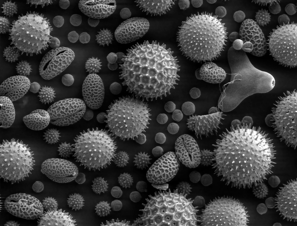
Pollen grains from a variety of common plants can cause hay fever.
Pronunciation
SpecialtyInfectious disease, allergy and immunology

Rhinitis, also known as coryza,[3] is irritation and inflammation of the mucous membrane inside the nose. Common symptoms are a stuffy nose, runny nose, sneezing, and post-nasal drip.[4]

The inflammation is caused by viruses, bacteria, irritants or allergens. The most common kind of rhinitis is allergic rhinitis,[5] which is usually triggered by airborne allergens such as pollen and dander.[6] Allergic rhinitis may cause additional symptoms, such as sneezing and nasal itching, coughing, headache,[7] fatigue, malaise, and cognitive impairment.[8][9] The allergens may also affect the eyes, causing watery, reddened, or itchy eyes and puffiness around the eyes.[7] The inflammation results in the generation of large amounts of mucus, commonly producing a runny nose, as well as a stuffy nose and post-nasal drip. In the case of allergic rhinitis, the inflammation is caused by the degranulation of mast cells in the nose. When mast cells degranulate, they release histamine and other chemicals,[10] starting an inflammatory process that can cause symptoms outside the nose, such as fatigue and malaise.[11] In the case of infectious rhinitis, it may occasionally lead to pneumonia, either viral or bacterial. Sneezing also occurs in infectious rhinitis to expel bacteria and viruses from the respiratory tract.

Rhinitis is very common. Allergic rhinitis is more common in some countries than others; in the United States, about 10–30% of adults are affected annually.[12] Mixed rhinitis (MR) refers to patients with nonallergic rhinitis and allergic rhinitis. MR is a specific rhinitis subtype. It may represent between 50 and 70% of all AR patients. However, true prevalence of MR has not been confirmed.[13]

Types

[edit]

Rhinitis is categorized into three types (although infectious rhinitis is typically regarded as a separate clinical entity due to its transient nature): (i) infectious rhinitis includes acute and chronic bacterial infections; (ii) nonallergic rhinitis[14] includes vasomotor, idiopathic, hormonal, atrophic, occupational, and gustatory rhinitis, as well as rhinitis medicamentosa (rebound congestion); (iii) allergic rhinitis, triggered by pollen, mold, animal dander, dust, Balsam of Peru, and other inhaled allergens.[15]

Infectious

[edit]

Rhinitis is commonly caused by a viral or bacterial infection, including the common cold, which is caused by Rhinoviruses, Coronaviruses, and influenza viruses, others caused by adenoviruses, human parainfluenza viruses, human respiratory syncytial virus, enteroviruses other than rhinoviruses, metapneumovirus, and measles virus, or bacterial sinusitis, which is commonly caused by Streptococcus pneumoniae, Haemophilus influenzae, and Moraxella catarrhalis. Symptoms of the common cold include rhinorrhea, sneezing, sore throat (pharyngitis), cough, congestion, and slight headache.[16][citation needed]

Nonallergic rhinitis

[edit]

Nonallergic rhinitis refers to rhinitis that is not due to an allergy. The category was formerly referred to as vasomotor rhinitis, as the first cause discovered was vasodilation due to an overactive parasympathetic nerve response. As additional causes were identified, additional types of nonallergic rhinitis were recognized. Vasomotor rhinitis is now included among these under the more general classification of nonallergic rhinitis.[17] The diagnosis is made upon excluding allergic causes.[18] It is an umbrella term of rhinitis of multiple causes, such as occupational (chemical), smoking, gustatory, hormonal, senile (rhinitis of the elderly), atrophic, medication-induced (including rhinitis medicamentosa), local allergic rhinitis, non-allergic rhinitis with eosinophilia syndrome (NARES) and idiopathic (vasomotor or non-allergic, non-infectious perennial allergic rhinitis (NANIPER), or non-infectious non-allergic rhinitis (NINAR).[19]

In vasomotor rhinitis,[20][21] certain nonspecific stimuli, including changes in environment (temperature, humidity, barometric pressure, or weather), airborne irritants (odors, fumes), dietary factors (spicy food, alcohol), sexual arousal, exercise,[22] and emotional factors trigger rhinitis.[23] There is still much to be learned about this, but it is thought that these non-allergic triggers cause dilation of the blood vessels in the lining of the nose, which results in swelling and drainage.

Non-allergic rhinitis can co-exist with allergic rhinitis, and is referred to as "mixed rhinitis".[24] The pathology of vasomotor rhinitis appears to involve neurogenic inflammation[25] and is as yet not very well understood. The role of transient receptor potential ion channels on the non-neuronal nasal epithelial cells has also been suggested. Overexpression of these receptors have influence the nasal airway hyper-responsiveness to non-allergic irritant environmental stimuli (e.g., extremes of temperature, changes in osmotic or barometric pressure).[26] Vasomotor rhinitis appears to be significantly more common in women than men, leading some researchers to believe that hormone imbalance plays a role.[27][28] In general, age of onset occurs after 20 years of age, in contrast to allergic rhinitis which can be developed at any age. Individuals with vasomotor rhinitis typically experience symptoms year-round, though symptoms may be exacerbated in the spring and autumn when rapid weather changes are more common.[17] An estimated 17 million United States citizens have vasomotor rhinitis.[17]

Drinking alcohol may cause rhinitis as well as worsen asthma (see alcohol-induced respiratory reactions). In certain populations, particularly those of East Asian countries such as Japan, these reactions have a nonallergic basis.[29] In other populations, particularly those of European descent, a genetic variant in the gene that metabolizes ethanol to acetaldehyde, ADH1B, is associated with alcohol-induced rhinitis. It is suggested that this variant metabolizes ethanol to acetaldehyde too quickly for further processing by ALDH2 and thereby leads to the accumulation of acetaldehyde and rhinitis symptoms.[30] In these cases, alcohol-induced rhinitis may be of the mixed rhinitis type and, it seems likely, most cases of alcohol-induced rhinitis in non-Asian populations reflect true allergic response to the non-ethanol and/or contaminants in alcoholic beverages, particularly when these beverages are wines or beers.[29] Alcohol-exacerbated rhinitis is more frequent in individuals with a history of rhinitis exacerbated by aspirin.[31]

Aspirin and other non-steroidal anti-inflammatory drugs (NSAIDs), particularly those that inhibit cyclooxygenase 1 (COX1), can worsen rhinitis and asthma symptoms in individuals with a history of either one of these diseases.[32] These exacerbations most often appear due to NSAID hypersensitivity reactions rather than NSAID-induced allergic reactions.[33]

The antihistamine azelastine, applied as a nasal spray, may be effective for vasomotor rhinitis.[34] Fluticasone propionate or budesonide (both are steroids) in nostril spray form may also be used for symptomatic treatment. The antihistamine cyproheptadine is also effective, probably due to its antiserotonergic effects.

A systematic review on non-allergic rhinitis reports improvement of overall function after treatment with capsaicin (the active component of chili peppers). The quality of evidence is low, however.[35]

Allergic

[edit]

Allergic rhinitis or hay fever may follow when an allergen such as pollen, dust, or Balsam of Peru[36] is inhaled by an individual with a sensitized immune system, triggering antibody production. These antibodies mostly bind to mast cells, which contain histamine. When the mast cells are stimulated by an allergen, histamine (and other chemicals) are released. This causes itching, swelling, and mucus production.

Symptoms vary in severity between individuals. Very sensitive individuals can experience hives or other rashes. Particulate matter in polluted air and chemicals such as chlorine and detergents, which can normally be tolerated, can greatly aggravate the condition.[citation needed]

Characteristic physical findings in individuals who have allergic rhinitis include conjunctival swelling and erythema, eyelid swelling, lower eyelid venous stasis, lateral crease on the nose, swollen nasal turbinates, and middle ear effusion.[37]

Even if a person has negative skin-prick, intradermal and blood tests for allergies, they may still have allergic rhinitis, from a local allergy in the nose. This is called local allergic rhinitis.[38] Many people who were previously diagnosed with nonallergic rhinitis may actually have local allergic rhinitis.[39]

A patch test may be used to determine if a particular substance is causing the rhinitis.

Rhinitis medicamentosa

[edit]

Rhinitis medicamentosa is a form of drug-induced nonallergic rhinitis which is associated with nasal congestion brought on by the use of certain oral medications (primarily sympathomimetic amine and 2-imidazoline derivatives) and topical decongestants (e.g., oxymetazoline, phenylephrine, xylometazoline, and naphazoline nasal sprays) that constrict the blood vessels in the lining of the nose.[40]

Chronic atrophic rhinitis

[edit]

Chronic rhinitis is a form of atrophy of the mucous membrane and glands of the nose.

Rhinitis sicca

[edit]

Chronic form of dryness of the mucous membranes.

Polypous rhinitis

[edit]

Chronic rhinitis associated with polyps in the nasal cavity.

Pathophysiology

[edit]
Pathological changes in non-allergic rhinitis[26]
Mechanism of non-allergic rhinitis: Imbalance between sympathetic and parasympathetic components in the nasal mucous membrane[26]

Most prominent pathological changes observed are nasal airway epithelial metaplasia in which goblet cells replace ciliated columnar epithelial cells in the nasal mucous membrane.[26] This results in mucin hypersecretion by goblet cells and decreased mucociliary activity. Nasal secretion are not adequately cleared with clinical manifestation of nasal congestion, sinus pressure, post-nasal dripping, and headache. Over-expression of transient receptor potential (TRP) ion channels, such as TRPA1 and TRPV1, may be involved in the pathogenesis of non-allergic rhinitis.[41]

Association between rhinitis and asthma

[edit]

Neurogenic inflammation produced by neuropeptides released from sensory nerve endings to the airways is a proposed common mechanism of association between both allergic and non-allergic rhinitis with asthma. This may explain higher association of rhinitis with asthma developing later in life.[42] Environmental irritants acts as modulators of airway inflammation in these contiguous airways. Development of occupational asthma is often preceded by occupational rhinitis. Among the causative agents are flours, enzymes used in processing food, latex, isocyanates, welding fumes, epoxy resins, and formaldehyde. Accordingly, prognosis of occupational asthma is contingent on early diagnosis and the adoption of protective measures for rhinitis.[43]

Diagnosis

[edit]

The different forms of rhinitis are essentially diagnosed clinically.[clarification needed] Vasomotor rhinitis is differentiated from viral and bacterial infections by the lack of purulent exudate and crusting. It can be differentiated from allergic rhinitis because of the absence of an identifiable allergen.[44]

Evidence has been published from a few health apps for mobile devices that show potential to assist in the diagnosis of rhinitis and rhinosinusitis and to evaluate management and treatment adherence. While this shows promise for clinical management, as of 2022 few had been validated in the scientific literature, and even fewer included considerations for multimorbidity.[45][46]

Management

[edit]

The management of rhinitis depends on the underlying cause.

For allergic rhinitis, intranasal corticosteroids are recommended.[47] For severe symptoms intranasal antihistamines may be added.[47]

Pronunciation and etymology

[edit]

Rhinitis is pronounced /raɪˈnaÉŖtÉŖs/,[48] while coryza is pronounced /kÉ™ĖˆraÉŖzə/.[49]

Rhinitis comes from the Ancient Greek ῄίς rhis, gen.: įæ„Ī¹Ī½ĻŒĻ‚ rhinos, "nose". Coryza comes through Latin from Ancient Greek ĪŗĻŒĻĻ…Ī¶Ī±. According to physician Andrew Wylie, "we use the term [coryza] for a cold in the head, but the two are really synonymous. The ancient Romans advised their patients to clean their nostrils and thereby sharpen their wits."[50]

References

[edit]
Revisions and contributorsEdit on WikipediaRead on Wikipedia
from Grokipedia
Rhinitis is an inflammation of the nasal mucosa, defined by one or more symptoms including nasal congestion, rhinorrhea (runny nose), sneezing, nasal itching, and nasal obstruction.[1] It represents a common global health issue that can impair quality of life, sleep, and daily functioning, often extending to associated conditions like sinusitis or conjunctivitis.[1] The condition is broadly classified into two main types: allergic rhinitis (AR), also known as hay fever, and non-allergic rhinitis (NAR).[2] Allergic rhinitis occurs due to an immunoglobulin E (IgE)-mediated immune response to inhaled allergens such as pollen, dust mites, animal dander, or mold spores, leading to the release of inflammatory mediators like histamine.[3] It is further subdivided into seasonal (intermittent, e.g., triggered by pollen) and perennial (persistent, e.g., from indoor allergens) forms based on exposure patterns.[1] In contrast, non-allergic rhinitis involves nasal inflammation without allergic sensitization, triggered by non-immunologic factors including irritants (e.g., tobacco smoke, perfumes), weather changes, viral infections, medications (e.g., aspirin), hormonal fluctuations, or occupational exposures.[4] Epidemiologically, rhinitis affects a substantial portion of the population worldwide, with allergic rhinitis prevalence estimated at 10% to 30% based on physician diagnosis and self-reporting, showing higher rates in industrialized regions and among individuals with a family history of atopy.[2] Non-allergic rhinitis prevalence is less precisely quantified but is believed to impact 12% to 19% of adults, often overlapping with other forms in mixed rhinitis cases.[5] Symptoms typically manifest as nasal blockage, clear or mucoid discharge, and sneezing bouts, with AR frequently accompanied by ocular itching, tearing, and throat irritation, while NAR may include more prominent postnasal drip.[2][4] Management focuses on trigger avoidance, pharmacotherapy (e.g., intranasal corticosteroids, antihistamines), and, for AR, allergen immunotherapy to achieve long-term symptom control.[6]

Overview

Definition

Rhinitis is defined as an inflammation of the mucous membrane lining the nasal passages, resulting in a heterogeneous group of symptoms primarily affecting the nose.[6] This condition manifests through nasal congestion, rhinorrhea (runny nose), sneezing, and itching of the nasal mucosa.[7] It arises from various triggers leading to nasal inflammation or dysfunction, but the core feature is localized to the nasal cavity without involvement of deeper structures unless specified otherwise.[1] Rhinitis must be distinguished from sinusitis, which involves inflammation of the paranasal sinuses in addition to or instead of the nasal passages, often presenting with facial pain or pressure as key differentiators.[8] It also differs from the common cold, an acute viral upper respiratory infection that typically resolves within 7-10 days and includes systemic symptoms like fever or sore throat, whereas rhinitis can persist beyond this timeframe without viral etiology.[9] Rhinitis is classified based on duration into acute forms, typically lasting up to 7-10 days and often linked to transient irritants or infections, and chronic forms, persisting for more than 12 weeks and requiring evaluation for underlying contributors.[10] This temporal distinction helps guide clinical assessment, though causes are explored separately in typological classifications.[10]

Epidemiology

Rhinitis is a common condition worldwide, affecting an estimated 10% to 40% of the global population as of 2023, with allergic rhinitis comprising the majority of cases. In industrialized regions such as Europe and the United States, prevalence rates reach up to 30% to 40%, particularly among children and adolescents, based on data up to 2023. A systematic review of studies reported median prevalences of 29.4% for unspecified rhinitis, 18.1% for allergic rhinitis, and 12.0% for nonallergic rhinitis, with wide variations (1% to 63%) depending on diagnostic criteria and populations studied.[11][12][13][5] The incidence of rhinitis has been increasing over recent decades, driven by factors including urbanization, air pollution, and climate change, which exacerbate allergen exposure and sensitization. In children, the annual incidence averages around 3%, with cumulative rates reaching up to 17% in the first five years of life and 33.6% from ages 8 to 19. These trends reflect broader rises in allergic diseases, with sensitization rates among schoolchildren approaching 40% to 50% globally.[14][15][16][17][18][19] Key risk factors for rhinitis include genetic predisposition, where a family history of allergic diseases increases the odds by 3- to 5-fold, with an odds ratio of 3.6 for parental allergic rhinitis. Environmental exposures such as pollens and dust mites further elevate risk, alongside age-related patterns peaking in childhood and young adulthood. Post-puberty, prevalence is slightly higher in females, potentially due to hormonal influences.[20][21][22][23][24] Regional variations show higher prevalence in urban areas compared to rural settings, with sensitization rates increasing with urbanization (e.g., 17.2% urban vs. 6.0% rural in some studies), attributed to greater pollution and allergen concentrations. The COVID-19 pandemic temporarily reduced reported acute rhinitis cases from 2020 to 2022, likely due to lockdowns limiting exposures and healthcare access. Economically, rhinitis imposes a substantial burden; as of 2021, total annual costs in the United States exceed $18 billion, encompassing direct medical expenses of about $4.6 billion and significant indirect costs from lost productivity.[25][26][27][28][29][30]

Signs and Symptoms

Nasal Symptoms

Nasal congestion, a hallmark symptom of rhinitis, manifests as a sensation of blockage or stuffiness in the nasal passages due to swelling of the nasal mucosa caused by inflammation.[31] This swelling often follows the physiological nasal cycle, leading to alternating congestion between the nostrils, which can exacerbate the feeling of obstruction in affected individuals.[32] Rhinorrhea refers to excessive nasal discharge, which is typically clear and watery in allergic rhinitis due to histamine-mediated vascular permeability.[2] In contrast, infectious rhinitis often produces purulent, thicker discharge resulting from bacterial or viral involvement.[33] Sneezing occurs in paroxysmal bursts, particularly prominent in allergic rhinitis, triggered by irritants that stimulate sensory nerves in the nasal mucosa.[34] These episodes can involve multiple consecutive sneezes, contributing to significant discomfort.[35] Nasal itching, or pruritus, arises from the irritation of the nasal lining and frequently prompts repetitive rubbing or sniffing behaviors to alleviate the sensation.[36] This symptom is especially characteristic of allergic forms, where it often accompanies sneezing.[2] Postnasal drip involves the perception of mucus accumulating and draining from the back of the nose into the throat, often resulting from increased mucus production in the nasal passages.[2] Reduced sense of smell (hyposmia) can also occur, particularly in chronic cases, due to inflammation affecting the olfactory epithelium.[37] The duration of nasal symptoms in rhinitis can vary, classified as intermittent if occurring fewer than four days per week or for less than four consecutive weeks, or persistent if more frequent or prolonged, typically influenced by ongoing allergen or irritant exposure.[38] In chronic rhinitis, symptoms are typically persistent and commonly include nasal congestion or stuffiness, runny nose often with clear discharge, postnasal drip, frequent throat clearing or cough, reduced sense of smell (hyposmia), and facial pressure or headache in some cases. Sneezing is generally less prominent compared to acute or allergic forms. Symptoms vary depending on the type of chronic rhinitis; allergic forms often feature more prominent sneezing and nasal itching, whereas non-allergic forms typically lack these prominent itching and sneezing symptoms.[37][2]

Extranasal and Associated Symptoms

Rhinitis, particularly the allergic form, often manifests with extranasal symptoms that extend beyond the nasal passages, significantly impacting patients' daily functioning. Ocular symptoms are among the most common extranasal manifestations, affecting up to 70% of individuals with allergic rhinitis. These include allergic conjunctivitis characterized by eye itching, redness, tearing, and swelling of the eyelids, resulting from the systemic release of inflammatory mediators like histamine that affect the conjunctival mucosa.[39] Throat and ear involvement further contributes to discomfort in rhinitis patients. Postnasal drip can lead to a sore or irritated throat, coughing, and a sensation of throat clearing; however, significant swelling in the throat that obstructs breathing is not a typical symptom of rhinitis.[40] While ear-related symptoms such as fullness, popping, or pain arise from Eustachian tube dysfunction caused by mucosal swelling and inflammation extending to the nasopharynx. These symptoms are reported in approximately 20-40% of cases and can exacerbate during allergen exposure.[41] Systemic effects of rhinitis include fatigue, headaches, and sleep disturbances, primarily driven by chronic nasal congestion that impairs breathing and rest. Headaches often stem from sinus pressure secondary to inflammation, while fatigue results from disrupted sleep and the overall inflammatory burden, with studies indicating that up to 50% of patients experience moderate to severe tiredness. Sleep disturbances, such as snoring or frequent awakenings, are prevalent in 40-60% of affected adults and children.[42] Rhinitis is associated with several comorbidities that heighten disease burden. It increases the risk of asthma exacerbations, with allergic rhinitis present in over 80% of asthma patients, creating a bidirectional inflammatory pathway that worsens respiratory symptoms.[43] Additionally, chronic rhinitis predisposes individuals to recurrent sinus infections (rhinosinusitis), as nasal obstruction hinders sinus drainage, leading to bacterial overgrowth. The quality of life impacts of these extranasal and associated symptoms are profound, often more challenging to manage than nasal ones alone. Patients report reduced productivity at work or school, with allergic rhinitis linked to 2-3 times higher rates of absenteeism in children compared to unaffected peers.[44] Overall, these symptoms contribute to decreased concentration, emotional distress, and impaired social activities, underscoring the need for holistic symptom assessment.

Types

Allergic Rhinitis

Allergic rhinitis is an immunoglobulin E (IgE)-mediated hypersensitivity reaction of the nasal mucosa to environmental allergens, characterized by symptoms such as sneezing, nasal itching, rhinorrhea, and congestion.[45] Common allergens include aeroallergens like pollen from trees, grasses, and weeds; house dust mites; animal dander; and molds.[46] This type I hypersensitivity involves sensitization where initial exposure leads to production of allergen-specific IgE antibodies, which bind to mast cells and basophils, priming them for subsequent reactions upon re-exposure. A subset known as local allergic rhinitis (LAR) involves nasal symptoms due to local production of specific IgE without detectable systemic sensitization, confirmed by nasal provocation testing.[47] The condition is subclassified into seasonal and perennial subtypes based on the timing and nature of allergen exposure. Seasonal allergic rhinitis, commonly known as hay fever, occurs during specific periods such as spring (tree pollen), summer (grass pollen), or fall (weed pollen), leading to episodic symptoms aligned with pollination seasons.[48] In contrast, perennial allergic rhinitis persists year-round, typically triggered by indoor aeroallergens including dust mites, pet dander, and indoor molds, resulting in chronic, ongoing nasal inflammation.[3] Sensitization to these triggers is typically confirmed through diagnostic tests such as skin prick testing or measurement of serum-specific IgE levels, though detailed testing protocols are addressed elsewhere.[49] Allergic rhinitis accounts for the majority of chronic rhinitis cases, with estimates indicating it comprises approximately 70-80% of such presentations in adults and children, depending on the population studied.[50] It most commonly begins in childhood, with about 80% of cases manifesting by age 20 years.[46] A strong genetic component underlies its development, particularly through atopy—a hereditary predisposition to produce IgE in response to allergens—which increases risk when combined with environmental exposures.[51] Family history of atopic diseases significantly elevates susceptibility, highlighting the interplay between genetics and allergen sensitization.[52]

Nonallergic Rhinitis

Nonallergic rhinitis (NAR) refers to a heterogeneous group of conditions characterized by chronic nasal symptoms lasting more than 12 weeks, including rhinorrhea, nasal congestion, sneezing, and postnasal drip, in the absence of IgE-mediated hypersensitivity or allergic sensitization.[53] Unlike allergic rhinitis, NAR is diagnosed through negative results on skin prick testing or serum-specific IgE assays for common aeroallergens, confirming the lack of an immune-mediated allergic response.[54] These symptoms arise from non-immune mechanisms, primarily involving neurogenic inflammation, vascular hyperreactivity, or direct irritation of nasal mucosa.[33] NAR encompasses several subtypes, each linked to distinct nonallergic triggers. Vasomotor rhinitis, the most common form also known as idiopathic rhinitis, is triggered by environmental factors such as sudden changes in temperature, humidity, or barometric pressure, as well as irritants like strong odors, perfumes, or tobacco smoke.[54] Occupational rhinitis develops in response to workplace exposures, including chemicals, dust, or fumes, leading to symptoms that may improve outside the work environment. Gustatory rhinitis manifests as profuse watery rhinorrhea shortly after eating, particularly spicy or hot foods, due to activation of the trigeminal nerve and parasympathetic reflexes.[55] Hormonal rhinitis occurs in association with physiological changes, such as during pregnancy or the menstrual cycle, where elevated estrogen levels may enhance nasal mucosal sensitivity.[56] Common triggers for NAR extend beyond subtype-specific ones and include a range of environmental irritants like cleaning agents, weather variations, and air pollution, alongside idiopathic cases where no clear precipitant is identified.[1] These factors provoke nasal symptoms through mechanisms like autonomic nervous system imbalance or sensory nerve stimulation, rather than inflammation driven by allergens. Symptoms of NAR often overlap with those of allergic rhinitis, such as congestion and rhinorrhea, but typically lack prominent sneezing and nasal itching, as well as seasonal patterns or positive allergy tests. Symptom variations in chronic rhinitis are detailed in the Signs and Symptoms section.[54] The prevalence of NAR varies widely but accounts for approximately 20-40% of chronic rhinitis cases in adults, with higher rates observed in individuals over 20 years of age compared to children.[57] In the United States, it affects about 7% of the general population, or roughly 22 million people, often presenting perennially rather than episodically.[58] This subtype is particularly prevalent among middle-aged adults and may be underdiagnosed due to its nonspecific triggers and similarity to other rhinitis forms.[5]

Infectious Rhinitis

Infectious rhinitis refers to inflammation of the nasal mucosa primarily caused by microbial pathogens, distinguishing it from allergic or non-infectious forms. The acute form, often manifesting as the common cold, is predominantly viral and represents the most frequent type of upper respiratory infection worldwide. According to the Global Burden of Diseases, Injuries, and Risk Factors Study, there were approximately 17.2 billion cases of the common cold globally in 2019, underscoring its immense public health impact.[59] The primary causative agents in acute infectious rhinitis are viruses, with human rhinoviruses accounting for 30-50% of cases, followed by coronaviruses at 10-20%. Other contributing viruses include influenza viruses (10-15%) and adenoviruses (5%). These infections typically last 7-10 days, with symptoms peaking around days 3-5 and resolving spontaneously in most immunocompetent individuals. Transmission occurs mainly through respiratory droplets generated by coughing, sneezing, or talking, with higher incidence during winter months due to increased indoor crowding, lower humidity, and environmental factors that enhance viral stability. Secondary bacterial infections can complicate the viral illness in 0.5-2% of cases, often involving Streptococcus pneumoniae or Haemophilus influenzae, leading to purulent nasal discharge and prolonged symptoms.[60][61][61][62][63][64][65] Chronic infectious rhinitis is rare and typically arises in the context of persistent sinusitis or underlying immune deficiencies, such as primary immunodeficiencies or conditions like cystic fibrosis that impair mucociliary clearance. In these scenarios, recurrent or prolonged infections may involve opportunistic pathogens, but pure chronic infectious rhinitis without contributing factors like structural abnormalities or immunosuppression is uncommon. Diagnosis often requires evaluation for immune status, as affected individuals experience more frequent and severe episodes compared to the general population.[66][67][68]

Rhinitis Medicamentosa

Rhinitis medicamentosa, also known as rebound rhinitis or rebound congestion, is a form of nonallergic rhinitis characterized by nasal mucosa inflammation resulting from the overuse of topical nasal decongestants.[69] It typically arises from prolonged use—exceeding 3 to 5 days—of alpha-adrenergic agonist sprays such as oxymetazoline or phenylephrine, which initially provide symptomatic relief but lead to dependency.[69] The underlying mechanism involves tachyphylaxis, where repeated exposure to these vasoconstrictive agents desensitizes alpha-adrenergic receptors in the nasal vasculature, diminishing their decongestive effect.[70] This receptor downregulation promotes vascular dysregulation, including increased vascular permeability, rebound vasodilation, and subsequent mucosal edema, exacerbating nasal congestion beyond the original symptoms.[71] Over time, chronic inflammation and structural changes in the nasal mucosa, such as glandular hyperplasia, further contribute to persistent obstruction.[71] Risk factors primarily include self-medication practices among adults treating acute conditions like colds, allergies, or sinusitis without professional guidance, often leading to extended use beyond recommended durations.[72] Individuals with more severe baseline nasal blockage, longer symptom duration, sleep disturbances, or higher body mass index are particularly susceptible to overuse and subsequent development of the condition.[73] The prevalence of rhinitis medicamentosa is estimated to affect 1% to 9% of patients in otolaryngology settings, though this may underestimate true incidence due to underreporting and self-treatment patterns.[69] It impacts a notable proportion of decongestant users, peaking in young and middle-aged adults with no significant gender disparity.[74] Onset typically follows an initial period of relief, with symptoms progressively worsening as tachyphylaxis develops, often resulting in severe bilateral nasal blockage that may become total and refractory to further decongestant use.[75] The timeline varies, potentially emerging as early as 3 days of continuous use or delaying up to 6 weeks in some cases, accompanied by minimal rhinorrhea or sneezing compared to other rhinitis forms.[75] Management generally requires supervised withdrawal of the offending agent, often supported by alternative therapies to mitigate rebound effects during discontinuation.[69]

Other Specific Types

Chronic atrophic rhinitis is characterized by progressive atrophy of the nasal mucosa and underlying bone, resulting in enlarged nasal cavities filled with crusts and a foul-smelling discharge known as ozaena.[76] This condition leads to symptoms including nasal obstruction, epistaxis, anosmia, and recurrent infections, often exacerbated by secondary bacterial overgrowth.[76] It predominantly affects women and is more prevalent in regions with dry, warm climates such as India and the Middle East, where primary idiopathic forms are linked to factors like heredity, hormonal imbalances, and chronic infections.[1] Secondary forms arise post-infection, trauma, surgery, or radiation, commonly in elderly patients.[77] Rhinitis sicca, also referred to as dry nose syndrome, involves dryness and inflammation of the anterior nasal mucosa, leading to crusting, epistaxis, and discomfort without significant discharge.[78] This variant is frequently associated with environmental factors such as low humidity or irritants like air conditioning, which impair mucociliary clearance and mucosal hydration.[79] It also occurs in autoimmune conditions, notably primary Sjƶgren's syndrome, where glandular dysfunction causes sicca symptoms affecting up to 40% of patients with nasal involvement.[80] These specific types (atrophic and sicca) collectively represent less than 1% of all rhinitis cases globally, with chronic atrophic rhinitis showing higher incidence (up to 1%) in specific populations like those in South Asia.[81] Structurally, they involve glandular atrophy in atrophic and sicca forms, reducing secretory function and mucosal resilience.[1][82]

Pathophysiology

Inflammatory Mechanisms

Rhinitis encompasses a range of inflammatory processes in the nasal mucosa, characterized by the activation of resident and recruited immune cells that release mediators such as histamine, leukotrienes, prostaglandins, and cytokines, resulting in vasodilation, increased vascular permeability, mucosal edema, and enhanced glandular secretion.[83] These mediators are primarily derived from mast cells, eosinophils, and epithelial cells upon stimulation by various triggers, perpetuating a cycle of local inflammation that distinguishes rhinitis from noninflammatory nasal conditions.[84] In allergic rhinitis, the core mechanism involves a type 2 immune response orchestrated by T helper 2 (Th2) cells, which secrete cytokines including interleukin-4 (IL-4), IL-5, and IL-13 to drive B-cell production of allergen-specific immunoglobulin E (IgE).[83] Upon re-exposure to the allergen, IgE binds to high-affinity receptors (FcεRI) on mast cells and basophils in the nasal mucosa, leading to cross-linking and rapid degranulation that releases preformed mediators like histamine and tryptase, alongside de novo synthesis of leukotrienes (e.g., LTC4, LTD4) and prostaglandins (e.g., PGD2).[84] This early-phase response manifests within minutes, causing immediate symptoms through smooth muscle contraction, vascular leakage, and nerve stimulation.[85] Hours later, the late-phase response emerges as chemokines (e.g., eotaxin) and cytokines recruit additional inflammatory cells, including eosinophils, basophils, and Th2 cells, which amplify tissue damage via eosinophil cationic protein and major basic protein, sustaining chronic inflammation.[86] Epithelial cells also contribute by producing thymic stromal lymphopoietin (TSLP) and IL-33, which further polarize the response toward Th2 dominance.[87] Nonallergic rhinitis features inflammation independent of IgE-mediated hypersensitivity, often driven by neurogenic mechanisms involving sensory nerves of the trigeminal system.[88] Triggers such as cold air, irritants, or changes in humidity activate afferent nerves, prompting antidromic release of neuropeptides like substance P, calcitonin gene-related peptide (CGRP), and vasoactive intestinal peptide (VIP) from nerve endings in the nasal mucosa.[89] These neuropeptides induce plasma extravasation, glandular secretion, and vasodilation through direct effects on endothelial and epithelial cells, as well as indirect mast cell activation without allergen involvement.[90] In some subtypes, such as vasomotor rhinitis, an imbalance in autonomic nervous system tone—favoring parasympathetic over sympathetic activity—exacerbates this neurogenic inflammation, leading to persistent mucosal hyperreactivity.[55] Infectious rhinitis arises from viral, bacterial, or fungal invasion of the nasal epithelium, eliciting an innate immune response characterized by a rapid cytokine storm.[31] Pathogens bind to pattern recognition receptors (e.g., Toll-like receptors) on epithelial cells and macrophages, triggering release of pro-inflammatory cytokines such as IL-1β, tumor necrosis factor-alpha (TNF-α), IL-6, and IL-8, which promote neutrophil recruitment and activation.[91] This acute inflammatory cascade enhances phagocytosis and antimicrobial peptide production but can lead to excessive tissue damage if unchecked, with elevated cytokine levels in nasal secretions correlating with symptom severity in acute viral infections like rhinovirus-induced common colds.[92] A common feature across rhinitis types is epithelial barrier dysfunction, where disruption of tight junctions increases paracellular permeability, facilitating allergen or pathogen entry and amplifying inflammation.[93] Tight junction proteins, including claudins (e.g., claudin-1, claudin-4), occludin, and zonula occludens-1 (ZO-1), are downregulated by inflammatory cytokines like TNF-α and IL-4, compromising the mucosal barrier and allowing deeper penetration of stimuli into subepithelial tissues.[94] This impaired barrier function not only perpetuates local inflammation but also contributes to systemic associations, such as with asthma in the united airway disease paradigm.[95]

Association with Other Conditions

Rhinitis is closely associated with asthma as part of the united airway disease concept, which posits a shared inflammatory process affecting both the upper and lower airways. Epidemiological studies indicate that up to 80% of patients with asthma also have rhinitis, and allergic rhinitis increases the risk of developing asthma by 2- to 3-fold.[96][97] In many cases, rhinitis precedes the onset of asthma, with longitudinal data showing this progression in 40% to 77% of comorbid patients depending on the cohort.[98][99] Rhinosinusitis frequently overlaps with rhinitis, as nasal inflammation typically precedes or accompanies sinus involvement. The term rhinosinusitis reflects this interconnection, with studies estimating that rhinitis contributes to over 90% of acute sinusitis cases through viral upper respiratory infections that involve both nasal and sinus mucosa.[100][101] Chronic rhinosinusitis prevalence is higher in individuals with rhinitis, reaching 20% to 30% in affected populations.[102] Rhinitis also links to other conditions, including eczema as part of the atopic march—a sequential progression of allergic diseases often starting with atopic dermatitis in infancy and advancing to rhinitis and asthma. Children with rhinitis face an elevated risk of otitis media, particularly otitis media with effusion, due to eustachian tube dysfunction from nasal obstruction, with allergic rhinitis identified as an independent risk factor.[103][104] Additionally, nasal obstruction from rhinitis contributes to obstructive sleep apnea, increasing its prevalence by 2- to 3-fold in affected adults and children through heightened airway resistance during sleep.[105][106] The relationship between rhinitis and asthma is bidirectional, with asthma exacerbations worsening rhinitis symptoms via mechanisms such as postnasal drip, which irritates the nasal mucosa and perpetuates inflammation. Epidemiological evidence from cohort studies supports these associations, demonstrating a 2- to 3-fold increased risk of asthma and elevated risk of rhinosinusitis in rhinitis patients, underscoring the need for integrated management approaches.[107][97]

Diagnosis

Clinical History and Examination

The diagnosis of rhinitis begins with a detailed clinical history to identify the nature, duration, and triggers of symptoms, which helps differentiate between allergic, nonallergic, infectious, and other forms. Patients typically report nasal congestion, rhinorrhea, sneezing, and itching, with the onset and persistence of these symptoms providing key clues; for instance, symptoms lasting less than four days are often indicative of acute infectious causes, while those persisting for weeks or recurring seasonally suggest allergic rhinitis.[2] Triggers are explored through questions about environmental exposures, such as pollen, dust mites, or animal dander for seasonal or perennial patterns, and irritants like smoke or temperature changes for nonallergic rhinitis.[108] A family history of atopy, including allergic rhinitis, asthma, or eczema, increases the likelihood of an allergic etiology, as genetic predisposition plays a significant role.[50] Medication use is also assessed, particularly over-the-counter decongestants, which may indicate rhinitis medicamentosa if rebound congestion is reported after prolonged use.[2] To quantify symptom severity, validated tools like the Total Nasal Symptom Score (TNSS) are employed during history taking. The TNSS assesses four primary nasal symptoms—rhinorrhea, nasal congestion, sneezing, and nasal itching—each rated on a 0-3 scale (0 indicating no symptoms and 3 severe symptoms interfering with daily activities), yielding a total score from 0 to 12.[109] This scoring aids in classifying rhinitis as mild, moderate, or severe and monitors response to interventions, with scores above 6 often denoting moderate-to-severe disease.[110] Physical examination focuses on anterior rhinoscopy, performed using a nasal speculum and light source to visualize the nasal cavity. In allergic rhinitis, the nasal mucosa often appears pale, boggy, and edematous with swollen inferior turbinates, while infectious rhinitis may show erythematous, inflamed mucosa with purulent discharge.[108] Turbinate hypertrophy is common across types, contributing to obstruction, and the presence of clear versus colored secretions helps in initial categorization.[111] Red flags in the history and examination prompt further evaluation to rule out non-rhinitis causes. Unilateral symptoms, such as persistent obstruction or bloody discharge, may suggest structural issues like nasal polyps, foreign bodies, or rarely tumors.[50] Purulent rhinorrhea, especially with fever or facial pain, points toward bacterial infection or sinusitis in the differential diagnosis, distinguishing it from the clear discharge typical of allergic or vasomotor rhinitis.[111] These findings guide the clinician in prioritizing allergic versus infectious etiologies based on clinical presentation alone.[108]

Laboratory and Imaging Tests

Laboratory and imaging tests play a crucial role in confirming the type of rhinitis, identifying underlying causes, and ruling out differential diagnoses such as structural abnormalities or infections, often guided by the patient's clinical history and guidelines such as the ARIA 2024-2025 recommendations.[112][113] Allergy testing is essential for distinguishing allergic rhinitis from other forms. Skin prick tests involve applying allergen extracts to the skin and pricking the surface, with a positive result defined as a wheal diameter of at least 3 mm greater than the negative control after 15-20 minutes.[114] These tests demonstrate high diagnostic accuracy for allergic rhinitis, with sensitivity and specificity often exceeding 80-90% for common inhalant allergens.[114] Serum-specific IgE testing, such as the ImmunoCAP assay, measures allergen-specific immunoglobulin E antibodies in the blood and serves as an alternative when skin testing is contraindicated, offering comparable accuracy to skin prick tests for diagnosing sensitization in rhinitis patients.[115] The older radioallergosorbent test (RAST) has largely been replaced by more sensitive fluorescence-based methods like ImmunoCAP, which provide quantitative IgE levels to assess clinical relevance.[116] Nasal cytology, obtained via scraping or swabbing of the nasal mucosa, evaluates inflammatory cell patterns to differentiate rhinitis subtypes. A predominance of eosinophils, often exceeding 20% of total cells, strongly suggests allergic rhinitis, reflecting type 2 inflammation driven by allergens.[117] In contrast, elevated neutrophils, often greater than 50% in nonallergic or infectious rhinitis, indicate neutrophilic inflammation associated with irritants, infections, or occupational exposures.[118] This non-invasive technique aids in precision diagnosis, particularly for mixed or nonallergic phenotypes, though it requires expertise for accurate cell identification.[118] Imaging modalities are reserved for cases suspecting complications or structural issues rather than routine uncomplicated rhinitis. Computed tomography (CT) scans of the sinuses provide detailed visualization of mucosal thickening, polypoid changes, or anatomical variants like a deviated septum, helping to exclude chronic rhinosinusitis or neoplasms.[119] However, CT is not recommended for simple rhinitis due to radiation exposure and cost, as symptoms alone often suffice for initial management.[120] Additional procedures include nasal endoscopy, which uses a flexible endoscope to directly inspect the nasal cavity for polyps, erosions, or foreign bodies, offering higher sensitivity than routine examination for detecting nasal polyps in rhinitis patients.[121] For nonallergic rhinitis subtypes like neutrophilic forms, pH-impedance monitoring of the nasopharynx can assess gastroesophageal reflux associations, with abnormal acid exposure time supporting a reflux-related etiology.[122] Limitations of these tests must be considered; for instance, skin prick tests carry a false-positive rate of approximately 10-20% due to factors like dermographism or cross-reactivity, necessitating correlation with clinical history for interpretation.[114]

Management

Pharmacological Treatments

Pharmacological treatments for rhinitis are selected based on the underlying etiology, such as allergic, non-allergic, or infectious causes, with the goal of alleviating symptoms like sneezing, itching, rhinorrhea, and nasal congestion.[2] In allergic rhinitis, second-generation oral antihistamines, such as loratadine at a dose of 10 mg once daily, effectively reduce itching, sneezing, and rhinorrhea by blocking histamine H1 receptors without significant sedation.[123] Intranasal antihistamines, like azelastine (1-2 sprays per nostril twice daily), provide rapid relief for similar symptoms and are particularly useful for ocular involvement in allergic rhinitis due to their local action and minimal systemic absorption.[124] These agents are recommended as monotherapy for mild intermittent symptoms or in combination for moderate to severe cases.[125] Intranasal corticosteroids represent the most effective first-line therapy for persistent allergic rhinitis, particularly for nasal congestion, by inhibiting inflammatory mediators and reducing mucosal edema.[2] Fluticasone propionate, administered as 1-2 sprays (50 mcg per spray) per nostril once or twice daily, demonstrates significant symptom improvement with an onset of action around 12 hours and maximal effects after 1-2 weeks of regular use.[126] These agents are preferred over oral alternatives due to their targeted delivery, lower systemic bioavailability, and superior efficacy in controlling all major rhinitis symptoms.[127] For severe, uncontrolled allergic rhinitis, biologic therapies targeting type 2 inflammation, such as dupilumab (anti-IL-4/13 monoclonal antibody) and omalizumab (anti-IgE monoclonal antibody), have shown efficacy in reducing nasal and ocular symptoms, particularly in patients with comorbidities like asthma or chronic rhinosinusitis with nasal polyps. These are recommended in recent guidelines for refractory cases as of 2025.[128][129] Oral decongestants like pseudoephedrine, dosed at 60 mg every 4-6 hours (not exceeding 240 mg daily), offer short-term relief from nasal congestion in both allergic and non-allergic rhinitis by stimulating alpha-adrenergic receptors to cause vasoconstriction.[130] However, their use should be limited to 3-5 days to prevent rebound congestion, especially in patients at risk for rhinitis medicamentosa.[125] For infectious rhinitis, which is predominantly viral, antibiotics are reserved for confirmed bacterial superinfection, such as in acute bacterial rhinosinusitis complicating viral illness.[131] Amoxicillin, at 500 mg three times daily for 7-10 days, serves as the first-line antibiotic due to its coverage of common pathogens like Streptococcus pneumoniae and Haemophilus influenzae.[131] Leukotriene receptor antagonists, such as montelukast at 10 mg once daily, are indicated for allergic rhinitis, particularly in patients with comorbid asthma, where they block cysteinyl leukotrienes to reduce inflammation, nasal symptoms, and bronchoconstriction.[132] This therapy is often used as an add-on to antihistamines or intranasal corticosteroids when symptoms persist.[2]

Non-Pharmacological and Surgical Interventions

Non-pharmacological interventions for rhinitis focus on environmental modifications, mechanical symptom relief, and immune modulation to reduce exposure to triggers and alleviate nasal inflammation without relying on medications. These approaches are particularly beneficial for allergic rhinitis but can also support management of nonallergic forms, serving as adjuncts to other therapies.[133] Allergen avoidance strategies aim to minimize contact with common triggers such as dust mites, pollen, mold, and pet dander. For dust mites, encasing mattresses, pillows, and duvets in allergen-proof covers reduces mite populations and allergen levels in bedding, complemented by frequent bed sheet changes such as weekly washing in hot water.[134] HEPA filters in air purifiers capture airborne particles, while reducing indoor humidity below 50% with dehumidifiers further limits mite proliferation.[135] To avoid pollen, outdoor allergens, and irritants like pollution, individuals should keep windows closed during high-pollen seasons, use air conditioning with clean filters, shower after outdoor exposure, and wear masks outdoors.[135] Steering clear of strong smells also helps minimize symptom exacerbation in allergic rhinitis. Pet removal from the home or at least from sleeping areas decreases dander exposure, and controlling indoor mold through ventilation and dehumidifiers prevents fungal allergen buildup.[136] Nasal filters or masks can provide additional barriers during unavoidable exposure.[135] Nasal irrigation involves flushing the nasal passages with saline solutions using devices like neti pots, squeeze bottles such as NeilMed irrigators, or pharmacy saline packets to remove mucus, allergens, and irritants. This method improves mucociliary clearance and reduces inflammation, leading to symptom relief in allergic rhinitis, with daily use 1-2 times recommended for optimal benefit. Studies show that regular saline irrigation decreases nasal symptoms and enhances quality of life.[137] Isotonic or hypertonic saline solutions are effective, with high-volume irrigations (e.g., 240 mL per side) outperforming low-volume sprays for clearing secretions and easing congestion.[138] Prepared with distilled or boiled water to avoid infections, irrigation can be performed daily or as needed.[139] Allergen immunotherapy modifies the immune response to specific allergens in allergic rhinitis through gradual exposure, available as subcutaneous injections (SCIT) or sublingual tablets/drops (SLIT). Treatment typically lasts 3 to 5 years to achieve long-term tolerance, with evidence indicating sustained benefits post-discontinuation. SCIT involves escalating doses injected under the skin, while SLIT is administered daily under the tongue for convenience and lower systemic risk. Both forms reduce symptoms and medication needs, with meta-analyses showing approximately 40-50% symptom reduction for SCIT and 20-35% for SLIT after 2-3 years.[140][141] These therapies are most effective for pollen or mite allergies confirmed by testing.[140] For nonallergic rhinitis, options like humidifiers and acupuncture offer symptomatic relief with varying evidence. Humidifiers maintain indoor humidity at 30-50% to prevent nasal dryness and irritation, particularly in arid environments, though clinical trials show modest benefits for dryness symptoms.[142] Acupuncture involves needle insertion at specific points and has demonstrated reductions in nasal symptoms and IgE levels in some studies, but overall evidence is limited and insufficient for strong recommendation.[143][144] Surgical interventions address structural issues contributing to chronic rhinitis symptoms, such as turbinate hypertrophy or nasal polyps, when conservative measures fail. Turbinate reduction procedures, including radiofrequency ablation or submucosal resection, shrink enlarged inferior turbinates to improve airflow, with long-term studies showing sustained relief in nasal obstruction for allergic rhinitis patients.[145] These are often performed endoscopically to minimize tissue removal and preserve function.[146] For polypoid rhinitis or associated sinusitis, polypectomy removes benign nasal polyps via endoscopic sinus surgery, restoring ventilation and reducing recurrence when combined with addressing underlying inflammation.[147] Endoscopic techniques allow precise intervention with low complication rates, typically on an outpatient basis.[148]

Prognosis and Complications

Prognosis

The prognosis of rhinitis depends on its etiology, with allergic, nonallergic, and infectious forms exhibiting distinct long-term trajectories. Allergic rhinitis is generally a chronic condition that can be effectively controlled through management strategies, allowing most patients to achieve symptom relief and maintain quality of life. Remission occurs in approximately 20-30% of cases by adulthood, particularly among those with milder sensitization or lower specific IgE levels, though persistence is common without intervention.[17][149] Nonallergic rhinitis tends to be persistent and may worsen over time, with 52% of patients experiencing disease progression and a 12% increase in symptom persistence during follow-up, often worsening with advancing age due to progressive mucosal changes or exacerbating triggers like environmental irritants.[150] In contrast, infectious rhinitis has a favorable outlook when acute, typically resolving within 1-2 weeks following viral clearance, with chronic variants being rare and usually linked to underlying structural or immune deficiencies.[151] Key factors influencing recovery include the timing and adequacy of intervention; early treatment, such as allergen immunotherapy, enhances clinical responses and improves long-term outcomes by reducing symptom severity and frequency in a substantial proportion of allergic cases. Poorly controlled rhinitis, however, increases the risk of comorbidities like asthma or sinusitis. Overall, prognosis remains good with consistent management, enabling symptom control in the majority of patients, though some patients may experience structural changes, such as nasal remodeling, potentially leading to airflow obstruction.[152][153]

Potential Complications

Untreated or severe rhinitis can lead to sinusitis through obstruction of the sinus ostia by swollen nasal mucosa, preventing normal mucus drainage and promoting bacterial overgrowth. In patients with allergic rhinitis, the incidence of sinusitis is approximately 22%, significantly higher than in those without rhinitis.[154][8] In children, rhinitis contributes to otitis media primarily via Eustachian tube dysfunction caused by nasal inflammation and edema, which impairs middle ear ventilation. Children with allergic rhinitis face roughly double the risk of developing otitis media compared to non-allergic peers.[104] Approximately 25% to 40% of upper respiratory infections, often linked to rhinitis, are accompanied by otitis media episodes in children under 3 years old.[155] Rhinitis and asthma are interconnected through the unified airway hypothesis, where inflammation in the upper airways can exacerbate lower airway symptoms bidirectionally. Up to 40% of patients with rhinitis also develop asthma, and uncontrolled rhinitis increases the frequency and severity of asthma exacerbations.[156] Nasal septal perforation is a rare complication, occurring in atrophic rhinitis due to progressive mucosal thinning and atrophy, or following surgical interventions for rhinitis-related issues like septal deviation. In advanced atrophic cases, perforation arises from chronic dryness and tissue breakdown, potentially leading to saddle nose deformity.[157][158] Chronic rhinitis can result in anosmia, the partial or complete loss of smell, due to prolonged inflammation affecting the olfactory epithelium. This may be temporary in acute flares but can become permanent in longstanding cases, particularly with associated polyposis or severe congestion.[159][160]

History and Etymology

Historical Development

The understanding of rhinitis dates back to ancient times, when Hippocrates (c. 460–370 BCE) described symptoms resembling hay fever, such as nasal discharge and congestion, under the term "coryza," attributing them to imbalances in bodily humors like phlegm flowing into the nasal passages.[161] This early characterization framed rhinitis as a catarrhal condition often linked to seasonal or environmental triggers, laying foundational observations for later medical inquiry without distinguishing allergic mechanisms. In the 19th century, clinical recognition advanced significantly with John Bostock's 1819 paper, which coined the term "hay fever" to describe a seasonal affliction involving paroxysmal sneezing, nasal irritation, and ocular symptoms, based on his personal experience and observations of affected individuals.[162] Building on this, Charles Blackley demonstrated in 1873 through self-experimentation, including skin tests and airborne exposure, that grass pollen was the primary causative agent, shifting perceptions from vague environmental factors to specific allergens and establishing pollen as a key trigger for what is now known as allergic rhinitis.[163] The 20th century marked a pivotal shift toward immunological insights, exemplified by the 1966 discovery of immunoglobulin E (IgE) by Kimishige and Teruko Ishizaka, who identified it as the reaginic antibody responsible for immediate hypersensitivity reactions in allergic conditions like rhinitis.[164] This breakthrough enabled the classification of rhinitis into allergic and non-allergic subtypes, with IgE-mediated mechanisms central to the former. Concurrently, therapeutic milestones emerged: the widespread availability of antibiotics in the 1940s, starting with penicillin, revolutionized management of infectious rhinitis by targeting bacterial etiologies such as those in acute rhinosinusitis.[165] By the 1970s, intranasal corticosteroids were introduced, providing targeted anti-inflammatory relief for allergic symptoms with minimal systemic effects, further refining treatment paradigms.[166] Modern classifications crystallized with the Allergic Rhinitis and its Impact on Asthma (ARIA) guidelines, first published in 2001, with updates including 2019 and 2024–2025, which integrated evidence-based strategies to categorize rhinitis severity and persistence while emphasizing integrated management of allergic rhinitis and associated asthma, including planetary health considerations in the latest revision.[167][128] These guidelines represent a synthesis of historical progress, prioritizing patient-centered, multidisciplinary approaches over earlier symptomatic descriptions.

Pronunciation and Etymology

The term rhinitis originates from the combination of the Greek root rhinos, meaning "nose," and the suffix -itis, indicating inflammation, forming a medical Latin term specifically denoting nasal inflammation. This neologism first appeared in medical literature in 1829, as recorded in early 19th-century translations and treatises on respiratory conditions.[168][169] In terms of pronunciation, rhinitis is typically rendered in American English as /raɪˈnaɪtɪs/ (rye-NY-tis), with emphasis on the second syllable and a short "i" sound in the final syllable. British English pronunciation follows a closely similar pattern, /raɪˈnaɪtɪs/, though it may feature a subtly crisper vowel in the stressed syllable. These phonetic conventions are standardized in major linguistic references to ensure consistency across English-speaking medical contexts.[170] Historically, the terminology for nasal inflammation evolved from the more general 18th-century concept of "catarrh," a term borrowed from Greek katarrhous (meaning "to flow down") and used in medical texts to describe any profuse mucous discharge, often from the nose or respiratory tract. By the early 19th century, rhinitis emerged as a more precise descriptor, supplanting catarrh for conditions centered on nasal mucosa. Additionally, "coryza" served as an archaic variant and synonym for acute rhinitis, stemming from Greek koryza (a catarrhal discharge from the nose) and frequently applied in older literature to viral upper respiratory infections like the common cold.[171][172][173]

References

User Avatar
No comments yet.