Second-harmonic imaging microscopy
Main page
1468456

Second-harmonic imaging microscopy

logo
Community Hub0 subscribers
Read side by side
from Wikipedia

Second-harmonic imaging microscopy (SHIM) is based on a nonlinear optical effect known as second-harmonic generation (SHG). SHIM has been established as a viable microscope imaging contrast mechanism for visualization of cell and tissue structure and function.[1] A second-harmonic microscope obtains contrasts from variations in a specimen's ability to generate second-harmonic light from the incident light while a conventional optical microscope obtains its contrast by detecting variations in optical density, path length, or refractive index of the specimen. SHG requires intense laser light passing through a material with a noncentrosymmetric molecular structure, either inherent or induced externally, for example by an electric field.[2]

Second-harmonic light emerging from an SHG material is exactly half the wavelength (frequency doubled) of the light entering the material. While two-photon-excited fluorescence (TPEF) is also a two photon process, TPEF loses some energy during the relaxation of the excited state, while SHG is energy conserving. Typically, an inorganic crystal is used to produce SHG light such as lithium niobate (LiNbO3), potassium titanyl phosphate (KTP = KTiOPO4), or lithium triborate (LBO = LiB3O5). Though SHG requires a material to have specific molecular orientation in order for the incident light to be frequency doubled, some biological materials can be highly polarizable, and assemble into fairly ordered, large noncentrosymmetric structures. While some biological materials such as collagen, microtubules, and muscle myosin[3] can produce SHG signals, even water can become ordered and produce second-harmonic signal under certain conditions, which allows SH microscopy to image surface potentials without any labeling molecules.[2] The SHG pattern is mainly determined by the phase matching condition. A common setup for an SHG imaging system will have a laser scanning microscope with a titanium sapphire mode-locked laser as the excitation source. The SHG signal is propagated in the forward direction. However, some experiments have shown that objects on the order of about a tenth of the wavelength of the SHG produced signal will produce nearly equal forward and backward signals.

Second-harmonic image of collagen (shown in white) in liver

Advantages

[edit]

SHIM offers several advantages for live cell and tissue imaging. SHG does not involve the excitation of molecules like other techniques such as fluorescence microscopy therefore, the molecules shouldn't suffer the effects of phototoxicity or photobleaching. Also, since many biological structures produce strong SHG signals, the labeling of molecules with exogenous probes is not required which can also alter the way a biological system functions. By using near infrared wavelengths for the incident light, SHIM has the ability to construct three-dimensional images of specimens by imaging deeper into thick tissues.

Difference and complementarity with two-photon fluorescence (2PEF)

[edit]

Two-photons fluorescence (2PEF) is a very different process from SHG: it involves excitation of electrons to higher energy levels, and subsequent de-excitation by photon emission (unlike SHG, although it is also a 2-photon process). Thus, 2PEF is a non coherent process, spatially (emitted isotropically) and temporally (broad, sample-dependent spectrum). It is also not specific to certain structure, unlike SHG.[4]

It can therefore be coupled to SHG in multiphoton imaging to reveal some molecules that do produce autofluorescence, like elastin in tissues (while SHG reveals collagen or myosin for instance).[4]

History

[edit]

Before SHG was used for imaging, the first demonstration of SHG was performed in 1961 by P. A. Franken, G. Weinreich, C. W. Peters, and A. E. Hill at the University of Michigan, Ann Arbor using a quartz sample.[5] In 1968, SHG from interfaces was discovered by Bloembergen [6] and has since been used as a tool for characterizing surfaces and probing interface dynamics. In 1971, Fine and Hansen reported the first observation of SHG from biological tissue samples.[7] In 1974, Hellwarth and Christensen first reported the integration of SHG and microscopy by imaging SHG signals from polycrystalline ZnSe.[8] In 1977, Colin Sheppard imaged various SHG crystals with a scanning optical microscope. The first biological imaging experiments were done by Freund and Deutsch in 1986 to study the orientation of collagen fibers in rat tail tendon.[9] In 1993, Lewis examined the second-harmonic response of styryl dyes in electric fields. He also showed work on imaging live cells. In 2006, Goro Mizutani group developed a non-scanning SHG microscope that significantly shortens the time required for observation of large samples, even if the two-photons wide-field microscope was published in 1996 [10] and could have been used to detect SHG. The non-scanning SHG microscope was used for observation of plant starch,[11][12] megamolecule,[13] spider silk[14][15] and so on. In 2010 SHG was extended to whole-animal in vivo imaging.[16][17] In 2019, SHG applications widened when it was applied to the use of selectively imaging agrochemicals directly on leaf surfaces to provide a way to evaluate the effectiveness of pesticides.[18]

Quantitative measurements

[edit]

Orientational anisotropy

[edit]

SHG polarization anisotropy can be used to determine the orientation and degree of organization of proteins in tissues since SHG signals have well-defined polarizations. By using the anisotropy equation:[19]
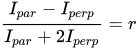
and acquiring the intensities of the polarizations in the parallel and perpendicular directions. A high value indicates an anisotropic orientation whereas a low value indicates an isotropic structure. In work done by Campagnola and Loew,[19] it was found that collagen fibers formed well-aligned structures with an value.

Forward over backward SHG

[edit]

SHG being a coherent process (spatially and temporally), it keeps information on the direction of the excitation and is not emitted isotropically. It is mainly emitted in forward direction (same as excitation), but can also be emitted in backward direction depending on the phase-matching condition. Indeed, the coherence length beyond which the conversion of the signal decreases is:

with for forward, but for backward such that >> . Therefore, thicker structures will appear preferentially in forward, and thinner ones in backward: since the SHG conversion depends at first approximation on the square of the number of nonlinear converters, the signal will be higher if emitted by thick structures, thus the signal in forward direction will be higher than in backward. However, the tissue can scatter the generated light, and a part of the SHG in forward can be retro-reflected in the backward direction.[20] Then, the forward-over-backward ratio F/B can be calculated,[20] and is a metric of the global size and arrangement of the SHG converters (usually collagen fibrils). It can also be shown that the higher the out-of-plane angle of the scatterer, the higher its F/B ratio (see fig. 2.14 of [21]).


Polarization-resolved SHG

[edit]

The advantages of polarimetry were coupled to SHG in 2002 by Stoller et al.[22] Polarimetry can measure the orientation and order at molecular level, and coupled to SHG it can do so with the specificity to certain structures like collagen: polarization-resolved SHG microscopy (p-SHG) is thus an expansion of SHG microscopy.[23] p-SHG defines another anisotropy parameter, as:[24]

which is, like r, a measure of the principal orientation and disorder of the structure being imaged. Since it is often performed in long cylindrical filaments (like collagen), this anisotropy is often equal to ,[25] where is the nonlinear susceptibility tensor and X the direction of the filament (or main direction of the structure), Y orthogonal to X and Z the propagation of the excitation light. The orientation ϕ of the filaments in the plane XY of the image can also be extracted from p-SHG by FFT analysis, and put in a map.[25][26]

Fibrosis quantization

[edit]

Collagen (particular case, but widely studied in SHG microscopy), can exist in various forms : 28 different types, of which 5 are fibrillar. One of the challenge is to determine and quantify the amount of fibrillar collagen in a tissue, to be able to see its evolution and relationship with other non-collagenous materials.[27]

To that end, a SHG microscopy image has to be corrected to remove the small amount of residual fluorescence or noise that exist at the SHG wavelength. After that, a mask can be applied to quantify the collagen inside the image.[27] Among other quantization techniques, it is probably the one with the highest specificity, reproductibility and applicability despite being quite complex.[27]

Others

[edit]

It has also been used to prove that backpropagating action potentials invade dendritic spines without voltage attenuation, establishing a sound basis for future work on Long-term potentiation. Its use here was that it provided a way to accurately measure the voltage in the tiny dendritic spines with an accuracy unattainable with standard two-photon microscopy.[28] Meanwhile, SHG can efficiently convert near-infrared light to visible light to enable imaging-guided photodynamic therapy, overcoming the penetration depth limitations.[29]

Materials that can be imaged

[edit]
Biological tissues imaged by second-harmonic generation (SHG) microscopy. (a) Transverse cut of a human cornea. (b) Skeletal muscle from zebrafish (myosin). (c) Adult mice-tail tendon. (d) Surface cartilage from a knee of a mature horse.

SHG microscopy and its expansions can be used to study various tissues: some example images are reported in the figure below: collagen inside the extracellular matrix remains the main application. It can be found in tendon, skin, bone, cornea, aorta, fascia, cartilage, meniscus, intervertebral disks...

Myosin can also be imaged in skeletal muscle or cardiac muscle.

Table 1: Materials visible by or that efficiently generate SHG.
Type Material Found in SHG signal Specificity
Carbohydrate Cellulose Wood, green plant, algae. Quite weak in normal cellulose,[18] but substantial in crystalline or nanocrystalline cellulose. -
Starch Staple foods, green plant Quite intense signal [30] chirality is at micro and macro level, and the SHG is different under right or left-handed circular polarization
Megamolecular polysaccharide sacran Cyanobactery From sacran cotton-like lump, fibers, and cast films signal from films is weaker [13]
Protein Fibroin and sericin Spider silk Quite weak [14]
Collagen[9] tendon, skin, bone, cornea, aorta, fascia, cartilage, meniscus, intervertebral disks; connective tissues Quite strong, depends on the type of the collagen (does it form fibrils, fibers ?) nonlinear susceptibility tensor components are , , , with ~ and / ~ 1.4 in most cases
Myosin Skeletal or cardiac muscle[3] Quite strong nonlinear susceptibility tensor components are , , with ~ but / ~ 0.6 < 1 contrary to collagen
Tubulin Microtubules in mitosis or meiosis,[31] or in neurites (mainly axons)[32] Quite weak The microtubules have to be aligned to efficiently generate
Minerals Piezoelectric crystals Also called nonlinear crystals Strong if phase-matched Different types of phase-matching, critical of non-critical
Polar liquids Water Most living organisms Barely detectable (requires wide-field geometry and ultra-short laser pulses [33]) Directly probing electrostatic fields, since oriented water molecules satisfy phase-matching condition [34]

Coupling with THG microscopy

[edit]

Third-Harmonic Generation (THG) microscopy can be complementary to SHG microscopy, as it is sensitive to the transverse interfaces, and to the 3rd order nonlinear susceptibility [35][36]

Applications

[edit]

Cancer progression, tumor characterization

[edit]

The mammographic density is correlated with the collagen density, thus SHG can be used for identifying breast cancer.[37] SHG is usually coupled to other nonlinear techniques such as Coherent anti-Stokes Raman Scattering or Two-photon excitation microscopy, as part of a routine called multiphoton microscopy (or tomography) that provides a non-invasive and rapid in vivo histology of biopsies that may be cancerous.[38]

Breast cancer

[edit]

The comparison of forward and backward SHG images gives insight about the microstructure of collagen, itself related to the grade and stage of a tumor, and its progression in breast.[39] Comparison of SHG and 2PEF can also show the change of collagen orientation in tumors.[40] Even if SHG microscopy has contributed a lot to breast cancer research, it is not yet established as a reliable technique in hospitals, or for diagnostic of this pathology in general.[39]

Ovarian cancer

[edit]

Healthy ovaries present in SHG a uniform epithelial layer and well-organized collagen in their stroma, whereas abnormal ones show an epithelium with large cells and a changed collagen structure.[39] The r ratio (see #Orientational anisotropy) is also used [41] to show that the alignment of fibrils is slightly higher for cancerous than for normal tissues.

Skin cancer

[edit]

SHG is, again, combined to 2PEF is used to calculate the ratio:

where shg (resp. tpef) is the number of thresholded pixels in the SHG (resp. 2PEF) image,[42] a high MFSI meaning a pure SHG image (with no fluorescence). The highest MFSI is found in cancerous tissues,[39] which provides a contrast mode to differentiate from normal tissues.

SHG was also combined to Third-Harmonic Generation (THG) to show that backward (see #Forward over backward SHG) THG is higher in tumors.[43]

Pancreatic cancer

[edit]

Changes in collagen ultrastructure in pancreatic cancer can be investigated by multiphoton fluorescence and polarization-resolved SHIM.[44]

Other cancers

[edit]

SHG microscopy was reported for the study of lung, colonic, esophageal stroma and cervical cancers.[39]

Pathologies detection

[edit]

Alterations in the organization or polarity of the collagen fibrils can be signs of pathology,.[45][46]

In particular, the anisotropic alignment of collagen fibers allowed the discrimination of healthy dermis from pathological scars in skin.[47] Also, pathologies in cartilage such as osteoarthritis can be probed by polarization-resolved SHG microscopy,.[48][49] SHIM was later extended to fibro-cartilage (meniscus).[50]

Tissue engineering

[edit]

The ability of SHG to image specific molecules can reveal the structure of a certain tissue one material at a time, and at various scales (from macro to micro) using microscopy. For instance, the collagen (type I) is specifically imaged from the extracellular matrix (ECM) of cells, or when it serves as a scaffold or conjonctive material in tissues.[51] SHG also reveals fibroin in silk, myosin in muscles and biosynthetized cellulose. All of this imaging capability can be used to design artificials tissues, by targeting specific points of the tissue : SHG can indeed quantitatively measure some orientations, and material quantity and arrangement.[51] Also, SHG coupled to other multiphoton techniques can serve to monitor the development of engineered tissues, when the sample is relatively thin however.[52] Of course, they can finally be used as a quality control of the fabricated tissues.[52]

Structure of the eye

[edit]

Cornea, at the surface of the eye, is considered to be made of plywood-like structure of collagen, due to the self-organization properties of sufficiently dense collagen.[53] Yet, the collagenous orientation in lamellae is still under debate in this tissue.[54] Keratoconus cornea can also be imaged by SHG to reveal morphological alterations of the collagen.[55] Third-Harmonic Generation (THG) microscopy is moreover used to image the cornea, which is complementary to SHG signal as THG and SHG maxima in this tissue are often at different places.[56]

See also

[edit]

Sources

[edit]

References

[edit]
Revisions and contributorsEdit on WikipediaRead on Wikipedia
from Grokipedia
Second-harmonic generation (SHG) imaging microscopy is a nonlinear optical imaging technique that leverages the second-harmonic generation process—a coherent scattering phenomenon where two photons of incident light at frequency ω combine to emit a single photon at twice the frequency (2ω)—to produce label-free, high-resolution images of non-centrosymmetric structures, such as fibrillar collagen, microtubules, and certain polysaccharides in biological samples.[1] This method requires materials lacking inversion symmetry to exhibit a second-order nonlinear susceptibility (χ(2)), enabling endogenous contrast without the need for staining or exogenous dyes, and typically employs near-infrared femtosecond lasers (700–1000 nm) for excitation to minimize photodamage and enhance tissue penetration.[2] The foundational demonstration of SHG for biological imaging occurred in 1986, when Freund and colleagues used it to assess collagen polarity in rat tail tendons, marking an early application of nonlinear optics to biomedicine.[1] Practical high-resolution SHG microscopy for tissue imaging emerged in the early 2000s, driven by advancements in ultrafast laser technology and confocal setups, as pioneered by researchers like Mohler and Campagnola in 2002.[1] Since then, the technique has evolved from basic wide-field imaging to sophisticated variants, including polarization-resolved SHG (P-SHG) for analyzing molecular orientation and multi-photon combinations for multimodal imaging.[3] SHG microscopy's key strengths include instantaneous signal generation with no photobleaching or phototoxicity, submicron lateral resolution, and imaging depths up to several hundred microns in scattering tissues, making it superior to traditional fluorescence methods for long-term studies.[2] It is particularly valuable in biomedical applications for quantifying extracellular matrix remodeling, such as collagen alignment in fibrosis, tumor microenvironments (e.g., tumor-associated collagen signatures in breast and ovarian cancers), and corneal disorders, where structural changes inform disease progression and diagnosis.[3] Recent integrations with machine learning have further enhanced its utility, achieving high accuracy (83–91%) in automated tissue classification for clinical pathology.[3]

Principles

Second-harmonic generation mechanism

Second-harmonic generation (SHG) is a second-order nonlinear optical process wherein two input photons, each with frequency ω\omega, interact coherently within a nonlinear medium to produce a single output photon at frequency 2ω2\omega. This frequency-doubling effect occurs when the medium's polarization responds quadratically to the intense electric field of the incident light, enabling the conversion of the fundamental wave into its harmonic without net absorption of energy by the material. The mechanism is governed by the second-order nonlinear susceptibility tensor χ(2)\chi^{(2)}, a third-rank tensor that quantifies the material's nonlinear response. The induced nonlinear polarization at the harmonic frequency is expressed as Pi(2)(2ω)=ϵ0j,kχijk(2)Ej(ω)Ek(ω)P_i^{(2)}(2\omega) = \epsilon_0 \sum_{j,k} \chi_{ijk}^{(2)} E_j(\omega) E_k(\omega), where ϵ0\epsilon_0 is the vacuum permittivity and E(ω)E(\omega) is the electric field amplitude of the fundamental wave. This polarization acts as a source term in Maxwell's equations, driving the generation of the second-harmonic field E(2ω)E(2\omega). The tensorial nature of χ(2)\chi^{(2)} imposes symmetry constraints on the process, with its components determining the efficiency and polarization dependence of the generated light. The intensity of the generated second-harmonic light depends on phase-matching conditions and is derived from the coupled wave equations for the fundamental and harmonic fields. Under the undepleted pump approximation and slowly varying envelope conditions, the second-harmonic intensity I2ωI_{2\omega} is proportional to
I2ωχ(2)2Iω2L2\sinc2(ΔkL2), I_{2\omega} \propto |\chi^{(2)}|^2 I_{\omega}^2 L^2 \sinc^2\left(\frac{\Delta k L}{2}\right),
where IωI_{\omega} is the fundamental intensity, LL is the interaction length in the medium, Δk=k2ω2kω\Delta k = k_{2\omega} - 2k_{\omega} is the wave-vector mismatch (with kk denoting the wave numbers), and \sinc(x)=sin(x)/x\sinc(x) = \sin(x)/x. Efficient generation requires Δk0\Delta k \approx 0 to avoid destructive interference over the propagation distance. This expression highlights the quadratic scaling with input intensity and the critical role of phase matching in maximizing conversion efficiency. From a quantum mechanical viewpoint, SHG is a parametric down-conversion process in reverse, where the nonlinear interaction Hamiltonian facilitates the annihilation of two fundamental photons and the creation of one harmonic photon, strictly conserving energy (2ω=2ω2\hbar\omega = \hbar \cdot 2\omega) and momentum (enforced by phase matching Δk=0\Delta \mathbf{k} = 0). This coherent scattering involves virtual intermediate states in the material, without real excitation or population transfer.[4] SHG exhibits distinct coherence properties compared to fluorescence: it produces phase-locked emission instantaneously with the input field, resulting in a narrow spectral linewidth and directional forward scattering, whereas fluorescence involves incoherent re-emission after absorption, leading to broader spectra, lifetime delays, and isotropic emission. These traits enable SHG to provide background-free contrast in imaging applications.

Molecular and structural requirements

Efficient second-harmonic generation (SHG) requires materials or structures that lack inversion symmetry, as the second-order nonlinear susceptibility tensor χ^(2) vanishes in centrosymmetric environments, prohibiting the process at the electric dipole level.[5] This non-centrosymmetry must exist at multiple scales: molecular (e.g., asymmetric charge distribution), crystalline (e.g., polar crystal classes), or supramolecular (e.g., ordered assemblies like fibrils). In biological contexts, such as collagen, the triple-helical motif breaks inversion symmetry, enabling coherent SHG emission from aligned peptide bonds.[6] The selection rules for SHG activity stem from parity conservation and the tensorial nature of χ^(2), which is forbidden under inversion operations. Structures lacking an inversion center—such as chiral molecules with helical or asymmetric geometries, or aligned polymers—satisfy these rules, allowing the second-order polarization response. For instance, SHG is active in left- or right-handed helical proteins but absent in symmetric globular proteins unless symmetry is broken by external fields or aggregation. Microscopically, this arises from the molecular first hyperpolarizability β, a measure of the induced dipole moment's quadratic dependence on the electric field; β contributes to the bulk χ^(2) through molecular orientation and density, with enhanced β in push-pull chromophores or biomolecules like collagen amplifying the signal.[7][8] Environmental conditions significantly modulate SHG efficiency in biomolecules by altering molecular alignment or secondary structure. In collagen hydrogels, pH variations during fibrillogenesis influence fiber diameter and organization: lower pH (e.g., 5.5) yields thicker fibrils with larger pore sizes and increased SHG intensity compared to higher pH (e.g., 8.5), which produces thinner, denser fibrils with reduced pore sizes and diminished SHG, due to differences in fibrillogenesis and stabilization of triple helices. Temperature affects polymerization similarly, with lower temperatures (e.g., 4°C) producing thicker fibrils and stronger SHG signals, while elevated temperatures (e.g., 37°C) result in finer networks and diminished emission. Hydration levels also play a role, as dehydration disrupts ordered hydrogen-bond networks in collagen, reducing SHG by up to 50% in air-dried samples versus hydrated ones, highlighting the need for physiological conditions in imaging.[9][10] Representative examples illustrate SHG-active versus inactive motifs. Centrosymmetric dyes, such as symmetric squaraines, typically exhibit negligible SHG due to zero β under inversion, but can generate signals if aggregated into non-centrosymmetric domains via techniques like Langmuir-Blodgett films. In contrast, the collagen type I triple helix is intrinsically SHG-active owing to its non-centrosymmetric pitch and aligned Gly-Pro-Hyp repeats, producing bright forward-directed emission, whereas non-fibrillar, centrosymmetric collagens like type IV show little to no signal.[11][6]

Instrumentation

Laser excitation and optical setup

Second-harmonic generation (SHG) microscopy relies on ultrafast femtosecond lasers to provide the high peak intensities necessary for efficient nonlinear optical processes while maintaining low average power to prevent sample photodamage. Typically, mode-locked titanium:sapphire (Ti:sapphire) lasers are employed, operating in the near-infrared range of 700–1000 nm with pulse durations around 100 fs and repetition rates of 80 MHz. Alternatives to Ti:sapphire lasers, such as ultrafast fiber lasers operating at fixed wavelengths like 780 nm, 920 nm, or 1050 nm, are increasingly employed for their compactness and robustness in clinical and portable setups.[12] These lasers, often pumped by a continuous-wave argon ion or frequency-doubled Nd:YVO₄ source, deliver peak powers on the order of gigawatts per square centimeter within the focal volume, enabling SHG without significant linear absorption in biological tissues.[2][13] The optical train for beam delivery begins with beam expansion using achromatic doublet lenses to match the laser output to the entrance pupil of the microscope, ensuring uniform illumination and maximizing photon throughput. Scanning is achieved via galvanometric mirrors or acousto-optic deflectors, which raster the focused beam across the sample in a confocal or multiphoton setup. High numerical aperture (NA > 1.2) objective lenses, such as oil-immersion types, are critical for tight focusing, concentrating the excitation light into a diffraction-limited spot that drives the quadratic SHG response. Polarization control elements, including half-wave plates and polarizers, are often integrated to probe molecular orientations, though they are optional for basic imaging.[2][6] SHG signal collection employs either forward (transmission) or backward (epifluorescence-like) geometries, selected based on sample thickness and scattering properties. In forward detection, a high-NA condenser collects the coherently propagating SHG signal, ideal for thick tissues where backward scattering is minimal; backward detection, using the same objective, suits thin samples or scattering media like skin. Confocal pinhole-based or non-descanned configurations reject out-of-focus light and the fundamental excitation beam via short-pass or bandpass filters (e.g., 390–450 nm for 800 nm excitation), ensuring clean SHG detection with photomultiplier tubes or GaAsP detectors.[2][13][6] The spatial resolution in SHG microscopy is diffraction-limited, arising from the nonlinear excitation volume akin to two-photon processes, yielding typical lateral resolutions of approximately 300 nm and axial resolutions of about 1 μm under standard conditions with 800 nm excitation and NA 1.2 objectives. This intrinsic three-dimensional confinement enhances sectioning without exogenous labels, though actual performance varies with wavelength and NA.[2][6]

Signal detection and imaging configurations

In second-harmonic generation (SHG) microscopy, signal detection relies on sensitive photodetectors capable of capturing the weak, coherent SHG emission in low-light conditions. Photomultiplier tubes (PMTs) are commonly employed for their high quantum efficiency and fast response times, enabling the collection of both forward- and backward-propagating SHG signals through epi- or transmission configurations.[6] For wide-field approaches, charge-coupled device (CCD) cameras provide parallel detection across larger areas, though they may require longer integration times compared to point-scanning PMTs.[2] To enhance sensitivity, photon counting modes are often integrated with PMTs, which generate shot-noise-limited signals by registering individual photon arrivals, thereby improving the signal-to-noise ratio for sparse emitters like collagen fibrils.[14] Spectral separation is essential to distinguish the SHG signal at frequency 2ω from the excitation laser at ω and confounding autofluorescence, which typically emits over broader spectra. This is achieved using dichroic mirrors to direct the SHG beam while reflecting residual excitation light, followed by narrow bandpass filters (e.g., 10 nm full width at half maximum) centered on the SHG wavelength.[6] Such optical components ensure clean isolation of the instantaneous SHG response, minimizing crosstalk in biological samples where endogenous fluorescence is prevalent.[2] Imaging configurations in SHG microscopy vary to balance resolution, speed, and depth penetration. Raster scanning, typically implemented with galvanometer-driven mirrors in laser scanning microscopes, enables high-resolution 2D and 3D imaging by sequentially illuminating pixels in a grid pattern, often using high numerical aperture objectives for tight focusing.[6] Alternatively, wide-field illumination excites the entire field of view simultaneously, allowing faster acquisition rates—up to 2–3 orders of magnitude quicker than scanning methods—but necessitates computational reconstruction to suppress out-of-focus contributions and achieve diffraction-limited resolution.[2] Image formation involves optimizing acquisition parameters to capture volumetric data without excessive photobleaching or damage. Pixel dwell times are generally set in the microsecond range (e.g., 20–200 μs) to accumulate sufficient photons per voxel while maintaining frame rates suitable for live imaging, with line scan rates around 1–10 kHz and full frame rates of 1–30 Hz depending on resolution.[2] For 3D reconstruction, z-stacking is performed by axially translating the sample or objective in increments matching the axial resolution (typically 1–2 μm), generating depth-resolved datasets that reveal structural anisotropy in tissues.[6] To mitigate common artifacts, post-processing and hardware corrections are applied during signal collection. Background subtraction removes non-specific signals, such as residual autofluorescence or dark counts, by acquiring reference frames from signal-free regions or using phase-sensitive techniques like interferometry.[15] Laser power normalization accounts for fluctuations in excitation intensity across scans or depths, ensuring quantitative comparability by dividing SHG intensities by measured input power, often via in-line power meters.[14] Aberration correction employs adaptive optics, including deformable mirrors, to compensate for refractive index mismatches in scattering media, thereby restoring signal intensity and resolution—enhancements of 2–3 fold have been reported at depths exceeding 200 μm.[6]

Advantages and limitations

Advantages in biological imaging

Second-harmonic imaging microscopy (SHIM) enables label-free visualization of biological samples by relying on the intrinsic nonlinear optical properties of endogenous molecules, eliminating the need for exogenous fluorescent dyes that can introduce toxicity and imaging artifacts in live cells and tissues.[6] This approach is particularly advantageous for long-term studies of dynamic processes, as it preserves the native state of samples without chemical perturbations.[16] The use of near-infrared excitation wavelengths in SHIM minimizes scattering and absorption by biological chromophores, allowing for deeper tissue penetration compared to traditional visible-light microscopy, with reported depths reaching up to 550 μm in mouse muscle tissue.[17] This enhanced depth facilitates in vivo imaging of intact organs and reduces the reliance on invasive sample preparation. Additionally, the nonlinear nature of second-harmonic generation provides intrinsic optical sectioning, confining signal generation to the focal plane and enabling three-dimensional reconstruction without physical sectioning, which supports non-destructive optical biopsies.[6] SHIM offers superior photostability, as the coherent, non-resonant process of second-harmonic generation avoids energy absorption and subsequent bleaching or phototoxicity that plague fluorescence-based techniques.[16] Unlike fluorescence microscopy, which often requires labels prone to degradation, SHIM maintains signal integrity over extended imaging sessions, making it ideal for monitoring live samples.[17] Furthermore, its specificity for non-centrosymmetric, ordered structures—such as type I collagen in the extracellular matrix—provides inherent contrast without staining, highlighting architectural features like fibrillar organization in tissues.[6] This selectivity is invaluable for studying fibrosis, wound healing, and tumor microenvironments where collagen remodeling plays a key role.[16]

Limitations and mitigation strategies

One primary limitation of second-harmonic imaging microscopy (SHIM) is the inherently weak signal intensity generated by second-harmonic generation (SHG), which is typically 3–4 orders of magnitude lower in biological samples compared to reference materials like quartz.[2] This necessitates the use of high laser powers to achieve detectable signals, which can lead to sample heating and potential photodamage, particularly in sensitive biological tissues. To mitigate these issues, strategies such as pulse shaping can optimize the laser pulse profile to enhance SHG efficiency while minimizing thermal effects, and employing lasers with lower repetition rates allows for higher peak powers at reduced average power levels, thereby decreasing heat accumulation without compromising signal quality. Another constraint is that SHG signals arise exclusively from non-centrosymmetric materials, excluding isotropic or centrosymmetric structures common in many biological contexts, such as certain cellular components or amorphous tissues. This selectivity limits the applicability of SHIM to specific ordered structures like collagen fibrils. Mitigation often involves integrating SHIM into multimodal imaging setups, combining it with techniques like two-photon excitation fluorescence (TPEF) to visualize complementary features and provide a more comprehensive view of the sample. In thicker samples, phase-matching sensitivity plays a critical role, as mismatches in the wave vectors reduce SHG conversion efficiency and lead to anisotropic signal directionality, particularly favoring forward-propagating emission over backward. This diminishes overall signal collection in bulk tissues. Approaches to address this include preparing thin-sectioned samples, which shortens the interaction length and relaxes phase-matching requirements, or utilizing engineered nonlinear crystals with quasi-phase-matching designs to improve efficiency in controlled setups, though the latter is more common in non-biological applications. Deep-tissue imaging with SHIM suffers from low signal-to-noise ratios (SNR) due to strong scattering of both excitation and emission light, which broadens the point spread function and attenuates signals at depths beyond a few hundred micrometers. Countermeasures include shifting to longer excitation wavelengths in the NIR-II range (1000–1300 nm), which experiences less scattering for greater penetration, and applying wavefront shaping techniques, such as adaptive optics or holographic compensation, to refocus light through scattering media and improve SNR in tissues like mouse hippocampus at depths around 500 μm.[18] Recent advancements as of 2025, including computational adaptive optics, have further enhanced deep-tissue performance by correcting aberrations in real-time for high-resolution imaging.[19] Finally, the reliance on ultrafast lasers, such as Ti:sapphire systems, introduces challenges related to high cost and operational complexity, including the need for precise alignment, water cooling, and specialized maintenance. More affordable alternatives, like mode-locked fiber lasers, offer compact, alignment-free designs with comparable femtosecond pulse durations and repetition rates suitable for SHIM, reducing overall system costs by an order of magnitude while maintaining performance for biological imaging.

Historical development

Early discoveries of SHG

The discovery of second-harmonic generation (SHG) marked a pivotal moment in nonlinear optics, beginning with the experimental observation by Peter A. Franken and colleagues in 1961. Using a pulsed ruby laser emitting at 694 nm, they focused the beam into a crystalline quartz sample, detecting the first SHG signal at 347 nm, confirming the nonlinear frequency doubling predicted by quantum electrodynamics.[20] This breakthrough, conducted at the University of Michigan, demonstrated that intense laser light could induce higher harmonics in transparent media, opening the field of nonlinear optical interactions.[21] Building on this observation, a comprehensive theoretical framework emerged in 1962 through the work of J.A. Armstrong, N. Bloembergen, J. Ducuing, and P.S. Pershan. Their analysis formalized SHG within the context of nonlinear polarization, introducing the second-order susceptibility tensor χ^(2) as the key parameter governing the process in non-centrosymmetric media.[22] This paper, published in Physical Review, provided the mathematical foundation linking macroscopic optical effects to microscopic material responses, enabling predictions of efficiency and phase relations in harmonic generation.[21] Further advancements in the late 1960s focused on optimizing SHG through phase-matching techniques, as detailed in experiments by G.D. Boyd and D.A. Kleinman in 1968. Using potassium dihydrogen phosphate (KDP) crystals, they demonstrated efficient SHG by aligning the beam focus and crystal orientation to satisfy birefringent phase-matching conditions, achieving conversion efficiencies that highlighted the role of Gaussian beam profiles in nonlinear interactions. These studies quantified the impact of walk-off and focusing geometry, establishing practical guidelines for high-efficiency harmonic generation in anisotropic crystals.[23] The 1970s saw the advent of tunable lasers, particularly dye lasers, which broadened SHG investigations across spectral ranges. Developed from the late 1960s but widely adopted in the 1970s, these sources allowed precise wavelength control, facilitating studies of dispersion in nonlinear susceptibilities and phase-matching bandwidths in various materials. Initial applications of SHG in spectroscopy emerged during this period, primarily for characterizing χ^(2) tensors and optical nonlinearities in crystals like KDP and quartz, providing insights into material symmetries and electronic structures that foreshadowed advanced spectroscopic tools.

Emergence as an imaging technique

The adaptation of second-harmonic generation (SHG) into a practical microscopy technique began in the late 1980s and 1990s, building on early demonstrations of the nonlinear optical process. In 1986, Freund and colleagues integrated SHG with scanning confocal microscopy to image endogenous collagen structures in rat-tail tendon tissues, achieving resolutions of approximately 50 μm and revealing fibril orientations without labels. This work marked the initial transition from bulk SHG measurements to spatially resolved imaging in biological samples, leveraging pulsed laser excitation to probe non-centrosymmetric structures like collagen fibrils. A pivotal advancement occurred in 2000, when Campagnola et al. demonstrated second-harmonic imaging microscopy (SHIM) for label-free visualization of biomolecular arrays in living cells and tissues. Using a titanium:sapphire femtosecond laser tuned to 830 nm, they imaged collagen in C. elegans and mammalian cells, highlighting SHIM's compatibility with multiphoton setups and its ability to provide high-resolution, photobleaching-free contrast. This established SHIM as a viable tool for cellular and tissue-level studies, expanding beyond static collagen imaging to dynamic biological processes. Between 2005 and 2010, significant improvements in imaging speed were achieved, particularly through the development of video-rate SHIM by So and colleagues. Their interferometric SHG approaches and resonant scanning techniques enabled real-time acquisition at up to 30 frames per second, facilitating dynamic studies of collagen remodeling in living tissues without compromising resolution. These innovations, integrated into multiphoton platforms, overcame limitations of slow galvanometer-based scanning, making SHIM suitable for observing physiological events like cell migration and tissue deformation.[24][25] Key reviews in 2011 by Campagnola synthesized these developments, outlining SHG principles such as phase-matching and polarization dependence while emphasizing applications in disease diagnostics, including fibrosis and cancer through collagen architecture analysis. By 2015, SHIM's potential for clinical translation had advanced, with explorations into non-invasive diagnostics for breast and ovarian cancers via label-free imaging of stromal collagen alterations, paving the way for FDA-evaluated optical biopsy tools.[26] Since 2015, further advancements have included the integration of machine learning for automated tissue classification and analysis, achieving accuracies of 83–91% in clinical pathology applications as of 2023, and explorations of quantum-enhanced SHG for improved signal efficiency reported in 2024–2025.[3][27]

Quantitative analysis

Orientational and polarization measurements

Second-harmonic imaging microscopy enables the extraction of molecular orientation and alignment information through polarization-resolved techniques, particularly polarization-in, polarization-out (PIPO) SHG, where the input laser polarization is varied systematically to probe the components of the second-order nonlinear susceptibility tensor χ^(2).[28] In PIPO SHG, a polarization state generator, such as a rotating half-wave plate, modulates the excitation polarization across multiple angles (typically 0° to 180° in 10°–22.5° increments), while the emitted SHG signal is analyzed through a polarization state analyzer, often a linear polarizer, to capture both parallel and perpendicular components.[29] This approach maps the anisotropic response of the sample, revealing the orientation of non-centrosymmetric structures like protein fibrils without exogenous labels.[28] The SHG intensity in these measurements follows the fundamental relation
I2ωeoutχ(2):einein2, I_{2\omega} \propto \left| \mathbf{e}_{\mathrm{out}} \cdot \boldsymbol{\chi}^{(2)} : \mathbf{e}_{\mathrm{in}} \mathbf{e}_{\mathrm{in}} \right|^2,
where ein\mathbf{e}_{\mathrm{in}} and eout\mathbf{e}_{\mathrm{out}} are the input and output polarization unit vectors, respectively, and χ(2)\boldsymbol{\chi}^{(2)} is the second-order susceptibility tensor that encodes the material's nonlinear optical properties and molecular arrangement.[28] By acquiring intensity data as a function of input polarization angle ψ and analyzer angle, the tensor elements are reconstructed, providing quantitative insights into the sample's symmetry and alignment.[29] In applications to collagen fibrils, which exhibit C∞v symmetry and serve as model uniaxial structures, PIPO SHG determines the azimuthal orientation angle φ of the fibril axis relative to the laboratory frame via the relation tan(2ϕ)=2χ15/χ31\tan(2\phi) = 2 \chi_{15} / \chi_{31}, where χ_{15} and χ_{31} are off-diagonal and diagonal tensor components in the molecular frame.[30] Additionally, the susceptibility ratio R=χzzz/χzxxR = \chi_{zzz} / \chi_{zxx} quantifies the relative strength of longitudinal versus transverse nonlinear responses in these uniaxial assemblies, with reported values around 1.4–1.6 indicating the helical pitch and alignment of collagen molecules within the fibril.[28][31] These parameters distinguish structural variations, such as differences in fibril maturity or disorder.[29] To extract these quantities, experimental polarization curves—plots of SHG intensity versus input angle—are fitted using least-squares optimization algorithms, such as trust-region reflective methods, to minimize residuals between observed data and the theoretical model derived from the χ^(2) tensor.[29] This fitting process typically involves 64–81 polarization combinations per image stack, yielding pixel-wise maps of φ and R with uncertainties below 5° for orientation and 0.1 for ratios in well-aligned samples.[28]

Directionality and intensity quantification

In second-harmonic generation (SHG) microscopy, the forward-to-backward signal ratio (F/B = I_forward / I_backward) serves as a key metric for assessing sample structure, particularly the size, density, and alignment of nonlinear scatterers like collagen fibrils. In aligned fibers, coherent forward propagation is favored due to phase-matching conditions that enhance constructive interference in the forward direction, leading to F/B ratios that can exceed 20 in mature collagen tissues such as tendons, compared to near-unity ratios in thinner or disordered structures. This directionality arises from the axial extent of the scatterers relative to the SHG wavelength, where larger fibrils (>λ/2) promote forward dominance while smaller ones (<λ/10) yield more isotropic emission.[32][33] Quantifying absolute SHG intensity requires normalization to enable comparisons across samples and instruments, often achieved using reference standards like urea crystals, which exhibit well-characterized nonlinear susceptibility (χ^(2)) values of approximately 2.4 pm/V along their principal axis.[34] By imaging urea microcrystals under identical excitation conditions, the system's response can be calibrated to derive absolute χ^(2) measurements in biological samples, accounting for factors such as excitation power, numerical aperture, and collection efficiency. This approach has been instrumental in standardizing SHG signals for quantitative structural analysis, ensuring reproducibility in studies of fibrillar assemblies. Phase-matching effects profoundly influence SHG directionality, governed by the wavevector mismatch Δk = 2k_ω - k_{2ω}, where k denotes the wavevector magnitude. In thin samples (< coherence length L_c = π/Δk ≈ 8–10 μm for typical biological dispersion), emission is nearly isotropic due to limited buildup of the nonlinear polarization, resulting in comparable forward and backward signals. Conversely, thick samples enable quasi-phase matching over multiple scatterers, enhancing forward efficiency in aligned structures (e.g., F/B > 10) while backward signals dominate in random assemblies where Δk fluctuations average to shorter coherence lengths. These Δk-dependent models, incorporating dispersion and fibril packing, predict observed morphologies and conversion efficiencies in tissues.[35] Ratio imaging of SHG to two-photon excited fluorescence (TPEF) intensities (SHG/TPEF) facilitates depth-independent mapping of collagen density, as both signals originate from two-photon absorption and exhibit similar exponential decay with depth due to scattering and absorption. In collagen-rich tissues, higher SHG/TPEF ratios indicate denser fibrillar organization, enabling quantitative density profiles without corrective algorithms for tissue heterogeneity. This method leverages the coherent, non-bleaching nature of SHG against the isotropic TPEF from endogenous fluorophores like elastin, providing robust structural contrast in volumetric imaging.[26][36] Automated quantification of F/B ratios from dual-detection setups is supported by Fiji (ImageJ) plugins and macros, such as custom scripts for pixel-wise ratio computation or extensions like CollagenFitJ for polarization-resolved SHG analysis. These tools process forward and backward image stacks to generate directional maps, applying corrections for signal decay and background noise, thereby streamlining workflows for large-scale structural assessments in biological specimens.[37][38]

Clinical and structural assessments

Second-harmonic generation (SHG) microscopy enables quantitative fibrosis scoring by leveraging intensity thresholds derived from collagen-specific signals, distinguishing fibrillar collagen accumulation in pathological tissues. A common metric, the fibrosis index, is calculated as log(ISHGIautofluorescence)\log \left( \frac{I_{\text{SHG}}}{I_{\text{autofluorescence}}} \right), where ISHGI_{\text{SHG}} represents the SHG intensity from collagen fibrils and IautofluorescenceI_{\text{autofluorescence}} accounts for background cellular fluorescence; this logarithmic ratio provides a sensitive measure of collagen content relative to non-fibrotic elements, with higher values indicating increased fibrosis severity.[39] Elevated values of this index correlate with advanced fibrosis stages in models of liver and kidney disease, allowing non-invasive staging without dyes.[40][39] Tumor margin detection benefits from entropy-based texture analysis of SHG images, which quantifies spatial irregularity in collagen organization to delineate boundaries between healthy and malignant tissue. By computing entropy as a measure of pixel intensity disorder within sub-regions (e.g., 128 × 128 pixels), this approach highlights chaotic fibril patterns at tumor edges, with higher entropy values indicating high irregularity compared to uniform stromal regions.[41] In lung carcinoma samples, unsupervised K-means clustering of these texture features, combined with polarimetric SHG parameters like degree of circular polarization, achieves silhouette scores >0.6 for accurate margin identification across multiple patient sections, enabling precise surgical guidance.[41] For ECM remodeling metrics, forward SHG intensity quantifies fibril density by capturing coherent signals from aligned, micron-scale collagen structures, where higher forward-to-backward ratios (>2:1) reflect dense packing in remodeled matrices.[42] Polarization-resolved SHG further assesses fibril alignment through anisotropy parameters, such as the ratio of maximum to minimum SHG intensity under rotated excitation, revealing reorientation in fibrotic diseases like idiopathic pulmonary fibrosis. These metrics provide insights into pathological remodeling without exogenous labels, with forward SHG density correlating to ultrastructural changes observed via electron microscopy.[43] Validation of SHG-based assessments against traditional histology demonstrates strong agreement in liver and kidney fibrosis models; for instance, qFibrosis scores from SHG/two-photon excitation fluorescence imaging yield high correlation for staging nonalcoholic steatohepatitis-induced liver fibrosis, accurately differentiating advanced stages from earlier ones.[44] In kidney obstruction models, SHG/autofluorescence ratios show strong correlation with Masson's trichrome staining for collagen quantification, confirming reliability across organ-specific pathologies.[39] These correlations underscore SHG's utility as a label-free surrogate for histological evaluation, reducing inter-observer variability in clinical diagnostics.[45] Recent advances in the 2020s incorporate AI-driven segmentation for automated structural scoring of SHG images, enhancing precision in fibrosis and ECM analysis. Convolutional neural networks, such as U-Net models trained on collagen-positive pixel patches, achieve high F1 scores for segmenting fibrillar structures in thick tissue stacks, outperforming manual thresholding in volume accuracy at depths >100 μm.[46] In metabolic dysfunction-associated steatohepatitis (MASH) cohorts, AI algorithms processing SHG/two-photon data enable continuous fibrosis quantification with high area under the curve (AUC) for stage prediction, facilitating real-time clinical assessment and treatment monitoring.[47] As of 2025, machine learning has further boosted imaging speed and quality in SHG microscopy, enabling detailed structural analysis with reduced sample damage.[48] These tools integrate texture and polarimetric features for holistic scoring, marking a shift toward scalable, objective pathology evaluation.[49]

Imagenable materials

Endogenous biological structures

Collagen, particularly types I, II, and III, represents the strongest endogenous source of second-harmonic generation (SHG) signals in biological tissues due to its triple-helix structure, which lacks inversion symmetry and enables efficient nonlinear optical response.[6] These fibrillar collagens are abundant in the extracellular matrix (ECM) of connective tissues, such as tendons, skin, and cartilage, where their organized, non-centrosymmetric arrangement produces bright, coherent SHG signals that allow label-free visualization of fibril orientation and density.[50] Type I collagen, the most prevalent, generates particularly intense forward-directed SHG, facilitating high-resolution imaging of ECM remodeling without exogenous labels.[2] In cellular structures, microtubules and actin filaments exhibit weaker SHG signals arising from their partial molecular alignment, which provides limited non-centrosymmetry compared to collagen.[51] Microtubules, composed of tubulin dimers with inherent polarity, produce detectable SHG in organized axonal arrays, enabling imaging of cytoskeletal dynamics in neurons.[52] Similarly, actin filaments in stress fibers or bundles yield faint SHG, useful for tracking cytoskeletal organization during cell migration or division, though signal strength often requires polarization-sensitive techniques for enhancement.[53] In hard tissues like bone and cartilage, SHG primarily arises from mineralized collagen fibrils, where the crystalline hydroxyapatite phase interfaces with the organic collagen matrix to enhance signal generation.[54] Bone's lamellar collagen organization produces anisotropic SHG patterns that reveal osteon structure and mineralization density, while in cartilage, SHG highlights zonal variations in type II collagen alignment for evaluating joint health.[55][56] SHG signals from endogenous collagen vary with age due to increased non-enzymatic crosslinking, which alters fibril packing and reduces signal intensity by disrupting the coherent molecular order.[57] In aged tissues, such as skin or bone, this crosslinking leads to more disorganized fibers and diminished forward SHG, providing a biomarker for age-related ECM degradation.[58] Polarimetric SHG analysis quantifies these changes, showing progressive loss of collagen alignment with advancing age.[59]

Exogenous and synthetic materials

Second-harmonic imaging microscopy (SHIM) has been extended to exogenous and synthetic materials, enabling visualization of engineered nanostructures and non-biological components that exhibit nonlinear optical responses due to their non-centrosymmetric structures. These materials provide enhanced contrast for tracking and functional imaging, often surpassing the capabilities of endogenous signals by offering tunable properties and higher signal intensities.[60] Nanoparticles, particularly gold nanorods, are prominent in SHIM for their plasmonic enhancement of second-harmonic generation (SHG), which amplifies local fields and facilitates deep-tissue tracking. Gold nanorods generate coherent SHG signals due to their anisotropic shape and surface plasmon resonances, allowing for orientation-sensitive imaging and multiplexed detection in complex environments. Studies have shown that SHG efficiency in these nanorods depends more on surface area than aspect ratio, enabling design optimizations for biomedical applications. Similarly, ZnO nanoparticles doped for biocompatibility exhibit strong SHG for in situ upconversion of near-infrared light, supporting label-free imaging of cellular interactions.[61] Polymers and aligned structures, such as liquid crystals and electrospun fibers, serve as scaffolds in SHIM, where molecular orientation produces directional SHG signals for structural assessment. Ferroelectric nematic liquid crystals, for instance, yield high SHG intensities due to their spontaneous polarization, enabling real-time monitoring of alignment in dynamic systems. Electrospun nanofibers incorporating nonlinear organic polymers demonstrate measurable SHG, particularly in hybrid systems with semi-organic components, which is useful for evaluating scaffold architecture in tissue models.[62][63] Although SHIM emphasizes label-free imaging, select SHG-active dyes and probes have been developed for specific enhancements, such as membrane potential sensing. Styryl dyes like FM1-43 and analogs (e.g., Di-4-ANEPPS) generate voltage-sensitive SHG signals through electro-optic mechanisms, where transmembrane potential alters dye orientation and hyperpolarizability, providing sub-millisecond temporal resolution in neuronal imaging. These probes are rare in routine use due to the preference for intrinsic contrasts but offer targeted augmentation when needed.[64][65] Crystalline inclusions like calcite and quartz integrate into biological hybrids for SHIM, leveraging their inherent birefringence for high-fidelity signal generation in composite materials. Calcite microcrystals, observed in engineered pineal gland models or otoconia analogs, produce SHG intensities up to 41 times stronger than pure calcite due to bio-mineral interfaces, enabling visualization of mineralization processes. Quartz crystals in synthetic-biological hybrids similarly contribute phase-matched SHG, aiding in the study of geological-inspired biomaterials.[66][67] Doping strategies in synthetic biomaterials allow wavelength-specific tuning of SHG, expanding SHIM's spectral versatility for multimodal imaging. For example, samarium-doped BaTiO3 nanoparticles exhibit enhanced SHG at tunable wavelengths through defect-induced nonlinearities, while graphene doping enables electrical control of SHG in 2D hybrid films. These approaches prioritize non-centrosymmetric doping to break inversion symmetry, achieving up to two orders of magnitude signal increase without altering bulk properties.[68][69]

Multimodal integrations

Coupling with third-harmonic generation

Third-harmonic generation (THG) is a third-order nonlinear optical process in which three incident photons at frequency ω coherently combine to produce a single emitted photon at 3ω, providing label-free contrast sensitive to refractive index mismatches at interfaces and in isotropic media where second-harmonic generation (SHG) is absent.[3] Unlike SHG, which requires non-centrosymmetric structures, THG arises from third-order susceptibility χ^(3) and is particularly effective for imaging lipid droplets, cell membranes, and tissue boundaries due to its dependence on local asymmetry in the optical response.[70] The intensity of THG signal follows the relation $ I_{3\omega} \propto \chi^{(3)} I_{\omega}^3 $, highlighting its cubic dependence on the excitation intensity, which necessitates high-peak-power femtosecond lasers for efficient generation.[3] In multimodal setups, SHG and THG are integrated using a single near-infrared femtosecond laser source, typically a Ti:sapphire oscillator tuned to wavelengths around 800-1200 nm, with the backward- or forward-scattered signals separated by dichroic mirrors and bandpass filters into distinct detection channels on photomultiplier tubes.[71] This configuration allows simultaneous acquisition of coherent SHG signals from ordered, anisotropic structures like collagen fibers and coherent THG signals from isotropic regions such as cellular cytoplasm or lipid-rich areas, enhancing overall tissue contrast without additional labeling.[72] The coupling of SHG and THG provides complementary structural information, with SHG mapping fibrillar extracellular matrix components and THG delineating cellular and interfacial features, enabling comprehensive label-free imaging of complex biological environments.[3] For instance, in skin imaging, SHG visualizes collagen organization in the dermis to assess fibrosis or aging, while THG highlights epidermal layers and lipid structures in keratinocytes, facilitating non-invasive evaluation of skin pathology.[73] This multimodal approach improves depth penetration and resolution in thick tissues, reducing phototoxicity compared to fluorescence-based methods.[71]

Combinations with fluorescence and other modalities

Second-harmonic generation (SHG) imaging microscopy is frequently combined with two-photon excitation fluorescence (2PEF) to provide complementary structural and molecular information in biological samples. This multimodal approach leverages the same femtosecond laser excitation for both modalities, enabling simultaneous acquisition where SHG highlights non-centrosymmetric structures like collagen fibers, while 2PEF reveals endogenous fluorophores such as NADH or flavins for metabolic insights. Channel merging in post-processing software, such as ImageJ plugins or custom MATLAB scripts, allows overlaying these signals to correlate fibrillar architecture with cellular activity, enhancing specificity without additional labeling.[74][6] Integration of SHG with coherent anti-Stokes Raman scattering (CARS) or stimulated Raman scattering (SRS) extends contrast to vibrational signatures, combining SHG's sensitivity to protein organization with Raman's chemical specificity for lipids and other biomolecules. In CARS-SHG setups, pump-probe configurations using synchronized lasers enable label-free imaging of muscle tissues, where SHG delineates sarcomere alignment and CARS maps lipid distribution in membranes. Similarly, SRS-SHG hybrids facilitate rapid intraoperative assessment, such as in gastrointestinal tissues, by distinguishing protein-rich collagen (via SHG) from lipid-rich regions (via SRS) with sub-micron resolution. These combinations reduce the need for exogenous stains and improve diagnostic throughput in clinical pathology.[75][76][77] For in vivo applications, fiber-optic delivery systems enable endoscopic and intravital SHG-fluorescence multimodal imaging, allowing deep-tissue access in living organisms. Multicore or hollow-core fibers transmit ultrashort pulses with minimal dispersion, supporting simultaneous SHG and fluorescence lifetime imaging microscopy (FLIM) for real-time monitoring of dynamic processes like tumor progression. In intravital setups, these probes achieve frame rates up to 10 Hz, providing label-free visualization of extracellular matrix remodeling alongside fluorescent reporters for vascular perfusion. Such systems have been demonstrated in rodent models for organ-specific imaging, overcoming scattering limitations in turbid media.[78][76] Recent advances post-2020 include hyperspectral SHG coupled with fluorescence for spectral unmixing of multiple signals, enabling separation of overlapping fluorophores and SHG contributions in complex tissues. Hyperspectral detection, using tunable filters or grating spectrometers, captures wavelength-resolved data to deconvolve collagen anisotropy (via SHG polarization) from autofluorescence spectra, as demonstrated in brain and skin imaging. Algorithms like non-negative matrix factorization or phasor analysis facilitate blind unmixing, revealing subtle metabolic variations with improved accuracy over broadband methods. As of 2025, machine learning algorithms have been integrated to accelerate unmixing and analysis in these hyperspectral SHG-fluorescence setups, boosting imaging speed and quality while minimizing sample damage.[79][48] This integration supports high-throughput screening in preclinical studies, with resolutions down to 300 nm.

Applications

Cancer detection and characterization

Second-harmonic imaging microscopy (SHIM) plays a pivotal role in cancer detection by visualizing alterations in the tumor microenvironment, particularly collagen remodeling in the extracellular matrix, which facilitates tumor invasion and progression. SHIM detects nonlinear optical signals from non-centrosymmetric structures like collagen fibrils, enabling label-free assessment of stromal desmoplasia—a fibrotic response characterized by dense, aligned collagen deposition around tumors. In invasive breast cancer, SHIM reveals increased forward-to-backward SHG signal ratios (F/B > 5) at the tumor-stroma interface compared to the tumor bulk (F/B ≈ 5.3), indicating thicker, more organized fibrils associated with desmoplastic reactions that promote metastasis.[80][81] In breast cancer subtypes, SHIM differentiates collagen architecture; for instance, within TNBC cohorts, longer total collagen fiber lengths (mean 4724 μm) and greater fiber thickness indicate denser networks compared to other groups. Polarization-resolved SHIM further quantifies fibril metrics, such as orientation and retardation, to distinguish subtypes based on stromal anisotropy. In ovarian cancer, SHIM highlights collagen reorganization during peritoneal spread, with aged omental tissues displaying longer, more linear fibers (anisotropy p=0.01) and increased SHG intensity, facilitating metastatic adhesion and invasion. For skin melanoma, SHIM delineates tumor margins by detecting abrupt collagen density drops (from 38% in normal tissue to <1% in lesions), correlating closely with histological borders (mean discrepancy <1 mm). In pancreatic ductal adenocarcinoma, SHIM images the desmoplastic stroma as highly aligned collagen fibers (alignment index ≥0.60 in 12% of cases), correlating with epithelial-mesenchymal transition markers and poorer survival (HR 2.25).[82][83][84][85][86] SHIM aids tumor grading by quantifying stromal heterogeneity, as seen in prostate cancer where the anisotropic-to-isotropic fiber ratio (A:I) from Fourier transform SHG increases with Gleason scores (from 2.22 in 3+3 to 3.56 in 4+5, p=0.043 vs. benign), reflecting disorganized collagen patterns indicative of aggressiveness. This metric captures intratumoral variability without labels, supporting non-invasive biopsy evaluation. Intraoperatively, SHIM enables real-time margin assessment, as demonstrated in melanoma excision where it rapidly identifies invasive borders, potentially improving surgical precision and reducing re-excision rates by enhancing collagen-based tumor delineation.[87][85] Recent advancements (2022–2025) integrate artificial intelligence with SHIM for enhanced diagnostics; for example, machine learning models applied to polarization SHG images of breast cancer tissues analyze collagen texture for subtype classification, achieving high accuracy. As of 2025, machine learning has further improved SHG imaging speed and quality for cancer diagnostics. These AI-enhanced approaches leverage fibril alignment and density features to complement traditional histopathology.[88][89][48]

Tissue pathology and fibrosis evaluation

Second-harmonic imaging microscopy (SHIM), also known as second-harmonic generation (SHG) microscopy, plays a crucial role in evaluating tissue pathology, particularly in fibrotic conditions, by providing label-free visualization of collagen-rich extracellular matrix (ECM) structures without the need for exogenous dyes or stains.[90] In fibrotic diseases, SHIM quantifies collagen deposition and organization, enabling non-invasive assessment of disease progression and response to therapy. This technique excels in detecting subtle ECM alterations that correlate with pathological remodeling, offering higher specificity than traditional histological methods like Masson's trichrome staining.[91] In liver and kidney fibrosis, SHIM generates intensity maps of collagen fibers, facilitating staging comparable to the METAVIR scoring system used in clinical pathology. For non-alcoholic fatty liver disease (NAFLD), automated SHG analysis accurately stages fibrosis from F0 (no fibrosis) to F4 (cirrhosis) by measuring collagen density and fiber alignment in unstained biopsies, with high accuracy (AUROCs 0.85–0.99 across stages) for distinguishing fibrosis levels, including early stages that are challenging to diagnose via conventional methods.[92] Similarly, in kidney interstitial fibrosis, SHG microscopy quantifies collagen content in human and murine tissues, revealing increased fiber density and disorganized patterns in advanced disease, which aids in distinguishing fibrotic from inflammatory components.[90] These maps support prognostic evaluations, such as predicting progression in chronic kidney disease models.[40] For cardiac fibrosis, SHIM assesses perimyocardial collagen networks to evaluate arrhythmogenic risk, particularly in conditions like atrial fibrillation and age-related ventricular remodeling. In human atrial myocardium, SHG imaging differentiates fibrotic patterns associated with atrial fibrillation, where increased collagen anisotropy correlates with conduction abnormalities and higher arrhythmia susceptibility.[93] In left ventricular tissues, SHG reveals age-dependent collagen reorganization, with heightened perimysial fibrosis linked to arrhythmogenic substrates in aging hearts, enabling risk stratification without labels.[94] In wound healing, dynamic SHG tracking monitors scar formation and resolution by visualizing real-time collagen remodeling in skin and corneal tissues. During murine dorsal skin repair, SHG signals track the transition from disorganized type III collagen deposition at early stages (days 3–7 post-injury) to aligned type I fibers by day 13, quantifying scar maturity and predicting hypertrophic outcomes.[95] In human skin wounds, SHG combined with two-photon fluorescence assesses mesenchymal stem cell contributions to ECM reorganization, showing reduced fibrosis in treated versus untreated sites through decreased collagen intensity over 14 days.[96] SHIM also evaluates ECM alterations in inflammatory diseases, such as rheumatoid arthritis (RA) synovium, where it highlights pathological collagen disorganization. In RA synovial tissues, nonlinear optical microscopy with SHG detects thickened and fragmented collagen fibers in the intimal layer, correlating with synovial hyperplasia and joint destruction, unlike the ordered ECM in healthy synovium.[97] Polarization-resolved SHG further quantifies fiber orientation changes in RA cartilage-synovium interfaces, revealing misalignment that exacerbates inflammation.[98]

Tissue engineering and regenerative medicine

Second-harmonic imaging microscopy (SHIM) plays a crucial role in tissue engineering by enabling label-free visualization of collagen organization in scaffolds, which guides cellular behavior during bone regeneration. In hydroxyapatite (HA) scaffolds implanted with bone marrow-derived stromal cells, SHG reveals polarized collagen deposition forming lamellar structures with concentric layers and aligned fibers, mimicking native bone architecture and promoting ordered matrix mineralization over 6 months in murine models.[99] Conversely, HA-collagen composites exhibit isotropic collagen orientation without preferential alignment, highlighting how scaffold composition influences fiber guidance for osteoconduction.[99] These insights allow engineers to optimize nanofiber alignment in electrospun or 3D-printed scaffolds to enhance cell adhesion and directional migration essential for bone tissue formation. In vitro, SHIM facilitates real-time monitoring of extracellular matrix (ECM) deposition by stem cells within hydrogels, providing quantitative metrics of collagen fibrillogenesis without exogenous labels. Multiphoton SHG imaging of collagen gels seeded with stromal cells detects microstructural changes during polymerization, where SHG intensity increases upon polymerization at 37°C compared to 4°C, correlating with fiber bundling and a rise in storage modulus from ~0.3 Pa to ~23 Pa.[100] In scaffold-free constructs derived from Hoffa's fat pad stromal cells under chondrogenic conditions, SHG quantifies collagen distribution, showing increased signal area in growth factor-treated spheroids compared to controls after 21 days, enabling assessment of ECM maturity for cartilage regeneration.[101] This noninvasive approach supports iterative hydrogel design by tracking fibril density and alignment in real time. For vascularization in tissue grafts, SHIM combined with fluorescence microscopy evaluates collagen remodeling and endothelial interactions, informing graft patency and perfusion. In oriented microchannel vascular grafts implanted subcutaneously, SHG signals at 870 nm excitation demonstrate circumferential collagen alignment in the outer media by 12 weeks, correlating with 74% endothelial coverage at 4 weeks and higher capillary density compared to non-oriented controls.[102] Although direct pericyte-collagen interactions are less documented, SHG visualizes fibrillar ECM deposition that stabilizes nascent vessels, as seen in macrophage-mediated remodeling where collagen fibers (blue SHG) colocalize with perivascular cells in healing tissues.[103] In wound healing models, SHIM quantifies granulation tissue maturity by mapping collagen architecture and remodeling dynamics. High-resolution SHG at 860 nm excitation in excisional wounds reveals fibrillar collagen type I organization, distinguishing immature granulation (disorganized fibers) from mature scar tissue (parallel alignment) in human and porcine dermis post-burn.[104] In nanofiber matrix-treated murine wounds, SHG measures ECM deposition, showing significantly higher collagen signals in RGD-coupled polycaprolactone scaffolds at 4 weeks (p=0.0029 versus controls), indicating accelerated granulation maturation and closure rates.[105] Recent trends from 2024-2025 emphasize SHIM integration for real-time feedback in 3D bioprinting, ensuring layer alignment in collagen-based constructs for regenerative applications. Extrusion-based printing of hyaluronan-collagen bioinks aligns type I fibers via shear forces, with SHG confirming parallel orientation (peak alignment width of 43°) across printed layers to mimic zonal tissue architecture and modulate cell migration.[106] Emerging protocols, such as dual-orientation printing at controlled velocities, use SHG-compatible inks to verify multidirectional fiber patterns post-printing, enhancing scaffold fidelity for complex tissues like bone and cartilage.[107]

Ophthalmic and other specialized imaging

Second-harmonic generation (SHG) imaging microscopy has emerged as a valuable tool for visualizing collagen structures in the cornea, particularly for diagnosing keratoconus, a progressive disorder characterized by corneal thinning and irregular curvature. In keratoconus-affected corneas, SHG reveals disrupted horizontal arrangements of stromal collagen bundles, with cross-correlation analysis of SHG images enabling differentiation from healthy tissue based on collagen inclination angles. Quantitative SHG assessments show that keratoconic corneas exhibit altered lamellar organization, with reduced bundle alignment in the anterior stroma, providing non-invasive metrics for early detection. For instance, in vivo SHG imaging of collagenase-induced keratoconus models demonstrates quantifiable changes in collagen fibril density and orientation, supporting its clinical utility for monitoring disease progression. As of 2025, SHG has advanced quantification of collagen changes in in vivo corneal keratoconus models.[108] SHG microscopy also facilitates detailed mapping of the Bowman layer, the acellular anterior limiting lamina of the cornea, by highlighting collagen lamellae just beneath it. Three-dimensional SHG scans from the Bowman layer surface to depths of 150 μm reveal oriented collagen fibers forming distinct lamellae, with forward-scattered SHG signals visualizing aligned bundles in the anterior stroma. This approach has been used to characterize sutural lamellae orientations in intact human corneas, offering insights into structural integrity without staining. In edematous corneas, SHG quantifies stromal disorganization near the Bowman layer, aiding in the evaluation of conditions like post-surgical edema. In retinal imaging, SHG targets the nerve fiber layer to assess glaucoma progression, where axonal degeneration leads to birefringence changes. SHG microscopy visualizes unlabeled axons in the mouse retina, detecting microtubule alterations in retinal nerve fibers as a marker of glaucomatous damage. In models of elevated intraocular pressure, SHG signals from the optic nerve head indicate structural remodeling, with reduced intensity correlating to axonal loss. This label-free technique complements traditional retinal nerve fiber layer assessments, providing high-resolution views of collagenous scaffolds in the lamina cribrosa for early glaucoma monitoring. Beyond ophthalmology, SHG imaging extends to non-medical applications, such as analyzing plant cell walls in agricultural biotechnology. SHG signals from cellulose and lignin in plant polysaccharides enable visualization of cell wall architecture, with three-photon excited fluorescence enhancing lignin contrast for studying wood formation and biomass recalcitrance. Combined two-photon excitation fluorescence and SHG mapping discriminates lignin from cellulose in 3D, supporting genetic engineering for improved crop lignification and biofuel production. In geological materials testing, SHG microscopy identifies non-centrosymmetric minerals like quartz in ore samples, enabling direct mineralogical imaging without preparation. Polarization-dependent SHG reveals crystal orientations in gems and minerals, facilitating 3D structural analysis for resource exploration and quality assessment. This technique maps polar phases in rocks, distinguishing economically viable minerals through their SHG efficiency. SHG integration with microfluidics supports dynamic studies in organ-on-chip platforms, where it images collagen organization under flow conditions. In intervertebral disc-on-chip models, SHG depicts aligned fibrils in the annulus fibrosus during perfusion, revealing biomechanical responses to simulated loading without labels. This approach enables real-time monitoring of extracellular matrix remodeling in microfluidic environments, advancing tissue mimicry for disease modeling.

References

User Avatar
No comments yet.