Hubbry Logo
GlioblastomaGlioblastomaMain
Open search
Glioblastoma
Community hub
Glioblastoma
logo
8 pages, 0 posts
0 subscribers
Be the first to start a discussion here.
Glioblastoma
Glioblastoma
from Wikipedia
Not found
Revisions and contributorsEdit on WikipediaRead on Wikipedia
from Grokipedia
Glioblastoma, also known as glioblastoma multiforme (GBM), is a highly aggressive grade IV astrocytic tumor that originates from glial cells, particularly , within the or, rarely, the spinal cord. It represents the most common primary malignant in adults, accounting for more than half of all gliomas and approximately 15% of all primary tumors. Characterized by rapid proliferation, extensive infiltration into surrounding healthy tissue, and resistance to , it typically affects individuals aged 45 to 70, with a higher incidence in males. Common symptoms of glioblastoma arise from increased and disruption of function, including persistent headaches (often worsening in the morning), and vomiting, seizures, cognitive impairments such as confusion or memory loss, personality changes, and neurological deficits like , vision or speech difficulties, and balance problems. The exact cause remains unknown in most cases, involving genetic mutations that lead to uncontrolled cell growth, though risk factors include prior exposure to and rare inherited syndromes such as Li-Fraumeni or Lynch syndrome. In the United States, an estimated 24,820 new cases of malignant and tumors are diagnosed annually, with glioblastoma comprising a significant portion, particularly among adults. 
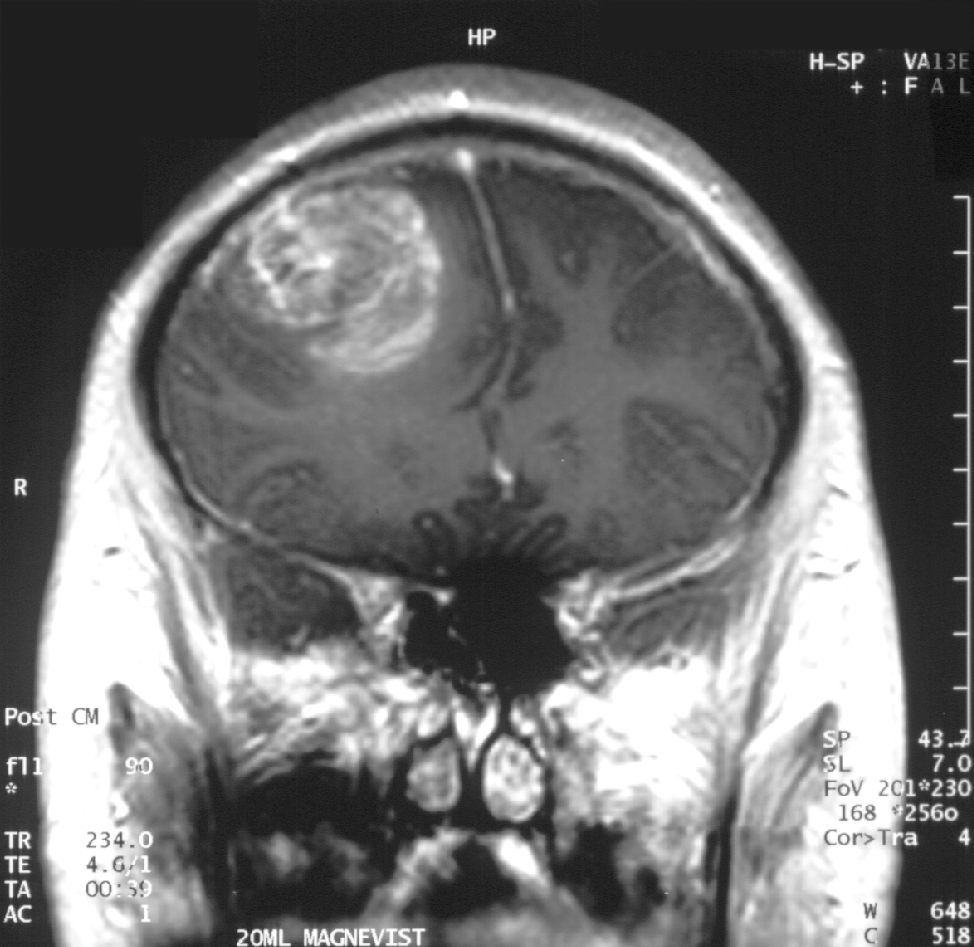
Diagnosis typically begins with a neurological examination to assess deficits in strength, coordination, reflexes, and sensory function, followed by imaging such as magnetic resonance imaging (MRI) with contrast or computed tomography (CT) scans to visualize the tumor's location, size, and characteristics. A definitive diagnosis requires a biopsy or surgical resection to examine tissue under a microscope, confirming the presence of malignant cells and determining the tumor's grade based on World Health Organization criteria. Treatment usually involves maximal safe surgical resection to remove as much tumor as possible, followed by radiation therapy and chemotherapy, often with temozolomide, to target residual cells; additional options may include tumor-treating fields (TTFields) or targeted therapies for specific molecular subtypes. Despite multimodal treatment, the prognosis for glioblastoma remains poor, with a median overall survival of approximately 14-16 months with standard treatment (maximal safe surgical resection, radiotherapy, and temozolomide chemotherapy). The addition of tumor-treating fields (TTFields) can extend median survival to around 20-21 months in some patients. The five-year survival rate is typically 5-10%. No major changes to these figures have been reported for 2025 or 2026. Survival is influenced by factors such as age, tumor location, extent of resection, and molecular markers like methylation status. The tumor's heterogeneity, ability to evade the blood-brain barrier, and high recurrence rate—often within the original field—underscore the need for ongoing research into , precision medicine, and novel methods to improve outcomes.

Clinical Presentation

Signs and Symptoms

Glioblastoma typically presents with a range of neurological symptoms that arise from the tumor's aggressive growth within the , leading to compression of surrounding structures and increased . Common initial manifestations include headaches, which are often persistent and more severe in the morning due to overnight accumulation of , affecting approximately 36% of patients at . Seizures occur in 30-62% of cases, with about two-thirds presenting at the time of , and represent one of the most frequent early signs. and vomiting, resulting from elevated , are also prevalent and may accompany headaches during the initial presentation. Focal neurological deficits depend on the tumor's location within the cerebral hemispheres, where most glioblastomas arise, and occur in about 64% of patients at discovery. For instance, tumors in the may cause or motor weakness on the contralateral side, while those in the can lead to or language difficulties; involvement often results in or , and occipital tumors may produce changes or hemianopia. These deficits typically develop gradually as the tumor infiltrates adjacent brain tissue. Cognitive and alterations are reported in around 46% of cases at and stem from disruption of higher brain functions by the expanding mass. Symptoms such as memory loss, , irritability, and behavioral changes, including or , are common, particularly with frontal or tumors. These changes can significantly impair daily functioning and . The onset of symptoms is often insidious, with subtle signs like mild headaches or cognitive fog progressing rapidly over weeks to months due to the tumor's high proliferative rate; more than 50% of patients have a clinical history shorter than three months before . This rapid deterioration frequently prompts to identify the underlying cause.

Diagnosis

Diagnosis of glioblastoma typically begins with clinical suspicion arising from neurological symptoms such as headaches, seizures, or focal deficits (e.g., mild facial numbness or stiffness), prompting neuroimaging evaluation. Focal symptoms like mild facial numbness or stiffness imply limited anatomical disruption sufficient to alter trigeminal pathway function, consistently producing detectable MRI changes such as signal abnormality or asymmetry; in contrast, occult tumors more often evade initial detection with non-localizing symptoms like headache. Neuroimaging plays a central role in the initial assessment, with magnetic resonance imaging (MRI) serving as the gold standard modality. On contrast-enhanced MRI, glioblastoma characteristically appears as an irregular, heterogeneous mass with ring-like enhancement, a hypointense necrotic core on T1-weighted images, and surrounding vasogenic edema visible as hyperintensity on T2-weighted and fluid-attenuated inversion recovery (FLAIR) sequences. Computed tomography (CT) is often used in acute settings, particularly when MRI is contraindicated or unavailable, revealing a hypodense lesion with possible hemorrhage or mass effect, though it is less sensitive for detecting subtle features like edema or necrosis compared to MRI. Advanced imaging techniques enhance diagnostic precision, particularly for differentiating glioblastoma from treatment-related changes or mimics. Perfusion-weighted MRI (PWI), often using dynamic susceptibility contrast (DSC) methods, measures relative cerebral (rCBV); glioblastoma typically shows elevated rCBV (e.g., >2.0) in enhancing regions due to neovascularization, aiding distinction from low-perfusion entities like abscesses. MR spectroscopy (MRS) assesses metabolic profiles, revealing elevated choline (Cho) peaks and reduced N-acetylaspartate (NAA) in glioblastoma, with a Cho/NAA ratio >1.11 supporting tumor presence over non-neoplastic processes. (PET) with tracers like 18F-fluoroethyltyrosine (FET) demonstrates high uptake in glioblastoma, improving tumor delineation and differentiation from radiation necrosis or lower-grade lesions. Definitive diagnosis requires histopathological confirmation through tissue sampling, guided by to target viable tumor areas. Stereotactic is preferred when surgical resection is not feasible, offering a minimally invasive approach with diagnostic yield >90% and low morbidity (<2% major complications), involving frame-based or frameless navigation to sample multiple sites if needed. Open surgical resection serves dual diagnostic and therapeutic purposes, allowing broader sampling for heterogeneous tumors. per (WHO) criteria identifies glioblastoma as a grade 4 diffuse astrocytic featuring cellular pleomorphism, mitotic activity, microvascular proliferation, and/or palisading . Molecular testing is integral to the integrated , mandated by WHO 2021 classification for adult cases. Glioblastoma is defined as IDH-wildtype (no mutations in IDH1/IDH2 genes, confirmed via and sequencing if needed), often with additional features like TERT promoter mutation or EGFR amplification; IDH-mutant cases are excluded and reclassified as . promoter methylation status is routinely assessed using methylation-specific PCR, as it predicts response to . EGFR amplification, present in ~40-50% of IDH-wildtype glioblastomas, further supports the when histological criteria are equivocal. Differential diagnosis involves distinguishing glioblastoma from mimics such as brain metastases, abscesses, and lower-grade gliomas, often requiring multimodal . Unlike the uniform enhancement and multiplicity (75% of cases) of metastases at the gray-white junction, glioblastoma exhibits irregular enhancement, non-enhancing infiltrative components, and frequent crossing of the midline via the . Abscesses show central restricted on diffusion-weighted imaging and low rCBV (<2.0) in the enhancing rim, contrasting with glioblastoma's high rCBV and peripheral metabolic activity on /PET. Lower-grade gliomas lack significant enhancement and , with lower Cho peaks on . Diagnostic challenges arise in tumors located in eloquent brain areas (e.g., , language centers), where biopsy risks neurological deficits, potentially limiting sampling to stereotactic methods and increasing sampling error in heterogeneous lesions. Multifocal disease, seen in up to 34% of cases with multiple enhancing foci connected by T2/FLAIR hyperintensity or distant lesions, complicates accurate biopsy targeting and radiological-pathological correlation due to infiltrative spread beyond visible enhancement.

Epidemiology and Risk Factors

Epidemiology

Glioblastoma, the most aggressive form of primary , has an annual age-adjusted incidence rate of approximately 3.27 per 100,000 population in the United States (as of 2018-2022), accounting for about 13.9% of all primary tumors and roughly 50% of malignant gliomas. Globally, incidence rates vary but generally range from 3 to 4 per 100,000 in developed regions, representing a significant portion of the overall burden of primary brain malignancies. The disease predominantly affects older adults, with incidence rates increasing sharply with age and peaking in the 65–74 age group, where rates can exceed 15 per 100,000. Approximately 48% of cases are diagnosed in individuals aged 65 years or older. There is a notable predominance, with a male-to-female ratio of about 1.6:1, reflected in higher incidence rates among (around 4.0 per 100,000) compared to females (2.5 per 100,000). Geographic variations show higher incidence in populations of European descent, with rates up to 3 times higher in Caucasians compared to Asian or African American groups. Incidence is generally elevated in developed countries and urban areas compared to lower-income or rural regions. Over time, rates have remained largely stable or shown a slight increase, attributed to aging populations and improved diagnostic capabilities, with no substantial rise in age-adjusted trends from 2000 to 2020.

Genetic Risk Factors

Glioblastoma, a highly aggressive form of brain cancer, has a limited role for inherited genetic factors in its etiology, with most cases arising sporadically; however, rare hereditary syndromes confer elevated susceptibility. Li-Fraumeni syndrome, characterized by germline mutations in the TP53 , significantly increases the risk of developing glioblastoma among other cancers, with brain tumors occurring in approximately 10-15% of affected individuals. Neurofibromatosis type 1, caused by germline mutations in the NF1 gene, is associated with optic pathway gliomas and other low-grade tumors, though higher-grade glioblastomas can emerge in a subset of patients. Turcot syndrome, encompassing both type 1 (germline mutations in mismatch repair genes such as MLH1, MSH2, MSH6, or PMS2) and type 2 (APC gene mutations), predisposes carriers to colorectal cancers alongside gliomas, including glioblastoma in some cases. Familial clustering accounts for about 5% of glioblastoma cases, where multiple family members develop the disease without an identifiable single-gene syndrome, suggesting contributions from polygenic inheritance. Emerging polygenic risk scores (PRS), which aggregate effects from multiple common variants, have shown promise in stratifying risk, with individuals in the top PRS facing up to an 8-fold increased for IDH-mutant gliomas. Specific variants outside of syndromes are rare but notable; for instance, TP53 mutations occur in about 13.6% of multifocal cases, while PTEN variants like Arg234Gln have been identified in isolated families with predisposition, though with low overall . Genome-wide association studies (GWAS) have identified common variants at key loci that modestly elevate glioblastoma risk, including rs2736100 at 5p15.33 near the TERT (odds ratio ~1.2-1.5) and variants at 7p11.2 in the EGFR locus ( ~1.2), which together explain a portion of the heritable component. These genetic risk factors may interact with environmental exposures to modulate susceptibility, as evidenced by higher glioma incidence in relatives exposed to potential carcinogens, though specific mechanisms remain under investigation.

Environmental and Other Risk Factors

Ionizing radiation is the only well-established environmental risk factor for glioblastoma, with exposure primarily occurring through therapeutic radiation treatments for other conditions, such as or . This risk is particularly pronounced following high-dose exposure to the head or neck during childhood, where studies have reported a 6- to 10-fold increase in glioma incidence compared to unexposed individuals. The latency period for tumor development is typically 10-15 years or longer, and the association is supported by cohort studies of pediatric patients treated with cranial . No strong evidence links sources, such as cell phones or electromagnetic fields, to increased glioblastoma risk. Chemical exposures have been investigated as potential risk factors, but the evidence remains inconsistent and inconclusive. Occupational or environmental contact with substances like , used in production, and certain pesticides has been associated with elevated risks in some cohort and case-control studies, though meta-analyses indicate no definitive causal relationship for glioblastoma specifically. Similarly, exposures to products, solvents, and herbicides show mixed results, with no proven link after adjusting for factors. Immune suppression, as seen in individuals with or organ transplant recipients on long-term immunosuppressive therapy, is associated with an elevated risk of glioblastoma development. In HIV-positive patients, the incidence of primary brain tumors, including gliomas, appears higher than in the general population, potentially due to impaired immune surveillance allowing . Transplant patients exhibit a similar pattern, with case series and registry data reporting increased glioblastoma occurrences attributed to chronic , though the absolute risk remains low. A history of allergies or atopic conditions is inversely associated with glioblastoma risk, consistent with the , which posits that reduced early-life microbial exposure leads to dysregulated immune responses favoring but protecting against certain cancers. Epidemiological studies, including large case-control analyses, report odds ratios around 0.78 for allergic conditions and , suggesting a protective effect possibly mediated by heightened IgE-mediated immunity or anti-tumor surveillance. This inverse relationship holds across various allergy types, such as eczema and hay fever. Occupational risks for glioblastoma are limited and show inconsistent evidence. Workers in synthetic rubber manufacturing have demonstrated slightly elevated brain tumor rates in some historical cohort studies, potentially linked to chemical exposures like aromatic amines, but meta-analyses conclude no overall increased risk after controlling for biases. Similarly, limited data suggest a possible association with aviation-related occupations, such as pilots or aircraft operators, due to cosmic radiation or solvents, though findings are not robust and require further confirmation.

Pathogenesis

Classification

Glioblastoma, also known as glioblastoma multiforme, is classified under the World Health Organization (WHO) 2021 Central Nervous System (CNS) tumor classification as a grade 4 diffuse astrocytic tumor, specifically IDH-wildtype astrocytoma. This classification emphasizes the integration of histopathological features with molecular markers to define the tumor precisely, moving away from purely morphological diagnoses. Key histological hallmarks include high cellularity, marked nuclear pleomorphism, frequent mitoses, microvascular proliferation (such as endothelial proliferation), and pseudopalisading necrosis, which distinguish it as a high-grade malignancy. These features reflect the tumor's aggressive infiltrative growth and poor prognosis, with the presence of necrosis and vascular proliferation being particularly indicative of grade 4 status. Traditionally, glioblastomas have been subdivided into primary (de novo) and secondary subtypes based on clinical progression. Primary glioblastomas arise without a precursor and account for approximately 90% of cases, typically presenting in older patients (over 50 years), while secondary glioblastomas progress from lower-grade astrocytomas and are more common in younger individuals. However, the WHO 2021 framework highlights that these subtypes are molecularly distinct, with primary tumors often harboring EGFR amplification, PTEN mutations, and TERT promoter mutations, whereas secondary ones frequently show TP53 mutations and alterations; nonetheless, the classification prioritizes IDH-wildtype status for glioblastoma over this historical dichotomy. The integration of molecular markers is central to the modern classification, requiring the absence of IDH1/IDH2 mutations (IDH-wildtype) and the presence of specific genetic alterations like combined TERT promoter mutation and or gain/ loss for definitive in adults. This molecular layering ensures more accurate categorization, especially in cases where alone might be ambiguous. In contrast to other gliomas, such as grade 3 (IDH-mutant), glioblastoma lacks the defining molecular features of lower-grade tumors and exhibits the full spectrum of aggressive histological elements like , which are absent in grade 3 lesions.

Molecular Alterations

Glioblastomas exhibit a complex landscape of somatic genetic and epigenetic alterations that drive tumor initiation and progression, primarily in primary tumors which constitute over 90% of cases. These changes often converge on key signaling pathways, leading to uncontrolled , survival, and . Comprehensive genomic analyses have identified recurrent , amplifications, and deletions that distinguish glioblastoma from lower-grade gliomas. Among the most frequent genetic alterations are amplifications of the epidermal growth factor receptor (EGFR) gene, occurring in 40-60% of primary glioblastomas, which promote aberrant receptor tyrosine kinase signaling. Loss-of-function mutations or deletions in the phosphatase and tensin homolog (PTEN) gene are seen in 25-40% of cases, leading to hyperactivation of the PI3K/AKT pathway. Mutations in the tumor suppressor TP53 are present in approximately 30% of tumors, disrupting DNA damage response and apoptosis. Inactivating alterations in neurofibromin 1 (NF1), a negative regulator of RAS signaling, affect 20-30% of glioblastomas. Isocitrate dehydrogenase 1 and 2 () mutations are rare in primary glioblastomas, occurring in less than 10% of cases, but are hallmark features of secondary glioblastomas that progress from lower-grade , with rates exceeding 70%. These result in the production of the oncometabolite 2-hydroxyglutarate, which alters epigenetic and cellular . In contrast, mutations in the TERT promoter are highly prevalent in 80% of primary glioblastomas, enabling telomerase reactivation and replicative . Chromosomal abnormalities are common, including of (gain of +7) in up to 70% of tumors, often co-occurring with EGFR amplification, and of (-10) in a similar proportion, which typically involves loss of PTEN. Deletions on (-9p), encompassing the locus, are also frequent, contributing to RB pathway deregulation. These aneuploidies underscore the genomic instability characteristic of glioblastoma. Epigenetic modifications play a critical role, with promoter hypermethylation of the O6-methylguanine-DNA methyltransferase () gene observed in 40-50% of glioblastomas, which sensitizes tumors to alkylating agents like by impairing . Additionally, global DNA hypomethylation affects millions of CpG sites across the , promoting activation and genomic instability. These epigenetic changes complement genetic hits in fostering tumor heterogeneity. The majority of glioblastomas harbor alterations in three core signaling pathways: (RTK)/RAS/PI3K in 88% of cases, which drives proliferation and survival; the pathway in 87%, impairing ; and the RB pathway in 78%, allowing unchecked progression through the . These pathway disruptions, often involving multiple concurrent alterations, highlight the of glioblastoma and inform targeted therapeutic strategies.

Cancer Stem Cells

Glioblastoma cancer stem cells (GSCs), also known as glioblastoma stem-like cells, represent a subpopulation of tumor cells characterized by self-renewal capacity, multipotency, and the ability to initiate and propagate tumors upon transplantation. These cells are typically identified by expression of specific markers, including the cell surface glycoprotein (prominin-1), the protein Nestin, and the SOX2, which collectively indicate their stem-like properties and distinguish them from differentiated tumor cells. GSCs can differentiate into neuronal, astrocytic, and oligodendroglial lineages, recapitulating the heterogeneity observed in glioblastoma tumors. The origin of GSCs remains debated but is thought to involve transformation of normal neural stem cells located in the or of more mature glial or neuronal progenitors under oncogenic pressure. Seminal studies have demonstrated that CD133-positive cells isolated from glioblastoma tissues exhibit properties akin to neural stem cells, supporting their derivation from this lineage. mechanisms, driven by genetic alterations, may also generate GSCs from non-stem tumor cells, contributing to intratumoral plasticity. GSCs play critical roles in tumor initiation by generating the bulk of the tumor mass through asymmetric division and differentiation. They promote invasion into surrounding brain parenchyma by upregulating matrix metalloproteinases and migrating along tracts. Additionally, GSCs drive by secreting (VEGF), which stimulates endothelial and new vessel formation to support tumor growth. GSCs contribute to therapy resistance through multiple intrinsic mechanisms, including entry into a quiescent state that shields them from cell cycle-targeted treatments like and . They express ATP-binding cassette (ABC) transporters, such as and ABCB1, which actively efflux chemotherapeutic agents like , reducing intracellular drug accumulation. Enhanced DNA repair pathways, including elevated levels of and RAD51, allow GSCs to efficiently repair - and chemotherapy-induced damage, further promoting survival post-treatment. Therapeutic strategies targeting GSCs focus on disrupting their self-renewal and resistance pathways, such as inhibition of the Notch signaling cascade with gamma-secretase inhibitors, which reduces GSC proliferation and sensitizes them to . Differentiation-inducing agents, like all-trans retinoic acid, promote GSC maturation into non-tumorigenic progeny, diminishing their stemness and tumorigenic potential. These approaches aim to overcome recurrence driven by GSC persistence. Evidence from orthotopic xenograft models underscores the enrichment of GSCs following standard therapies; for instance, treatment selects for + cells, leading to increased tumor engraftment and recurrence in immunodeficient mice. Such models confirm that surviving GSCs maintain tumor-initiating capacity, highlighting their role in post-treatment relapse.

Metabolic Features

Glioblastoma (GBM) cells exhibit profound metabolic reprogramming to support their rapid proliferation, survival, and to nutrient-poor environments, diverging from normal cellular to favor anabolic processes. This includes a shift toward aerobic , known as the Warburg effect, where glucose is converted to lactate even in the presence of oxygen, providing energy and biosynthetic intermediates. (LDHA), a key in this pathway, is upregulated in GBM, facilitating lactate production and contributing to tumor progression by maintaining an acidic microenvironment that promotes . Suppression of LDHA has been shown to downregulate the Warburg effect, compromising tumor growth and enhancing sensitivity to therapies. In addition to , GBM cells display glutamine addiction, relying heavily on as a carbon and source for synthesis, production, and balance. The glutaminase (GLS) hydrolyzes to glutamate, which is essential for replenishing α-ketoglutarate in the tricarboxylic acid cycle and supporting biosynthetic demands in proliferating cells. The ASCT2 transporter (SLC1A5) facilitates uptake, and its inhibition disrupts tumor . Targeting GLS with inhibitors like CB-839 induces metabolic stress in GBM cells, reducing proliferation and viability, particularly under glutamine restriction. Lipid metabolism is also dysregulated in GBM to meet the demands of membrane biogenesis and signaling. (FASN) is overexpressed, catalyzing of fatty acids from and , which supports rapid and tumor growth. Inhibition of FASN attenuates GBM and stemness, highlighting its role in maintaining tumor-initiating properties. This pathway is particularly active in hypoxic regions, linking synthesis to environmental adaptation. Hypoxia within the GBM tumor core drives further metabolic shifts through stabilization of hypoxia-inducible factor 1α (HIF-1α), a that reprograms energy production to favor and limit . HIF-1α upregulates glycolytic enzymes and glucose transporters, enhancing survival under low-oxygen conditions and promoting . This adaptation not only sustains energy needs but also contributes to therapeutic resistance by altering nutrient utilization. In secondary GBM, which arises from lower-grade gliomas, (IDH) mutations predominate, leading to the production of the oncometabolite D-2-hydroxyglutarate (2-HG). Mutant IDH1/2 enzymes convert α-ketoglutarate to 2-HG, which competitively inhibits α-ketoglutarate-dependent dioxygenases, disrupting epigenetic regulation via and . This results in altered that favors gliomagenesis and blocks . 2-HG accumulation is a hallmark of IDH-mutant tumors, distinguishing them from primary GBM. Therapeutic strategies targeting these metabolic vulnerabilities show promise, particularly for IDH-mutant cases. IDH inhibitors such as , which targets mutant IDH1, reduce 2-HG levels, reverse epigenetic alterations, and induce differentiation in cells. Clinical trials have demonstrated prolonged in patients with IDH1-mutant advanced gliomas treated with , with a favorable safety profile. Similarly, inhibitors of GLS, LDHA, and FASN are under investigation to exploit the Warburg effect and lipid dependency, potentially synergizing with standard therapies like .

Tumor Microenvironment

The (TME) in glioblastoma (GBM) comprises non-tumor cells, (ECM), and soluble factors that foster tumor progression, invasion, and resistance to therapy. This ecosystem supports GBM cells through dynamic interactions that create an immunosuppressive and hypoxic niche, distinct from intrinsic tumor . Key components include vasculature, immune cells, ECM, and stromal elements, which collectively enable tumor adaptation and recurrence. GBM vasculature is highly abnormal, characterized by excessive angiogenesis driven by vascular endothelial growth factor (VEGF) secretion from tumor and stromal cells, resulting in leaky, tortuous vessels that promote hypoxia and nutrient delivery inefficiencies. Hypoxia-inducible factors (HIFs) further upregulate VEGF under low oxygen conditions, exacerbating pseudopalisading necrosis and tumor invasion around vessels. These fragile vessels contribute to perivascular niches that shield tumor cells from therapies. Immune infiltration in the GBM TME is dominated by microglia and tumor-associated macrophages (TAMs), which comprise up to 50% of the non-neoplastic cellular component and predominantly adopt an M2-polarized, pro-tumorigenic phenotype that secretes immunosuppressive cytokines like IL-10 and TGF-β. T-cell infiltration remains sparse due to physical barriers and suppressive signals, with exhausted CD8+ T cells expressing PD-1; concurrently, PD-L1 upregulation on tumor and immune cells inhibits T-cell activation, correlating with poor prognosis. The ECM in GBM is enriched with hyaluronan (HA), a that interacts with receptors on tumor cells to enhance migration, invasion, and stem-like properties while forming a physical barrier that limits and immune cell penetration. Matrix metalloproteinases (MMPs), such as MMP-2 and MMP-9, remodel this HA-rich ECM, increasing stiffness and facilitating tumor dissemination along tracts. Stromal cells, including reactive and , reinforce the GBM TME by providing structural and signaling support. at tumor margins undergo reactive transformation, releasing factors like VEGF and to promote and . proliferate abnormally around vessels, stabilizing the leaky vasculature while contributing to the perivascular niche that sustains tumor quiescence. Metabolic crosstalk within the TME involves lactate export from glycolytic tumor cells via monocarboxylate transporters, acidifying the and suppressing and T-cell function to maintain . This lactate-mediated signaling also influences stromal cells, promoting a protumoral metabolic . Therapeutic strategies targeting the GBM TME focus on anti-angiogenic agents like , a against VEGF, which normalizes vasculature and extends in recurrent GBM by reducing and permeability, though it fails to improve overall survival and can induce hypoxia-driven resistance. Emerging approaches combine with inhibitors to counter compensatory .

Prevention

Primary Prevention Strategies

Primary prevention of glioblastoma focuses on minimizing exposure to established environmental risk factors and addressing genetic predispositions where possible, as the disease's remains largely idiopathic. The only well-confirmed environmental risk factor is , which underscores the importance of limiting unnecessary exposures. Other potential risks, such as certain occupational exposures, show inconsistent associations but warrant precautionary measures. To mitigate radiation-related risks, adherence to the ALARA (As Low As Reasonably Achievable) principle is essential in medical imaging and occupational settings, prioritizing justification of procedures and optimization of doses. Specifically, unnecessary computed tomography (CT) scans should be avoided, particularly in children, where even low-dose exposures have been linked to a small but significant increase in brain tumor incidence, including gliomas.60815-0/fulltext) For instance, a large cohort study found that cumulative radiation doses from CT scans in childhood were associated with an excess risk of brain tumors, with odds ratios increasing linearly with dose. Lifestyle modifications offer limited but supportive strategies for risk reduction. Cigarette has a weak and inconsistent association with glioblastoma, with meta-analyses generally showing no significant elevation in risk, though cessation is recommended for overall health benefits. A emphasizing organic produce and thorough washing of fruits and may help reduce incidental exposure, as some occupational studies suggest a possible link between high-level use and risk, particularly among farmers exposed to herbicides like . However, evidence for dietary pesticides and sporadic glioblastoma remains inconclusive. Occupational safety measures are critical in industries with potential exposure. Workers in rubber manufacturing and production should use , such as respirators and gloves, to limit inhalation and skin contact with solvents and fumes, as case-control studies have reported modest elevations in risk in these sectors. policies play a key role, including stringent regulations on —a chemical used in (PVC) production—enforced by the U.S. Environmental Protection Agency (EPA) and (OSHA), which limit workplace exposures to below 1 ppm after evidence linked cumulative high-level exposure to increased brain cancer mortality. These standards have contributed to declining exposure levels since the . For individuals with familial risk, is a vital preventive step. Families with Li-Fraumeni syndrome, caused by TP53 mutations, face a substantially elevated lifetime risk of gliomas, including glioblastoma, prompting recommendations for counseling to discuss surveillance and risk-modifying behaviors. Clinical guidelines advocate early and counseling for those with suggestive family histories to enable informed and enhanced monitoring.

Secondary Prevention Approaches

Secondary prevention of glioblastoma focuses on early detection and in individuals at elevated risk due to genetic syndromes or prior therapeutic exposures, aiming to identify tumors at a stage where intervention may alter progression. For patients with hereditary predisposition syndromes such as Li-Fraumeni syndrome (LFS), characterized by germline TP53 mutations, annual brain MRI is recommended starting from birth or upon diagnosis of the variant to facilitate early identification of brain tumors, including gliomas. The initial scan incorporates intravenous contrast, with subsequent non-contrast MRIs if prior imaging is normal, and this dedicated brain MRI cannot be replaced by whole-body MRI protocols. In neurofibromatosis type 1 (NF1), where there is an increased lifetime risk of high-grade gliomas including glioblastoma, routine is not advised for individuals; instead, clinical monitoring for neurological symptoms such as seizures or headaches guides targeted MRI . Individuals exposed to prior cranial , such as survivors of childhood cancers or those treated for benign conditions like , represent another high-risk cohort for secondary glioblastoma, with risks elevated 5- to 10-fold depending on dose and age at exposure. Long-term surveillance in these patients typically involves annual clinical assessments and symptom-directed , as routine MRI screening is not universally recommended due to limited evidence of benefit in asymptomatic cases, though enhanced protocols may be considered for those with very high prior doses. Emerging biomarker-based approaches include tests detecting TERT promoter or EGFRvIII variants in circulating tumor DNA or extracellular vesicles, which have shown promise in identifying glioblastoma alterations with sensitivities up to 71-100% in research settings, but these remain investigational and are not incorporated into routine screening. Challenges in implementing secondary prevention strategies include the absence of validated non-invasive markers with sufficient specificity for population-level use, as current blood-based assays often require known tumor mutations for detection and face issues with low levels in early-stage disease. Cost-effectiveness is another barrier, particularly for frequent MRI in pediatric or high-risk groups, where cumulative avoidance and psychological burden must be balanced against potential gains from early detection. Guidelines from organizations like the (NCCN) emphasize and tailored surveillance for familial cancer syndromes, incorporating protocols such as those for LFS within broader hereditary frameworks, though specific recommendations for glioblastoma remain integrated into general CNS tumor management rather than standalone screening pathways.

Treatment

Symptomatic Management

Symptomatic management in glioblastoma focuses on alleviating common symptoms such as seizures, headaches, , and neurological deficits to improve . These interventions are adjunctive and do not target tumor growth directly. Seizures occur in approximately 30-50% of glioblastoma patients and are managed primarily with antiepileptic drugs. is recommended as the first-line agent due to its favorable profile and lack of significant interactions with agents like . Enzyme-inducing antiepileptics such as and are generally avoided because they can accelerate the of chemotherapeutic drugs, potentially reducing efficacy. Prophylactic use of antiepileptics is not routinely recommended unless seizures are present. Patients at risk for seizures should moderate their intake of caffeine-containing substances, as caffeine may lower the seizure threshold in susceptible individuals. Moderate consumption of chocolate is generally acceptable following surgery, as there are no specific dietary prohibitions against it in standard guidelines for glioblastoma patients, unless a patient's physician advises otherwise due to individual factors such as seizure risk from caffeine or medication interactions. Cerebral edema, a frequent cause of headaches and neurological symptoms, is treated with corticosteroids, most commonly dexamethasone. Initial dosing typically ranges from 4 to 16 mg per day, depending on symptom severity, with the goal of reducing peritumoral swelling and improving neurological function. Once symptoms stabilize, dexamethasone should be tapered gradually to the lowest effective dose—often reducing by 2 mg every 3-5 days—to minimize side effects, avoiding abrupt discontinuation to prevent rebound . Headaches and pain are managed with analgesics, starting with non-opioid options like acetaminophen or nonsteroidal anti-inflammatory drugs for mild to moderate symptoms. For more severe pain, opioids such as may be used, titrated to effect while monitoring for . and , often related to increased or treatment side effects, are controlled with antiemetics like or metoclopramide. Supportive care plays a crucial role in maintaining function and emotional well-being, including physical and to address motor deficits and weakness, as well as speech therapy for communication impairments. Psychological support, provided by counselors or teams, helps patients and caregivers cope with anxiety, depression, and existential distress. For causing symptoms like severe or altered mental status, palliative ventriculoperitoneal shunting can divert and provide symptom relief in approximately 78% of cases (95% CI 66-88%). Management of side effects is essential for long-term tolerability. Steroid-induced requires regular , with insulin therapy initiated if levels exceed 180 mg/dL or in patients with preexisting ; dietary modifications and short-acting agents like metformin may also be employed. Proximal , characterized by in the limbs, is mitigated by dose reduction when possible and incorporation of to preserve strength and mobility. Other common issues, such as or mood changes, are addressed through supportive measures like or low-dose anxiolytics.

Surgical Resection

Surgical resection is the cornerstone of initial treatment for glioblastoma, aiming to achieve maximal safe removal of the tumor to improve survival and while preserving neurological function. The primary goal is gross total resection (GTR), defined as the removal of more than 98% of the contrast-enhancing tumor volume as assessed on preoperative MRI, which correlates with significantly better outcomes compared to subtotal resection (). Multiple studies have demonstrated that GTR extends median overall survival by 3 to 5 months, with reported medians ranging from 15 to 20 months versus 12 to 15 months for , independent of other factors like age or . To maximize the extent of resection, particularly in tumors adjacent to eloquent brain areas such as motor, , or sensory regions, awake is a preferred technique. During awake , the patient remains conscious and cooperative, enabling direct cortical and subcortical mapping through tasks like naming objects or moving limbs to identify functional boundaries in real time. Intraoperative MRI (iMRI) complements this by providing updated during surgery to visualize residual tumor and adjust the resection trajectory, often increasing the rate of GTR by 15-20%. Preoperative functional MRI (fMRI) further informs surgical planning by delineating critical areas, though iMRI offers dynamic guidance. Advanced navigation tools enhance precision in tumor localization and margin delineation. Neuronavigation systems integrate preoperative MRI, CT, or tensor imaging into a stereotactic frame, providing real-time three-dimensional guidance to track surgical instruments relative to the tumor. Fluorescence-guided surgery using 5-aminolevulinic acid (5-ALA), administered orally 3 hours preoperatively, induces accumulation in malignant cells, causing visible pink-to-red under blue light that highlights tumor tissue beyond standard white-light visualization. This technique has been shown to increase GTR rates by up to 20% and is particularly useful for defining infiltrative margins. Despite these advancements, surgical resection is associated with notable risks. New or worsened neurological deficits, such as , , or , occur in 10-20% of cases, with higher rates in eloquent-area resections, though approximately 70-80% resolve within months. Postoperative infections, including surgical site or , affect 4-7% of s, while hemorrhage occurs in about 5-6%, potentially requiring reoperation. These complications underscore the need for careful patient selection and multidisciplinary perioperative care. The extent of resection is quantitatively correlated with through volumetric assessment on early postoperative MRI, typically performed within 48-72 hours to minimize artifacts from or blood products. This involves segmenting preoperative and postoperative tumor volumes using software to calculate the percentage removed, with residuals under 1-2 cm³ often defining successful GTR. Such assessments confirm that even small differences in residual volume—e.g., less than 5 cm³ versus greater—impact by several months. Limitations of surgical resection are pronounced in certain tumor presentations. Multifocal glioblastomas, involving multiple discrete lesions, often preclude GTR due to their disseminated nature, with surgery typically limited to or of the dominant focus to confirm diagnosis and alleviate symptoms. Similarly, deep-seated tumors in structures like the or pose high risks of irreversible deficits, favoring over extensive resection in many cases. Debulking in these scenarios can still provide symptomatic relief from , such as reduced . Following surgical resection, a balanced diet is recommended to support patient recovery. Standard medical guidelines do not include specific dietary restrictions or prohibitions against chocolate consumption for glioblastoma patients after surgery. Patients can generally eat chocolate in moderation as part of a balanced diet, unless a patient's doctor advises otherwise due to individual factors such as seizure risk related to caffeine content or potential interactions with medications.

Radiotherapy

Radiotherapy plays a central role in the management of glioblastoma, typically administered following maximal safe surgical resection to target residual tumor cells and improve local control. The standard regimen, established by the Stupp protocol, involves delivering 60 Gy of external beam radiation in 30 fractions over 6 weeks, with the clinical target volume encompassing the postoperative tumor bed and surrounding edema plus a 2-3 cm margin. This approach has been shown to extend median overall survival by approximately 2.5 months when combined with compared to radiotherapy alone (14.6 months versus 12.1 months). Advanced techniques such as intensity-modulated radiotherapy (IMRT) are widely used to achieve conformal dose distribution, sparing critical structures like the hippocampus and while delivering therapeutic doses to the tumor. For recurrent glioblastoma, stereotactic radiosurgery (SRS) offers a non-invasive option for focal treatment of small, well-defined lesions, potentially improving in select cases without increasing toxicity.30003-0/fulltext) In elderly or frail patients unsuitable for standard , hypofractionated regimens provide a shorter treatment course with comparable efficacy. A common option is 40 Gy delivered in 15 fractions over 3 weeks, which has demonstrated non-inferiority to conventional radiotherapy in terms of survival and for patients over 65 years. serves as a radiosensitizer when given concurrently with radiotherapy at a dose of 75 mg/m² daily, enhancing DNA damage in tumor cells and synergizing with effects, as validated in the pivotal EORTC/NCIC trial. Acute side effects of radiotherapy include , skin , and transient worsening of neurological symptoms due to peritumoral , which typically resolve within weeks post-treatment. Late complications, occurring months to years later, encompass cognitive decline, such as impairment, and , affecting up to 10-20% of patients and often requiring management or surgical intervention.

Chemotherapy

Chemotherapy plays a central role in the management of glioblastoma, with temozolomide (TMZ) established as the standard cytotoxic agent due to its alkylating properties that induce DNA damage in rapidly dividing tumor cells. Administered orally, TMZ crosses the blood-brain barrier effectively, making it suitable for central nervous system malignancies. The standard regimen, known as the Stupp protocol, integrates TMZ with radiotherapy for newly diagnosed glioblastoma. During the concurrent phase, patients receive TMZ at 75 mg/m² daily for up to 49 days alongside radiotherapy, followed by an adjuvant phase of 150-200 mg/m² on days 1-5 of each 28-day cycle for six cycles. This approach synergizes with radiation by enhancing DNA damage and has demonstrated significant survival benefits; in a pivotal phase III trial, median overall survival was 14.6 months with radiotherapy plus TMZ compared to 12.1 months with radiotherapy alone. The response to is strongly influenced by O6-methylguanine-DNA methyltransferase () promoter status, a assessed via tumor tissue analysis. silences expression, reducing the enzyme's ability to repair -induced DNA at the O6 position of , thereby predicting greater treatment efficacy. Patients with methylated achieve approximately 50% better outcomes, with median survival extending to 21.7 months versus 15.3 months in the methylated subgroup receiving plus radiotherapy compared to radiotherapy alone ( 0.51). Resistance to TMZ arises primarily through MGMT-mediated repair of alkylated DNA and deficiency in mismatch repair pathways, which fail to recognize and excise damaged DNA strands, allowing tumor cells to survive. Unmethylated tumors exhibit intrinsic resistance, while acquired resistance in responsive cases often involves upregulated or mismatch repair defects. For recurrent glioblastoma, (CCNU), another oral alkylating agent, serves as an alternative, typically dosed at 110 mg/m² every six weeks. This compound similarly targets DNA but is used cautiously due to cumulative toxicity. Common side effects of TMZ include myelosuppression, such as neutropenia and thrombocytopenia, occurring in 7-14% of patients at grade 3/4 severity, alongside nausea and vomiting. Monitoring involves regular complete blood counts (CBC) to detect hematologic toxicity early and adjust dosing as needed.

Targeted Therapy

Targeted therapies for glioblastoma (GBM) focus on inhibiting specific molecular pathways dysregulated in the tumor, such as epidermal growth factor receptor (EGFR) signaling, vascular endothelial growth factor (VEGF)-mediated angiogenesis, isocitrate dehydrogenase (IDH) mutations, phosphoinositide 3-kinase (PI3K)/mammalian target of rapamycin (mTOR) activation, and DNA repair mechanisms via poly (ADP-ribose) polymerase (PARP). These approaches leverage the tumor's genetic heterogeneity, identified through mutations like EGFR amplification prevalent in up to 40% of primary GBM cases, to select patients via biomarker testing. However, challenges including blood-brain barrier (BBB) penetration, rapid resistance due to pathway crosstalk, and intratumoral heterogeneity have limited overall efficacy compared to standard chemotherapy. EGFR inhibitors, such as and , target amplified or mutated EGFR, a common alteration in primary GBM. Phase II trials of gefitinib monotherapy in recurrent GBM demonstrated good tolerability but minimal radiographic responses and no significant (PFS) benefit, with median PFS around 8-10 weeks. Similarly, erlotinib in first-relapse GBM showed limited efficacy, with objective response rates under 10% and median PFS of 1.8-2.4 months, attributed to poor CNS penetration and resistance mechanisms like PTEN loss. Combination strategies, such as erlotinib with or , have not yielded meaningful overall survival (OS) improvements in phase II studies. VEGF inhibitors like address tumor by blocking VEGF-A, earning FDA approval in 2009 for recurrent GBM based on phase II data showing rapid symptom relief and improved PFS. In pivotal trials, monotherapy extended median PFS to 4 months versus 1.5 months with , though OS remained similar at 9-10 months across arms, highlighting pseudoprogression risks and lack of curative impact. It is commonly used for recurrent disease to manage and progression, often combined with , but does not alter long-term outcomes. For IDH-mutant gliomas that may progress to secondary GBM, the IDH1/2 inhibitor vorasidenib represents a breakthrough in delaying progression from lower grades. Approved by the FDA in August 2024 and by the in September 2025 for grade 2 IDH-mutant astrocytomas or oligodendrogliomas, it delays progression in these precursors to GBM; the phase III trial reported a of 0.39 for PFS, with median time to progression nearly quadrupled to 27.7 months versus . Its oral formulation and brain penetration make it suitable for patients with IDH1 R132H mutations, detected in about 10% of GBMs overall but more common in secondary forms; however, data for established grade 4 secondary GBM remain limited, with phase 1 trials showing median PFS of 3.6 months. PI3K/mTOR inhibitors, including , target the hyperactive PI3K/AKT/mTOR pathway, altered in over 80% of GBMs via upstream signaling. In phase II trials for newly diagnosed GBM, combined with and radiotherapy achieved a PFS of 11.3 months but no OS advantage over standard , with toxicities like infections and limiting tolerability. Dual PI3K/mTOR inhibitors like voxtalisib have shown preclinical promise but similar clinical constraints due to off-target effects. PARP inhibitors, such as niraparib, exploit deficiency (HRD) in GBM subsets with BRCA or other alterations. A phase 0/2 "trigger" in newly diagnosed GBM demonstrated niraparib's superior penetration, achieving tumor concentrations exceeding those of other and inducing pharmacodynamic PARP inhibition in non-enhancing tissue. Ongoing phase III trials (e.g., Gliofocus) compare niraparib to in MGMT-unmethylated GBM, focusing on HRD-positive patients, with preliminary data suggesting potential PFS benefits in biomarker-selected cohorts. Biomarker-driven selection is essential for optimizing targeted therapies, with next-generation sequencing (NGS) enabling identification of actionable alterations like EGFR amplification, IDH mutations, or HRD signatures in 50-70% of GBMs. biopsies using (ctDNA) detect EGFRvIII or IDH1 variants in up to 55% of patients, facilitating real-time eligibility assessment and adaptive trial designs like INSIGhT, which match therapies to molecular profiles for personalized treatment. This approach underscores the shift toward precision oncology in GBM, though only a minority of patients currently access matched therapies due to limited approved options.

Immunotherapy

Immunotherapy represents a promising approach to treating glioblastoma by leveraging the patient's to target tumor cells, though its efficacy remains limited by the tumor's unique microenvironment. Key strategies include inhibitors, chimeric antigen receptor () T-cell therapies, and tumor vaccines, which aim to overcome the immunosuppressive barriers in the . Despite challenges, ongoing trials continue to refine these methods, with recent highlighting potential benefits in specific subsets of patients. Immune checkpoint inhibitors, such as nivolumab (anti-PD-1) and (anti-CTLA-4), have been evaluated in multiple phase III trials for glioblastoma but have shown limited overall survival (OS) improvements. In the 548 trial involving 716 patients with newly diagnosed glioblastoma and methylated promoter, the addition of nivolumab to standard radiotherapy and resulted in a median progression-free survival (PFS) of 10.6 months compared to 10.3 months with placebo, indicating no statistically significant benefit but a modest numerical difference in this subgroup. Similarly, the 498 trial in unmethylated cases reported a median OS of 13.4 months with nivolumab plus radiotherapy versus 14.9 months with plus radiotherapy, underscoring the lack of broad efficacy. Dual checkpoint blockade with nivolumab and has been tested in phase I/II settings, showing tolerable safety but no substantial PFS or OS gains over standard care in recurrent disease. CAR-T cell therapies target specific glioblastoma antigens, with intracranial delivery strategies addressing the blood-brain barrier. Therapies targeting interleukin-13 receptor alpha 2 (IL13Rα2) have demonstrated safety in phase I trials, including a 2024 cohort of 65 recurrent high-grade glioma patients receiving combined intracavitary and intraventricular infusions, which extended median OS to 10.2 months from a historical 7.7 months. CAR-T cells against (EGFRvIII) showed transient tumor regression in a phase I trial of 10 patients but no OS benefit, prompting refinements like the CARv3-TEAM-E approach in ongoing studies. These localized delivery methods aim to enhance T-cell persistence in the brain, though durable responses remain rare. Tumor vaccines, particularly dendritic cell-based approaches, seek to prime the against glioblastoma antigens. The DCVax-L vaccine, using autologous tumor lysate-pulsed dendritic cells, was assessed in a phase III trial (NCT00045968) of 331 patients with newly diagnosed or recurrent glioblastoma, reporting a median OS of 19.3 months for newly diagnosed cases versus 16.5 months in controls, and 13.2 months versus 7.8 months for recurrent disease; however, high crossover rates complicate interpretation, yet it suggests OS extension in personalized settings. This 's approval status remains under review, with 2025 analyses affirming its role in extending survival for select patients. Oncolytic viruses, such as PVSRIPO (a poliovirus-rhinovirus chimera), are under investigation in phase II trials for recurrent glioblastoma, showing immune activation and partial responses when delivered intratumorally, though detailed outcomes are explored in dedicated virotherapy studies. Major challenges in glioblastoma include the blood-brain barrier, which restricts immune cell and drug access, and the highly immunosuppressive featuring regulatory T cells, myeloid-derived suppressor cells, and expression that dampen T-cell activity. These factors contribute to low response rates, often below 10% in trials. As of 2025, combinations of with radiotherapy show promise in priming the ; for instance, a phase II trial of single-dose radiotherapy with anti-PD-1 inhibitors demonstrated enhanced antitumor immunity compared to fractionated regimens, while NRG BN007 confirmed safety of dual checkpoint blockade with radiotherapy in unmethylated cases, though PFS was not improved. These synergies suggest radiotherapy's abscopal effects could boost efficacy moving forward.

Emerging Therapies

Tumor treating fields (TTFields) represent a noninvasive therapeutic approach utilizing low-intensity, intermediate-frequency alternating electric fields, typically at 200 kHz, delivered via the Optune device to disrupt cancer cell division in glioblastoma. Clinical trials have demonstrated that TTFields, when added to standard temozolomide chemotherapy following maximal surgical debulking and radiotherapy, extend median overall survival by approximately 4.9 months, from 16 months to 20.9 months in newly diagnosed patients. Real-world data further confirm improved survival outcomes, with pooled median overall survival reaching 22.2 months for TTFields-treated patients compared to 17.3 months without, alongside a favorable safety profile characterized by low toxicity and preserved quality of life. Laser interstitial thermal therapy (LITT) offers a minimally invasive option for ablating inoperable or recurrent glioblastoma tumors, employing MRI-guided laser probes to induce localized and tissue . This technique is particularly suited for patients unsuitable for open resection due to tumor location or comorbidities, achieving comparable survival and local control rates to those of resectable cases in early studies. Safety data indicate LITT is well-tolerated, with common adverse events limited to transient or hemorrhage, and feasibility confirmed in both newly diagnosed unresectable and recurrent settings. Proton beam therapy emerges as an advanced radiotherapy modality for glioblastoma, leveraging the phenomenon to deliver precise radiation doses that spare surrounding healthy tissue more effectively than conventional photon-based radiotherapy. Dosimetric analyses show proton therapy reduces integral dose to uninvolved parenchyma and contralateral structures like the hippocampus, potentially lowering risks of and cognitive decline. Comparative studies report lower estimated probabilities of neurocognitive impairment with protons versus photons, though overall survival benefits remain under investigation in ongoing trials. Metabolic inhibitors targeting glutaminase, such as CB-839 (telaglenastat), aim to disrupt glutamine-dependent tumor metabolism in glioblastoma by blocking the enzyme essential for glutaminolysis, a pathway hyperactive in these cancers. Phase II trials have evaluated CB-839 in combination regimens, demonstrating feasibility and safety with robust glutaminase inhibition, though efficacy varies and some studies note metabolic adaptations in tumor cells. Early-phase data highlight its tolerability when paired with standard therapies, supporting further exploration despite mixed monotherapy outcomes. Combination strategies integrating TTFields with show promise in enhancing antitumor responses for glioblastoma, as electric fields may increase tumor immunogenicity and immune cell infiltration. In phase II trials, TTFields combined with and achieved improved and tumor control in newly diagnosed patients, with the regimen well-tolerated and evidence of enhanced immune activation. Feasibility and safety profiles support these approaches, with preliminary data indicating potential to convert immunologically "cold" tumors to more responsive states.

Prognosis

Survival Outcomes

Glioblastoma remains one of the most lethal primary brain tumors, with standard treatment consisting of maximal safe surgical resection followed by concurrent radiotherapy and yielding a median overall survival (OS) of 14-16 months. The 5-year survival rate is typically 5-10%, reflecting the disease's aggressive nature and limited therapeutic efficacy. The addition of tumor treating fields (TTFields) to standard therapy can extend median OS to approximately 20-21 months in some patients. These survival figures remain consistent as of 2025-2026, with no major changes reported. In favorable cases, such as younger patients achieving total resection combined with and radiotherapy, median OS can extend to 20-24 months. For recurrent disease following initial therapy, the median (PFS) typically ranges from 6-9 months, underscoring the challenges in managing tumor regrowth. Long-term survivors, defined as those living beyond 5 years, comprise fewer than 5% of patients and are often characterized by molecular features such as promoter methylation or IDH mutations, though these remain exceptional outcomes. Quality-adjusted life years are notably limited, as neurocognitive decline, , and other symptoms significantly impair daily functioning and overall during the survival period. Historically, has improved with the adoption of combined radiotherapy and regimens; prior to 2005, median OS was approximately 12 months with radiotherapy and non- chemotherapy.

Prognostic Factors

Prognostic factors in glioblastoma encompass a range of clinical, , and treatment-related variables that influence patient outcomes, allowing for stratification and personalized management. These factors help explain the variability in beyond average statistics, with favorable indicators generally associated with longer overall (OS) and (PFS). Key determinants include patient age, , surgical intervention extent, and specific genetic alterations, which are integrated into clinical decision-making and trial eligibility. Among clinical factors, younger age at (particularly under 50 years) has been associated with improved based on earlier , with hazard ratios (HR) increasing progressively with age (e.g., HR 1.67 for 50–59 years, 3.54 for 60–69 years, and 5.03 for 70+ years compared to <50 years); however, recent 2025 studies suggest age may not be an independent predictor in IDH-wildtype cases with optimal surgical and adjuvant treatment. A high Karnofsky Performance Status (KPS) score greater than 70 also correlates with better , reflecting better functional status and tolerance to . Extent of surgical resection is critical, as gross total resection or significantly enhances compared to alone (median OS 14.9 months vs. 8 months; HR 0.54, 95% CI 0.41–0.70). Molecular markers provide robust prognostic insights, particularly in the context of standard therapy. promoter methylation status is a key favorable factor, conferring sensitivity to () and reducing mortality risk (HR 0.69, 95% CI 0.52–0.93 for OS in patients receiving radiotherapy plus ). IDH1/2 , more common in secondary glioblastomas arising from lower-grade tumors, are associated with longer survival in univariate (HR 0.64, 95% CI 0.46–0.89), especially in younger patients, though not independently prognostic in multivariate models. Conversely, EGFRvIII expression or amplification indicates poorer outcomes, linked to aggressive tumor behavior and resistance to therapy. Imaging characteristics further refine , with larger preoperative tumor volume and multifocality signaling worse survival due to increased and challenges in complete resection. Patients with multifocal tumors exhibit shorter OS compared to those with unifocal disease, often reflecting more disseminated aggressive biology. Treatment response, notably the ability to complete adjuvant cycles as part of standard chemoradiotherapy, is a positive prognostic indicator, with full adherence yielding OS of 16.9 months versus 9.2 months for non-standard regimens (HR 0.09, 95% CI 0.06–0.13). Composite scoring systems like the Radiation Therapy Oncology Group (RTOG) recursive partitioning analysis (RPA) classes integrate age, KPS, surgical extent, and to categorize patients into prognostic groups, with Class III (younger age, high KPS, good resection) showing OS of 17.8 months, Class IV 14.7 months, and Class V 10.7 months. Emerging molecular combinations, such as concurrent TERT promoter mutation and EGFR amplification, are linked to particularly poor in primary glioblastomas, highlighting aggressive telomerase-driven growth and oncogenic signaling.

History

Early Discoveries

The earliest descriptions of what is now recognized as glioblastoma emerged in the through histopathological examinations of brain tumors. In 1865, provided the first comprehensive histomorphological description of s, identifying them as tumors originating from glial cells and distinguishing them from other cerebral neoplasms based on their cellular characteristics. Virchow's work, including references to sarcomatous variants termed "glioma sarcomatosum," laid the groundwork for understanding the aggressive nature of these lesions, though the specific entity of glioblastoma was not yet delineated. By the early , glioblastoma was increasingly recognized as a highly aggressive tumor distinct from lower-grade due to its rapid growth, , and pleomorphic . This distinction became formalized in the 1920s through the seminal classification efforts of Percival Bailey and Harvey Cushing, who in 1926 introduced the term "glioblastoma multiforme" to describe the most malignant form of astrocytic , characterized by its heterogeneous cellular morphology and poor . Their histogenetic approach emphasized the tumor's origin from primitive glial precursors, setting it apart from other and highlighting its invariably fatal course without intervention. Diagnostic capabilities advanced modestly in this era, enabling better preoperative identification of glioblastoma. Pneumoencephalography, introduced by Walter Dandy in 1919, allowed visualization of ventricular displacement caused by mass effects from tumors like glioblastoma, though it was invasive and often painful. This was followed by , pioneered by Egas Moniz in 1927, which demonstrated vascular abnormalities such as tumor staining and neovascularization specific to high-grade gliomas, improving localization prior to surgery. Initial treatments for glioblastoma were limited to crude surgical resections, often performed without precise localization, resulting in high morbidity and average survival of approximately 9 to 10 months. Pioneering neurosurgeons like Cushing advocated for maximal safe removal, but radiotherapy and were not available until the mid-20th century, leaving as the sole option. Percival Bailey's contributions extended beyond classification; his 1926 textbook, co-authored with Cushing, synthesized these early observations into a prognostic framework, influencing generations of neuropathologists.

Key Milestones in Understanding and Treatment

In the 1970s, the advent of computed tomography (CT) scanning marked a pivotal advancement in diagnosing glioblastoma, enabling precise, non-invasive imaging of brain tumors for the first time. Developed by in 1971, CT technology rapidly disseminated and by the mid-decade had transformed neurosurgical practice by improving tumor localization, reducing reliance on invasive procedures, and facilitating earlier detection of glioblastoma's characteristic infiltrative masses. The 1980s saw the formalization of the (WHO) grading system for tumors, first published in 1979, which classified glioblastoma as a grade IV astrocytic based on histological features like nuclear atypia, mitoses, microvascular proliferation, and . This standardization enhanced diagnostic consistency and prognostic accuracy across global institutions. Concurrently, clinical trials established carmustine (BCNU, a ) as a cornerstone of for malignant gliomas, with the Brain Tumor Study Group's randomized trial demonstrating that radiotherapy combined with carmustine prolonged median survival to 51 weeks compared to 36 weeks with radiotherapy alone in patients with glioblastoma. During the 1990s, molecular research unveiled critical genetic drivers of glioblastoma's aggressiveness, notably the amplification of the (EGFR) gene in up to 50% of primary tumors, which promotes uncontrolled through aberrant signaling pathways. These insights, building on earlier 1985 reports linking EGFR to oncogenesis, spurred investigations into targeted therapies and highlighted glioblastoma's molecular heterogeneity. A landmark in 2005 was the establishment of the Stupp protocol, which integrated (TMZ) with radiotherapy as the standard treatment for newly diagnosed glioblastoma. In a phase III trial, this regimen—TMZ at 75 mg/m² daily during 60 Gy radiotherapy followed by adjuvant TMZ cycles—extended median overall survival to 14.6 months from 12.1 months with radiotherapy alone, with a of 0.63, fundamentally reshaping first-line . The 2010s brought further therapeutic and genomic milestones, including the 2009 FDA accelerated approval of for recurrent glioblastoma, based on phase II data showing a 6-month rate of 42.6% as monotherapy versus 20.9% with alone, though overall survival benefits remained modest. Complementing this, (TCGA) glioblastoma project, initiated in 2008, delivered comprehensive genomic profiling of over 500 tumors, identifying core altered pathways—such as RTK/RAS/PI3K (66% of cases), (87%), and Rb (78%)—that informed subtype classification and precision medicine approaches. In 2021, the WHO classification of tumors underwent a major revision, incorporating to define glioblastoma exclusively as an IDH-wildtype, grade 4 diffuse astrocytic glioma, often with EGFR amplification, +7/-10 chromosomal changes, or TERT promoter mutations, thereby emphasizing over pure for and prognosis. More recently, in 2015, tumor treating fields (TTFields)—a noninvasive device delivering alternating electric fields to disrupt tumor —gained FDA approval for newly diagnosed glioblastoma when added to , following the EF-14 phase III trial that reported a median overall survival of 20.9 months versus 16.0 months with TMZ alone ( 0.63). The decade also featured extensive trials, including checkpoint inhibitors like nivolumab, which in phase III studies for recurrent disease achieved objective response rates of around 6-11% but failed to significantly extend overall survival in unselected patients due to the tumor's immunosuppressive microenvironment.

Research Directions

Gene and Cell-Based Therapies

Gene therapy approaches for glioblastoma (GBM) primarily involve the delivery of suicide s using (AAV) vectors to induce selective tumor cell death. One seminal strategy employs AAV-mediated transfer of the (HSV-TK) , which sensitizes GBM cells to the prodrug (GCV). Upon administration, HSV-TK phosphorylates GCV into its toxic triphosphate form, incorporating into DNA and halting replication, thereby triggering in transduced cells and nearby non-transduced cells via the . Preclinical studies have demonstrated prolonged survival in GBM animal models with AAV-HSV-TK/GCV, highlighting its potential despite challenges in vector across the blood-brain barrier. CRISPR/Cas9-based gene editing has emerged as a precise tool for targeting key oncogenic drivers in GBM preclinical models, such as (EGFR) amplification and phosphatase and tensin homolog (PTEN) loss. and in vivo studies have used to knock out EGFR variants like EGFRvIII, reducing tumor proliferation and in patient-derived GBM cell lines and orthotopic models. These approaches underscore CRISPR's role in addressing GBM's molecular alterations, though translation to clinical settings remains limited by delivery efficiency. Neural stem cell (NSC)-based delivery systems leverage the tumor-homing properties of NSCs to transport therapeutic payloads directly to GBM lesions. Engineered NSCs expressing tumor necrosis factor-related apoptosis-inducing ligand () have shown targeted killing of GBM cells by binding death receptors DR4 and DR5, inducing caspase-mediated selectively in malignant cells while sparing healthy tissue. In preclinical orthotopic models, -secreting NSCs extended median survival by migrating to disseminated tumors and eliciting a bystander antitumor response, with induced pluripotent stem cell-derived NSCs enhancing . Chimeric antigen receptor (CAR)-natural killer (NK) cell therapies offer an alternative to T-cell approaches, particularly for HER2-overexpressing GBM, with distinctions between off-the-shelf allogeneic and autologous formulations. Off-the-shelf -NK cells, derived from blood or induced pluripotent stem cells, avoid patient-specific manufacturing delays and reduce risks of , while autologous -NK uses the patient's own cells for HLA matching. Preclinical data indicate HER2-targeted - cells infiltrate intracranial GBM xenografts, lysing HER2-positive cells via perforin/granzyme pathways and synergizing with checkpoint inhibitors to overcome the immunosuppressive microenvironment. A phase I trial demonstrated of intracranially injected HER2--NK cells in recurrent GBM, with transient tumor regression observed in some patients. Despite promise, gene and cell-based therapies for GBM face significant hurdles, including immune rejection of allogeneic cells and off-target edits. Allogeneic CAR-NK cells may trigger host immune responses leading to rapid clearance, necessitating immunosuppressive regimens or engineering for evasion, while 's off-target effects risk unintended genomic alterations in neural tissue, potentially causing . Phase I/II trials of EGFRvIII-targeted CAR-T cells, such as those evaluating intravenous or intratumoral infusions in recurrent GBM, have reported manageable and partial responses in 20-30% of patients as of 2024, informing ongoing optimizations for antigen heterogeneity.

Oncolytic Virotherapy

Oncolytic virotherapy represents a promising approach for treating glioblastoma by employing genetically engineered viruses that selectively infect and lyse malignant cells while sparing healthy tissue. These viruses exploit the disrupted cellular pathways in glioblastoma, such as defects in the (RB) signaling, to preferentially replicate within tumor cells, leading to direct oncolysis and the release of tumor antigens. This strategy not only destroys cancer cells but also transforms the immunosuppressive into an immunogenic one, potentially eliciting a systemic antitumor . Key examples include DNX-2401, a conditionally replicating adenovirus (Delta-24-RGD) engineered with a 24-base-pair deletion in the E1A that restricts replication to cells with RB pathway alterations, prevalent in over 90% of glioblastomas, and an RGD motif for retargeting to αvβ3 and αvβ5 overexpressed on tumor vasculature and cells. Another notable agent is PVSRIPO, a recombinant nonneurovirulent (Sabin serotype 1) modified by replacing the P1 region with that of human type 2, enabling selective binding to (nectin-1), a receptor highly expressed on glioblastoma cells but minimally on normal neurons. These modifications enhance tumor specificity, allowing the viruses to propagate within the neoplastic tissue and induce cell death through viral replication and . Beyond direct lysis, oncolytic viruses stimulate immunity by triggering immunogenic cell death, which releases damage-associated molecular patterns (DAMPs) and tumor-associated antigens, recruiting CD8+ and CD4+ T-cells to the tumor site and upregulating MHC class I expression for improved antigen presentation to cytotoxic T-lymphocytes. This immune activation can overcome glioblastoma's immunosuppressive barriers, such as PD-L1 upregulation, by promoting dendritic cell maturation and cytokine release like IFN-γ. Delivery of these agents typically involves stereotactic intratumoral injection to ensure precise localization within the , often combined with convection-enhanced delivery (CED), a catheter-based method that uses positive pressure gradients to distribute the virus uniformly through the interstitial spaces, bypassing the blood-brain barrier and achieving broader tumor coverage than simple . CED has been employed in trials for both DNX-2401 and PVSRIPO, with infusion rates adjusted (e.g., 500 μl/hour over 6.5 hours for PVSRIPO) to minimize and maximize penetration. Clinical investigations have demonstrated encouraging efficacy. In a phase I dose-escalation of DNX-2401 in 37 patients with recurrent glioblastoma, intratumoral administration led to radiographic responses in 20% of cases (complete or partial), with responders achieving a overall of over 13 months compared to 7.8 months for non-responders, and evidence of in tumor tissue. Similarly, a phase I of PVSRIPO in 61 patients with recurrent glioblastoma reported a of 12.5 months, with 21% of patients alive at 36 months, highlighting prolonged in a subset responsive to the . Safety profiles have been favorable, with most adverse events being transient and manageable, such as fever, , and attributed to immune activation rather than , and no evidence of uncontrolled viral dissemination or permanent neurological deficits in these trials. Combination strategies amplify these effects; for instance, DNX-2401 paired with the PD-1 inhibitor in a phase I trial for recurrent glioblastoma was well-tolerated and yielded a 12-month overall of 59% in injected lesions, suggesting synergistic immune enhancement through checkpoint blockade of virus-induced T-cell exhaustion. Ongoing phase II studies continue to explore such integrations to improve response durability.

Novel Delivery Methods

Novel delivery methods for glioblastoma therapeutics aim to circumvent the blood-brain barrier (BBB), which limits drug penetration into the and contributes to treatment resistance. These approaches include direct techniques, temporary BBB disruption, and alternative administration routes, enabling higher local concentrations of agents like chemotherapeutics and immunotherapies while minimizing systemic exposure. The blood-brain barrier (BBB) poses a significant challenge in delivering therapeutics to glioblastoma tumors, contributing to treatment resistance. Convection-enhanced delivery (CED) involves the stereotactic placement of catheters into the tumor or peritumoral tissue to enable direct, pressure-driven infusion of drugs, bypassing the BBB and achieving widespread distribution within the brain parenchyma. In a phase 1b trial for recurrent glioblastoma, chronic CED of —a I inhibitor—demonstrated feasibility and safety, with the drug detected in tumor tissue up to 6 cm from the infusion site and a median of 2.7 months. Earlier phase I studies confirmed CED's ability to deliver effectively, with maximum tolerated doses up to 0.67 μg/min and evidence of antitumor activity in some patients. Focused ultrasound (FUS), often combined with intravenously administered microbubbles, induces temporary, localized BBB permeability through acoustic , allowing enhanced drug extravasation into the tumor. Preclinical models of glioblastoma have shown that FUS-mediated BBB opening increases delivery by up to 3-fold, leading to reduced tumor growth and prolonged survival compared to alone. Multiple FUS sessions with liposomal in rat models further improved outcomes, with treated animals exhibiting 50% longer survival and decreased tumor volumes. The intranasal route leverages the olfactory pathway to bypass the BBB, facilitating direct nose-to-brain transport of therapeutics via perineural and vascular connections. In glioblastoma models, intranasal administration of ()-conjugated gold nanoparticles targeted EphA3 receptors on tumor cells, resulting in enhanced cellular uptake, increased , and improved survival rates compared to free TMZ. Similarly, TMZ-loaded poly(lactic-co-glycolic acid) nanoparticles delivered intranasally achieved brain concentrations 2-3 times higher than intravenous dosing, with selective accumulation in tissue and reduced systemic toxicity. Osmotic disruption of the BBB using intra-arterial creates transient opening, permitting chemotherapeutic influx, though its efficacy remains limited by risks of and inconsistent penetration. Clinical phase 2 studies in high-grade gliomas, including glioblastoma, combined -induced BBB opening with agents like , yielding median overall survival of 15.6 months but highlighting complications such as seizures in 20% of patients. Historical series reported modest tumor responses, with only 15-20% of patients achieving partial remission, underscoring the approach's constraints in widespread adoption. Polymer implants, such as Gliadel wafers, provide localized, controlled release of carmustine (BCNU) directly into the resection cavity, avoiding BBB traversal altogether. Implanted post-surgery, these biodegradable wafers release the alkylating agent over 2-3 weeks, extending median survival in newly diagnosed glioblastoma from 11.6 to 13.9 months in pivotal trials. Systematic reviews confirm a 20-30% survival benefit in high-grade gliomas, though benefits diminish in recurrent cases due to heterogeneous drug distribution. Preclinical investigations into FUS combined with have advanced toward early clinical evaluation, with phase I trials initiated in 2024-2025 to assess and immune modulation in glioblastoma. In murine models, low-intensity FUS with microbubbles enhanced intratumoral infiltration of inhibitors, boosting + T-cell responses and reducing tumor burden by 40-60%. These findings support ongoing phase I studies evaluating FUS-augmented delivery, focusing on tolerability and preliminary efficacy in recurrent disease.

Molecular and Nanotechnology Approaches

Nanoparticles have emerged as promising vehicles for targeted drug delivery in glioblastoma treatment, leveraging their ability to cross the blood-brain barrier and enhance therapeutic specificity. Gold and silica nanoparticles, in particular, are engineered for high drug-loading capacity and surface functionalization to enable precise targeting. For instance, gold nanoparticles conjugated with cetuximab, an antibody against epidermal growth factor receptor (EGFR)—which is overexpressed in up to 60% of glioblastoma cases—facilitate selective accumulation in tumor cells, improving the delivery of chemotherapeutic agents like temozolomide and reducing off-target effects in preclinical models. Similarly, silica-based nanoparticles coated with EGFR-targeting ligands have demonstrated enhanced tumor penetration and photothermal ablation potential when loaded with drugs, showing significant reduction in tumor burden in orthotopic mouse models without systemic toxicity. Enhancer RNAs (eRNAs), non-coding transcripts derived from enhancer regions, act as key regulators of expression in glioblastoma by facilitating looping and transcriptional activation. These eRNAs are upregulated in tissues and correlate with aggressive phenotypes, such as increased proliferation and invasion, by modulating super-enhancers associated with genes like EGFR and PDGFR. Preclinical studies have explored eRNA inhibitors, such as BET bromodomain inhibitors (e.g., ), which disrupt eRNA-mediated activation, leading to reduced tumor growth in patient-derived xenograft models. In glioblastoma, specific eRNAs like those linked to HOXD cluster genes drive malignant progression, and their targeted silencing via CRISPR-based approaches has shown promise in halting oncogenic programs in vitro.00775-5) Small interfering RNAs (siRNAs) and microRNAs (miRNAs) delivered via lipid nanoparticles represent a molecular to silence key oncogenic pathways in glioblastoma. Lipid nanoparticles encapsulating siRNAs targeting —an oncoprotein that promotes tumor survival and immune evasion—have achieved efficient knockdown in glioblastoma stem cells, suppressing proliferation and inducing in preclinical orthotopic models. For PTEN, a tumor suppressor frequently lost in glioblastoma, lipid nanoparticle delivery of antisense oligonucleotides against miR-26a (which downregulates PTEN) restores PTEN expression, inhibits PI3K/AKT signaling, and reduces tumor invasion in cell lines and animal models. These approaches capitalize on the nanoparticles' and endosomal escape properties to achieve therapeutic levels in the microenvironment. Quantum dots, nanocrystals with superior optical properties, enable imaging-guided therapy for glioblastoma by providing real-time visualization of tumor margins during resection or treatment. Functionalized quantum dots, such as CdTe variants targeted to glioblastoma markers, accumulate in tumor vasculature and exhibit bright for intraoperative navigation, improving surgical precision and reducing recurrence risk in models. When combined with therapeutic payloads, these dots support multimodal applications, such as , where near-infrared excitation activates drug release precisely at the tumor site. Exosome-based delivery systems, particularly those derived from tumor cells, facilitate miRNA transfer to modulate glioblastoma progression. Glioblastoma-derived exosomes naturally carry oncogenic miRNAs (e.g., miR-21), which promote tumor growth by suppressing ; however, engineered exosomes can be repurposed to deliver tumor-suppressive miRNAs, such as miR-512-5p, to recipient cells, inhibiting and in coculture models. This biomimetic approach exploits exosomes' inherent ability to cross the blood-brain barrier and target cells selectively. In 2025, advances in eRNA modulation have utilized organoids to model therapeutic interventions, revealing stage-specific eRNA rewiring that drives recurrence. Modulators targeting eRNAs like TMZR1-eRNA in systems have disrupted enhancer-promoter interactions, reducing expression and tumor growth, offering insights into personalized therapies. These -based studies highlight eRNAs' role in transcriptional heterogeneity, with inhibitors showing enhanced efficacy in mimicking patient tumor responses.

Recent Clinical Advances

In 2025, a pilot phase 2 trial demonstrated promising results for a chemotherapy-free regimen combining nogapendekin alfa inbakicept (Anktiva), an IL-15 superagonist that activates natural killer (NK) cells, with ex vivo expanded NK cells and tumor treating fields (TTFields) in patients with recurrent glioblastoma. All five patients achieved disease control, with two showing near-complete responses and no severe adverse events reported, suggesting enhanced NK cell-mediated tumor killing as a viable approach for recurrent cases. A study published in 2025 highlighted the synergistic potential of TTFields combined with , specifically , in enhancing T-cell infiltration into glioblastoma tumors. In a phase 2 analysis of 26 patients with bulky, inoperable tumors, the combination increased median overall survival by approximately 70% compared to historical controls, attributed to TTFields disrupting tumor cell while promoting immune cell recruitment and activation across the blood-brain barrier. This approach addresses 's historical limitations in solid brain tumors by boosting T-cell penetration and anti-tumor immunity. Researchers at the University of Florida's Wertheim UF Scripps Institute reported in 2025 that inhibitors targeting cellular motors, specifically myosins essential for tumor and , effectively eliminated aggressive glioblastoma tumors in preclinical models. The experimental drug MT-125 sensitized tumors to and kinase inhibitors like , resulting in prolonged disease-free survival and complete tumor regression in resistant subtypes, paving the way for FDA-approved clinical trials as a first-line adjunct . At , a 2025 study introduced a short-course hypofractionated proton beam radiotherapy regimen, completed in 1-2 weeks and integrated with standard chemotherapy, for elderly patients (aged 65+) with newly diagnosed glioblastoma. Using advanced 18F-DOPA PET imaging for precise targeting, the approach yielded a median overall survival of 13.1 months—up from 6-9 months in prior elderly cohorts—with 56% of patients alive at 12 months and favorable toxicity profiles, particularly benefiting those with favorable methylation status. A phase 2 trial led by the , evaluated and nivolumab in patients with recurrent glioblastoma, building on prior inhibitor data to assess benefits in this challenging population. While full 2025 results are pending completion, interim observations indicate tolerable safety and potential PFS extension in select hypermutated or mismatch repair-deficient subsets, informing ongoing multi-arm designs for refractory disease. Multi-site phase 2 trials for vaccines in glioblastoma advanced in 2025, aiming to prime antigen-specific T-cell responses against tumor-associated antigens, with enrollment completion marking a step toward randomized assessment in improving progression-free and overall survival beyond alone.

References

Add your contribution
Related Hubs
User Avatar
No comments yet.