Hubbry Logo
Liver diseaseLiver diseaseMain
Open search
Liver disease
Community hub
Liver disease
logo
8 pages, 0 posts
0 subscribers
Be the first to start a discussion here.
Liver disease
Liver disease
from Wikipedia

Liver disease
Other namesHepatic disease
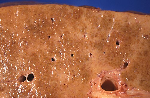
A gross pathology specimen of liver metastases caused by pancreatic cancer
SpecialtyHepatology, gastroenterology
TypesFatty liver disease, Hepatitis (and several more)[1]
Diagnostic methodLiver function tests[2]
TreatmentDepends on type (See types)

Liver disease, or hepatic disease, is any of many diseases of the liver.[1] If long-lasting it is termed chronic liver disease.[3] Although the diseases differ in detail, liver diseases often have features in common.

Signs and symptoms

[edit]

Some of the signs and symptoms of a liver disease are the following:

Types

[edit]

There are more than a hundred different liver diseases. Some of the most common are:[8]

Mechanisms

[edit]

Liver diseases can develop through several mechanisms:

DNA damage

[edit]

One general mechanism, increased DNA damage, is shared by some of the major liver diseases, including infection by hepatitis B virus or hepatitis C virus, heavy alcohol consumption, and obesity.[24]

Viral infection by hepatitis B virus, or hepatitis C virus causes an increase of reactive oxygen species. The increase in intracellular reactive oxygen species is about 10,000-fold with chronic hepatitis B virus infection and 100,000-fold following hepatitis C virus infection.[25] This increase in reactive oxygen species causes inflammation[25] and more than 20 types of DNA damage.[26] Oxidative DNA damage is mutagenic[27] and also causes epigenetic alterations at the sites of DNA repair.[28] Epigenetic alterations and mutations affect the cellular machinery that may cause the cell to replicate at a higher rate or result in the cell avoiding apoptosis, and thus contribute to liver disease.[29] By the time accumulating epigenetic and mutational changes eventually cause hepatocellular carcinoma, epigenetic alterations appear to have an even larger role in carcinogenesis than mutations. Only one gene, TP53, is mutated in more than 20% of liver cancers while 41 genes each have hypermethylated promoters (repressing gene expression) in more than 20% of liver cancers.[30]

Alcohol consumption in excess causes a build-up of acetaldehyde. Acetaldehyde and free radicals generated by metabolizing alcohol induce DNA damage and oxidative stress.[31][32][33] In addition, activation of neutrophils in alcoholic liver disease contributes to the pathogenesis of hepatocellular damage by releasing reactive oxygen species (which can damage DNA).[34] The level of oxidative stress and acetaldehyde-induced DNA adducts due to alcohol consumption does not appear sufficient to cause increased mutagenesis.[34] However, as reviewed by Nishida et al.,[28] alcohol exposure, causing oxidative DNA damage (which is repairable), can result in epigenetic alterations at the sites of DNA repair. Alcohol-induced epigenetic alterations of gene expression appear to lead to liver injury and ultimately carcinoma.[35]

Obesity is associated with a higher risk of primary liver cancer.[36] As shown with mice, obese mice are prone to liver cancer, likely due to two factors. Obese mice have increased pro-inflammatory cytokines. Obese mice also have higher levels of deoxycholic acid, a product of bile acid alteration by certain gut microbes, and these microbes are increased with obesity. The excess deoxycholic acid causes DNA damage and inflammation in the liver, which, in turn, can lead to liver cancer.[37]

Other relevant aspects

[edit]

Several liver diseases are due to viral infection. Viral hepatitides such as Hepatitis B virus and Hepatitis C virus can be vertically transmitted during birth via contact with infected blood.[38][39] According to a 2012 NICE publication, "about 85% of hepatitis B infections in newborns become chronic".[40] In occult cases, Hepatitis B virus is present by hepatitis B virus DNA, but testing for HBsAg is negative.[41] High consumption of alcohol can lead to several forms of liver disease including alcoholic hepatitis, alcoholic fatty liver disease, cirrhosis, and liver cancer.[42] In the earlier stages of alcoholic liver disease, fat builds up in the liver's cells due to increased creation of triglycerides and fatty acids and a decreased ability to break down fatty acids.[43] Progression of the disease can lead to liver inflammation from the excess fat in the liver. Scarring in the liver often occurs as the body attempts to heal and extensive scarring can lead to the development of cirrhosis in more advanced stages of the disease.[43] Approximately 3–10% of individuals with cirrhosis develop a form of liver cancer known as hepatocellular carcinoma.[43] According to Tilg, et al., gut microbiome could very well have an effect, be involved in the pathophysiology, on the various types of liver disease which an individual may encounter.[44] Insight into the exact causes and mechanisms mediating pathophysiology of the liver is quickly progressing due to the introduction new technological approaches like Single cell sequencing and kinome profiling [45]

Air pollutants

[edit]

Particulate matter or carbon black are common pollutants. They have a direct toxic effect on the liver; cause inflammation of liver caused by and thereby impact lipid metabolism and fatty liver disease; and can translocate from the lungs to the liver.[46]

Because particulate matter and carbon black are very diverse and each has different toxicodynamics, detailed mechanisms of translocation are not clear. Water-soluble fractions of particulate matter are the most important part of translocation to the liver, through extrapulmonary circulation. When particulate matter gets into the bloodstream, it combines with immune cells and stimulates innate immune responses. Pro-inflammatory cytokines are released and cause disease progression.[46]

Diagnosis

[edit]

A number of liver function tests are available to test the proper function of the liver. These test for the presence of enzymes in blood that are normally most abundant in liver tissue, metabolites or products. serum proteins, serum albumin, serum globulin, alanine transaminase, aspartate transaminase, prothrombin time, partial thromboplastin time.[2]

Imaging tests such as transient elastography, ultrasound and magnetic resonance imaging can be used to show the liver tissue and the bile ducts. Liver biopsy can be performed to examine liver tissue to distinguish between various conditions; tests such as elastography may reduce the need for biopsy in some situations.[47]

In liver disease, prothrombin time is longer than usual.[6] In addition, the amounts of both coagulation factors and anticoagulation factors are reduced as a diseased liver cannot productively synthesize them as it did when healthy.[48] Nonetheless, there are two exceptions in this falling tendency: coagulation factor VIII and von Willebrand factor, a platelet adhesive protein.[48] Both inversely rise in the setting of hepatic insufficiency, thanks to the drop of hepatic clearance and compensatory productions from other sites of the body.[48] Fibrinolysis generally proceeds faster with acute liver failure and advanced stage liver disease, unlike chronic liver disease in which concentration of fibrinogen remains unchanged.[48]

A previously undiagnosed liver disease may become evident first after autopsy.[citation needed] Following are gross pathology images:

Treatment

[edit]
Ursodeoxycholic acid

Anti-viral medications are available to treat infections such as hepatitis B.[49] Other conditions may be managed by slowing down disease progression, for example:

  • By using steroid-based drugs in autoimmune hepatitis.[50]
  • Regularly removing a quantity of blood from a vein (venesection) in the iron overload condition, hemochromatosis.[51]
  • Wilson's disease, a condition where copper builds up in the body, can be managed with drugs that bind copper, allowing it to be passed from the body in urine.[52]
  • In cholestatic liver disease, (where the flow of bile is affected due to cystic fibrosis[53]) a medication called ursodeoxycholic acid may be given.[54]

Epidemiology

[edit]
Deaths from liver disease

Liver diseases, including conditions such as non-alcoholic fatty liver disease (NAFLD), alcohol-related liver disease (ALD), and viral hepatitis, are significant public health concerns worldwide. In the United States, NAFLD is the most common chronic liver condition, affecting approximately 24% of the population, with the prevalence rising due to increasing rates of obesity and metabolic syndrome.[55][56] Alcohol-related liver disease accounts for about 4.5% of liver-related deaths globally, underscoring the substantial burden of alcohol misuse.[57] Viral hepatitis, primarily hepatitis B and hepatitis C, remains a leading cause of liver cirrhosis and liver cancer worldwide, despite advances in antiviral therapies and vaccination efforts.[58] Additionally, recent studies have highlighted lean steatotic liver disease (SLD), a subset of NAFLD, affecting over 12% of U.S. adults even in the absence of obesity.[59] These data emphasize the importance of early detection and targeted interventions to manage liver disease and its associated complications effectively.

New research reports the prevalence of lean steatotic liver disease (SLD) in the United States using data from the National Health and Nutrition Examination Survey (2017-2023), researchers estimated the age-adjusted prevalence of lean SLD at 12.8%.[60] This includes 9.3% for lean metabolic dysfunction-associated steatotic liver disease, 1.3% for metabolic dysfunction and alcohol-related steatotic liver disease, and 1.0% for alcohol-related liver disease.[61]

See also

[edit]

References

[edit]

Further reading

[edit]
[edit]
Revisions and contributorsEdit on WikipediaRead on Wikipedia
from Grokipedia
Liver disease encompasses a wide range of conditions that damage the liver, an organ essential for filtering toxins from the , producing to aid , storing energy, and supporting blood clotting. Located under the on the right side and weighing up to four pounds in adults, the liver performs over 500 vital functions, making any impairment potentially life-threatening if untreated. These diseases can be acute, developing rapidly, or chronic, progressing over months or years, and often lead to complications such as (scarring of the liver) or . Common types of liver disease include (inflammation caused by , B, or C viruses), metabolic dysfunction-associated steatotic liver disease (MASLD; formerly nonalcoholic fatty liver disease), involving buildup of fat in liver cells unrelated to alcohol, (damage from excessive alcohol consumption), (the immune system attacking liver cells), and genetic disorders like hemochromatosis ( in the liver). Other notable forms involve cancer, such as , and metabolic conditions like , which causes copper accumulation. Globally, liver diseases contribute significantly to morbidity, with chronic forms affecting approximately 1.5 billion people and serving as a leading cause of death, responsible for about 2 million deaths annually through complications like and failure. The primary causes of liver disease vary by type but commonly include infections from viruses, excessive alcohol use, leading to fat accumulation, exposure to toxins or certain medications, and autoimmune or genetic factors. For instance, viruses spread through contaminated food, water, or blood, while metabolic dysfunction-associated steatotic liver disease is linked to , including and high . Risk factors encompass heavy alcohol consumption, unprotected sex, sharing needles, , and family history of liver conditions, with prevention often involving for and B, moderate alcohol intake, and healthy choices. Symptoms of liver disease may not appear in early stages, allowing silent progression, but advanced cases often present with (yellowing of the skin and eyes), and swelling, swelling in the legs and ankles, itchy , dark urine, pale stools, chronic fatigue, , loss of , and easy bruising or bleeding. Complications can include (high blood pressure in the liver's ), (fluid buildup in the abdomen), (brain dysfunction from toxin buildup), and increased risk of infections or bleeding due to impaired clotting factor production. typically involves tests for liver function, imaging such as , and sometimes , with treatment ranging from lifestyle changes and medications to in severe cases.

Liver Anatomy and Physiology

Structure of the Liver

The liver is situated in the upper right quadrant of the , beneath the diaphragm and above the right , extending slightly across the midline to the left upper quadrant. It is the largest solid organ in the body, weighing approximately 1.2 to 1.5 kg in adults, and constitutes about 2% of total body weight. The organ is partially protected by the and has a soft, wedge-shaped structure with a convex superior surface conforming to the diaphragm and a concave inferior surface that contacts the , right , and colon. Externally, the liver is enveloped by a thin capsule of called Glisson's capsule, which extends inward as supporting septa. The liver is divided into four lobes: the larger right lobe, the smaller left lobe, and the caudate and quadrate lobes on the posterior and inferior surfaces, respectively. These divisions are primarily based on external morphology, with the right and left lobes separated by the , a sickle-shaped fold of that anchors the liver to the anterior and contains the remnant of the fetal , known as the ligamentum teres. Additional ligaments, including the coronary and triangular ligaments, attach the liver to the diaphragm, stabilizing its position. The quadrate lobe, located anteriorly on the inferior surface, and the caudate lobe, posterior to the , are functionally distinct and supplied by separate vascular branches. The liver receives a dual blood supply, with approximately 25% from the hepatic artery, which delivers oxygenated , and 75% from the , which carries nutrient-rich, deoxygenated from the . These vessels enter the liver at the , a fissure on the inferior surface, and branch into smaller vessels that converge to form sinusoids—wide, fenestrated capillaries lined by endothelial cells that allow exchange between and hepatocytes. from the sinusoids drains into central veins, which coalesce into emptying into the . At the microscopic level, the liver is organized into hexagonal lobules, each approximately 1-2 mm in diameter, serving as functional units. , the primary parenchymal cells, are polyhedral epithelial cells arranged in single-cell-thick plates or cords radiating from the central vein toward the periphery of the lobule. At the corners of each lobule are portal triads, consisting of a branch of the , , and , surrounded by . The space between hepatocyte plates and sinusoids is the space of Disse, containing stellate cells (also called Ito cells) that store and fat. Kupffer cells, specialized macrophages, line the sinusoids and phagocytose debris and pathogens. The biliary system begins at the microscopic level with bile canaliculi, narrow channels formed by tight junctions between adjacent hepatocytes that collect secreted by the liver cells. These canaliculi merge into progressively larger ductules and ducts within the portal triads, forming that converge into right and left hepatic ducts exiting the liver. The right and left hepatic ducts unite to form the , which joins the from the to create the ; this duct transports to the via the major duodenal papilla, where a regulates flow for fat digestion. The , attached to the inferior surface of the liver, stores and concentrates until release into the .

Functions of the Liver

The liver serves as a central organ in maintaining physiological through diverse functions that encompass , , synthesis of essential biomolecules, immune surveillance, and nutrient storage. These roles are primarily executed by hepatocytes, the predominant cell type in the liver, which process incoming blood from the and hepatic artery to regulate systemic nutrient levels, eliminate toxins, and support other organs. In metabolism, the liver plays a pivotal role in handling carbohydrates, lipids, and proteins. For carbohydrates, it stores excess glucose as in hepatocytes following meals and releases it via during to maintain blood glucose levels; additionally, it performs , synthesizing glucose from non-carbohydrate precursors like and lactate to prevent . Regarding lipids, the liver synthesizes , which is essential for formation and production, and assembles it into lipoproteins such as very low-density lipoproteins (VLDL) for transport to peripheral tissues. For proteins, hepatocytes produce , the primary plasma protein that maintains and transports molecules, as well as clotting factors like fibrinogen and prothrombin to ensure . Detoxification is another critical function, where the liver neutralizes harmful substances entering via the bloodstream. enzymes, a superfamily expressed predominantly in hepatocytes, catalyze the oxidation of drugs, toxins, and xenobiotics, making them more water-soluble for and preventing cellular damage. Complementing this, the —a series of enzymatic reactions localized in the liver—converts toxic , derived from protein breakdown, into for safe elimination by the kidneys, thereby averting . The liver's synthetic capabilities extend to producing bile and regulating hormones. , secreted by hepatocytes into the bile canaliculi and stored in the , contains bile salts, phospholipids, and that emulsify dietary fats in the intestine, facilitating their and absorption. In hormone regulation, the liver produces (IGF-1) under stimulation from , which promotes cell growth and metabolism, and metabolizes hormones like insulin and to control their circulating levels. Immune functions are mediated by resident cells and secretory products. Kupffer cells, specialized macrophages lining the hepatic sinusoids, perform to clear pathogens, debris, and senescent red blood cells from the portal circulation, acting as a frontline defense against infections. Hepatocytes also synthesize acute phase proteins, such as and , in response to inflammatory signals, which help modulate systemic immune responses and limit tissue damage. Finally, the liver functions as a storage depot for key nutrients. It stockpiles fat-soluble vitamins including A (for vision and immunity), D (for calcium ), and B12 (for formation), releasing them as needed; it also stores iron in form within s for synthesis and maintains reserves for .

Types of Liver Disease

Acute Liver Diseases

Acute liver diseases are characterized by sudden onset of liver dysfunction occurring within days to weeks, often resulting from identifiable triggers and potentially reversible with prompt intervention. Unlike chronic conditions, these disorders typically involve rapid injury leading to and impaired liver function, with recovery possible if the underlying cause is addressed early. Common manifestations include abrupt rises in serum transaminases, such as (ALT) and aspartate aminotransferase (AST) levels exceeding 1000 IU/L, alongside hyperbilirubinemia causing and potential due to reduced synthesis of clotting factors. In severe cases, progression to (ALF) may occur, defined by (international normalized ratio >1.5) and within eight weeks of symptom onset in a without prior liver disease. Major types of acute liver diseases encompass viral, toxic, ischemic, and alcohol-related etiologies. Acute , particularly from , B, and E viruses, represents a primary category, where leads to self-limited liver inflammation in most cases. For instance, virus spreads via the fecal-oral route, with an of 2 to 6 weeks, causing symptoms like fatigue, nausea, and that resolve within months without chronic sequelae in over 99% of immunocompetent individuals. Similarly, acute , transmitted through blood or sexual contact, can present with high and transaminitis, though a subset progresses to ALF requiring transplantation. Drug-induced liver injury (DILI) is another prevalent form, exemplified by acetaminophen () overdose, which accounts for nearly 50% of ALF cases in Western countries; toxicity arises from the metabolite N-acetyl-p-benzoquinone imine () depleting and causing centrilobular . , also known as shock liver, results from hypoperfusion during systemic hypotension, such as in , leading to massive elevations that peak within 24-48 hours and often normalize rapidly with hemodynamic stabilization. Acute , triggered by heavy , features and neutrophil infiltration, with a distinct clinical syndrome of fever, , and tender . These conditions share clinical hallmarks that demand urgent evaluation to prevent complications like multiorgan failure. typically emerges 1-2 weeks after enzyme peaks, reflecting impaired bilirubin conjugation, while signals synthetic dysfunction and heightened bleeding risk. , graded from subtle confusion to , marks ALF and correlates with poor , with survival rates varying by —over 60% for acetaminophen-induced ALF with supportive care, but lower for indeterminate causes. Management focuses on etiology-specific therapies, such as N-acetylcysteine for acetaminophen toxicity or antiviral agents for certain viral infections, underscoring the importance of early recognition for reversibility.

Chronic Liver Diseases

Chronic liver disease refers to a progressive deterioration of liver functions lasting more than six months, often involving impaired synthesis of clotting factors, proteins, and other essential substances, and frequently leading to or . This prolonged duration distinguishes it from acute conditions, allowing for gradual structural changes in the liver , including , scarring, and nodular regeneration. The major categories of chronic liver diseases encompass a range of etiologies, including chronic viral hepatitis caused by (HBV) or (HCV), which lead to persistent infection and damage; non-alcoholic fatty liver disease (NAFLD), now termed metabolic dysfunction-associated steatotic liver disease (MASLD), characterized by excessive fat accumulation in the liver unrelated to alcohol; , an immune-mediated attack on liver cells; (PBC), an autoimmune destruction of intrahepatic bile ducts; (PSC), involving inflammation and fibrosis of bile ducts; and genetic disorders such as hemochromatosis, involving iron overload, and , marked by copper accumulation. These conditions share a progressive trajectory but differ in their underlying mechanisms, with viral and metabolic types being the most prevalent globally. Chronic liver diseases typically exhibit an insidious onset with nonspecific symptoms or even progression, accompanied by gradual elevations in liver enzymes such as (ALT) and aspartate aminotransferase (AST), reflecting ongoing low-grade . In advanced stages, emerges as a hallmark complication due to increased intrahepatic from , leading to complications like and variceal bleeding. NAFLD/MASLD, in particular, is strongly associated with and , serving as a hepatic manifestation of , with a global exceeding 30%. Illustrative examples highlight the progressive nature of these diseases: chronic HCV infection advances to in approximately 20-30% of cases over 20-30 years, influenced by factors like age and alcohol use. Similarly, PBC involves autoimmune-mediated progressive destruction of small , resulting in and eventual .

Causes and Risk Factors

Infectious Causes

Infectious causes of liver disease primarily involve pathogens that directly invade hepatic tissue or trigger inflammatory responses leading to damage. Viral infections, particularly those caused by viruses, are among the most significant contributors globally. virus (HAV) and virus (HEV) are transmitted via the fecal-oral route, often through contaminated food or water, resulting in acute self-limiting without chronic sequelae. In contrast, (HBV) spreads through bloodborne or sexual contact, with perinatal transmission carrying a 90% risk of chronic infection in neonates compared to about 5% in adults, affecting an estimated 254 million chronic carriers worldwide as of 2022. (HCV) is predominantly bloodborne, leading to chronic infection in up to 85% of cases and progressive liver damage over decades. Hepatitis D virus (HDV) requires HBV co-infection for replication and exacerbates liver injury in those already infected with HBV. Other viruses, such as Epstein-Barr virus (EBV) and cytomegalovirus (CMV), can cause primarily in immunocompromised individuals, manifesting as elevated liver enzymes and potential progression to severe involvement. Bacterial infections contribute to liver disease through direct invasion or secondary complications. Leptospirosis, caused by species, is a zoonotic acquired via contact with contaminated water or soil, leading to acute with in severe cases known as Weil's disease. Q fever, resulting from exposure in livestock environments, can produce granulomatous with relapsing fevers and . Pyogenic liver abscesses often arise from portal vein spread of bacteria from intra-abdominal sources like or , involving pathogens such as or , and require drainage alongside antibiotics for management. Parasitic infections are prevalent in endemic regions and cause chronic liver pathology through mechanical obstruction or immune-mediated damage. , induced by species transmitted via freshwater snails, leads to periportal from deposition in the liver, potentially progressing to . Liver flukes, including and , are acquired by ingesting undercooked fish or aquatic plants, resulting in inflammation, cholangitis, and increased risk of . Fungal causes are uncommon but significant in vulnerable populations. , primarily from Candida species, occurs in liver transplant recipients due to , often disseminating to the liver and causing abscesses or fulminant with high mortality if untreated.

Non-Infectious Causes

Non-infectious causes of liver disease encompass a range of endogenous metabolic disturbances, genetic disorders, autoimmune processes, and exposure to environmental toxins, all of which can lead to hepatic injury without microbial involvement. These factors often result in progressive damage, including , , , and , depending on the duration and severity of exposure or underlying defect. Unlike infectious etiologies, non-infectious causes are frequently linked to lifestyle, occupational hazards, or inherited traits, and they account for a substantial proportion of chronic liver conditions worldwide. Toxic causes primarily involve substances that directly impair function or induce . Alcohol consumption is a leading toxic factor, where is metabolized by to , a highly reactive intermediate that generates and promotes in the liver. Chronic intake exceeding 30 grams per day for men or 20 grams per day for women significantly elevates the risk of , progressing from to and . In Western countries, alcohol-attributable comprises approximately 50% of cases. Drug-induced liver injury (DILI) represents another major toxic pathway, often idiosyncratic rather than dose-dependent; for instance, statins can cause hepatocellular injury through mitochondrial dysfunction, while leads to phospholipidosis and via inhibition of lysosomal degradation. Industrial and environmental toxins, such as used in production, induce hepatic and by forming reactive metabolites that damage DNA and promote fibrosis. Similarly, aflatoxins—mycotoxins from species contaminating foodstuffs—exert hepatocarcinogenic effects by alkylating DNA, particularly increasing risk in susceptible populations. Metabolic causes arise from dysregulated lipid and glucose homeostasis, frequently tied to systemic conditions. Non-alcoholic fatty liver disease (NAFLD), now termed metabolic dysfunction-associated steatotic liver disease (MASLD), develops due to and central , leading to excessive hepatic fat accumulation independent of significant alcohol use. This condition affects up to 25-30% of adults in high-income regions and can progress to non-alcoholic (NASH) with and . , an inherited metabolic disorder, causes liver disease through accumulation of misfolded protein in hepatocytes, triggering endoplasmic reticulum stress and ; it manifests in 10-15% of affected individuals with . Genetic and autoimmune causes involve inherited defects in metal homeostasis or aberrant immune responses targeting hepatic antigens. Wilson's disease results from mutations in the ATP7B gene, impairing biliary copper excretion and causing toxic copper accumulation in hepatocytes, which induces oxidative damage and fulminant hepatitis or chronic cirrhosis. Hereditary hemochromatosis, linked to HFE gene mutations, leads to excessive intestinal iron absorption and parenchymal iron overload, resulting in fibrosis and increased hepatocellular carcinoma risk. Autoimmune hepatitis, particularly type 1, is characterized by autoantibodies such as anti-nuclear antibodies (ANA) and anti-smooth muscle antibodies (ASMA), which mediate T-cell driven hepatocyte destruction and interface hepatitis. Other non-infectious causes include hemodynamic and nutritional factors. Ischemic , or "shock liver," occurs in the setting of acute or profound , where reduced impairs hepatic and causes centrilobular . , exemplified by in protein-deficient states, promotes hepatic through impaired synthesis and oxidation, leading to an enlarged, fatty liver in severely affected children.

Signs and Symptoms

Early Manifestations

Early manifestations of liver disease are often subtle and nonspecific, making early detection challenging but crucial for preventing progression. These initial symptoms can mimic common ailments, yet their persistence warrants medical evaluation to identify underlying hepatic involvement. Constitutional symptoms frequently herald the onset of liver dysfunction, including profound , generalized , low-grade fever, and unintentional . , a hallmark , affects 50% to 85% of patients with and varies by etiology, reported in about 50% of those with chronic C and up to 90% with chronic , often impairing daily activities without correlating directly to disease severity. contributes to an overall sense of unwellness, while low-grade fever may accompany inflammatory processes in early stages, particularly in viral etiologies. arises from reduced caloric intake and metabolic shifts, exacerbating debility. Gastrointestinal disturbances represent another early indicator, encompassing anorexia, nausea, mild right upper quadrant discomfort, and alterations in taste or smell perception. Anorexia and nausea diminish appetite, leading to nutritional deficits; nausea is particularly predominant in acute viral hepatitis cases, often accompanied by vomiting. Right upper quadrant discomfort reflects hepatic capsule irritation from subtle inflammation or enlargement. Altered taste (dysgeusia) and smell often stem from zinc deficiency associated with early liver impairment, further suppressing intake and promoting weight loss. Cutaneous and mucosal changes can emerge as initial vascular or cholestatic signs, such as pruritus, spider angiomata, and . Pruritus, driven by accumulation, affects the skin and mucous membranes, causing intense itching that disrupts sleep and quality of life. Spider angiomata, small dilated arterioles resembling spiders, and , reddish palm discoloration due to excess, signal early alterations in chronic liver conditions. Hematologic abnormalities in early liver disease may manifest as mild or easy bruising, attributable to impaired factor synthesis or nutrient . Mild results from chronic or iron dysregulation, contributing to , while easy bruising occurs from subtle deficiencies in clotting factors like prothrombin, even before overt develops.

Advanced Complications

In advanced liver disease, particularly , jaundice arises from impaired hepatic conjugation and of , leading to its accumulation in the blood and tissues, manifesting as yellowish discoloration of the skin and when serum levels exceed 3 mg/dL. , a hallmark of decompensated disease, further exacerbates this by obstructing flow, resulting in elevated conjugated , which causes dark urine due to renal and pale, clay-colored stools from reduced delivery to the intestines. Prolonged can also promote the deposition of and acids in the skin, forming pruritic xanthomas, especially in chronic cases. Portal hypertension, driven by increased resistance in the portal venous system, represents a critical complication that redirects blood flow and elevates pressure in collateral vessels. This commonly leads to ascites, the accumulation of fluid in the peritoneal cavity due to hypoalbuminemia and splanchnic vasodilation, often causing abdominal distension and discomfort. Peripheral edema, or swelling in the legs and ankles, can also occur due to low albumin levels and fluid retention. Esophageal varices develop as portosystemic collaterals, posing a high risk of rupture and life-threatening hemorrhage, with mortality rates for acute variceal bleeding ranging from 15% to 20% in cirrhotic patients. Splenomegaly frequently accompanies this, resulting from congestion and sequestration of blood cells in the enlarged spleen. Hepatic encephalopathy emerges from the liver's inability to detoxify and other neurotoxins, exacerbated by portosystemic shunting that bypasses clearance. Symptoms progress from mild confusion and sleep disturbances to severe disorientation, (a flapping tremor of the hands), and , graded clinically from I (subtle cognitive changes) to IV (deep coma) using the West Haven criteria. buildup, derived from gut bacterial metabolism, contributes significantly to and altered mental status in these patients. Coagulopathy in advanced liver disease stems from reduced synthesis of clotting factors and anticoagulants, leading to prolonged (PT) and international normalized ratio (INR), which reflect hepatic synthetic dysfunction. often coexists, primarily due to splenic sequestration from and decreased thrombopoietin production by the damaged liver, increasing risks despite a rebalanced but fragile hemostatic system. Hepatorenal syndrome constitutes a severe form of in patients with advanced and , characterized by functional renal triggered by splanchnic and reduced renal . This leads to and rising without structural kidney damage, often progressing rapidly in the context of and circulatory failure.

Pathophysiology

Cellular Injury Mechanisms

Cellular injury in liver disease primarily involves damage to , the main functional cells of the liver, through various interconnected mechanisms that initiate disease onset. These processes include pathways, inflammatory responses, oxidative damage, lipid accumulation, and immune-mediated attacks, often triggered by toxins, viruses, or metabolic stressors. and represent two key forms of hepatocyte death, with necrosis leading to uncontrolled cell lysis and inflammation, while apoptosis is a more regulated process that minimizes inflammatory spillover. In many liver pathologies, such as those induced by alcohol or viruses, these mechanisms overlap, amplifying injury and setting the stage for further hepatic dysfunction. Necrosis occurs when hepatocytes are exposed to severe insults like toxins or viral infections, causing rapid ATP depletion and membrane rupture, which releases damage-associated molecular patterns (DAMPs) that exacerbate local inflammation. In contrast, is mediated by activation, where extrinsic pathways involving death receptors like Fas and (TNFR1) or intrinsic mitochondrial pathways converge to dismantle the cell without immediate inflammation. For instance, in drug- or virus-induced , cleave cellular proteins, leading to DNA fragmentation and formation of apoptotic bodies that are typically cleared by . This distinction is critical, as excessive in chronic conditions can still contribute to progressive liver damage by reducing mass. Inflammation plays a central role in amplifying cellular injury, driven by the release of pro-inflammatory cytokines such as tumor necrosis factor-alpha (TNF-α) and interleukin-6 (IL-6) from activated Kupffer cells and infiltrating immune cells. TNF-α, produced in response to lipopolysaccharides from gut bacteria in , binds to TNFR1 on hepatocytes, promoting caspase-8 activation and while also recruiting . In alcoholic , neutrophil infiltration is particularly prominent, where these cells release proteases and (ROS) that directly damage membranes. IL-6, while aiding in acute-phase responses and hepatocyte regeneration, can sustain chronic inflammation when dysregulated, as seen in where it correlates with elevated liver levels. Oxidative stress arises from an imbalance in ROS production and defenses, prominently featured in alcohol metabolism where cytochrome P450 2E1 (CYP2E1) generates ROS during ethanol oxidation. These ROS, including and , damage and proteins, impairing energy production and triggering via cytochrome c release. In alcoholic , this mitochondrial dysfunction leads to and further ROS amplification, creating a vicious cycle of damage. like are often depleted in these scenarios, heightening vulnerability to ongoing oxidative insults. Steatosis, or fat accumulation in hepatocytes, contributes to cellular injury by mechanically stressing cells and promoting , particularly in non-alcoholic fatty liver disease (NAFLD) where impaired beta-oxidation of s plays a key role. In NAFLD, reduces peroxisome proliferator-activated receptor alpha (PPARα) activity, hindering mitochondrial and peroxisomal breakdown, which leads to buildup and stress. This accumulation sensitizes hepatocytes to further injury from ROS or cytokines, as lipid droplets interfere with cellular signaling and increase susceptibility to . Enhanced de novo further exacerbates steatosis, creating an environment conducive to and cell death. Immune-mediated mechanisms involve T-cell recognition and attack on hepatocytes, as seen in viral and autoimmune hepatitis. In viral hepatitis, cytotoxic CD8+ T cells target infected cells via Fas ligand or perforin/granzyme pathways, inducing to limit viral spread but causing collateral liver injury. Autoimmune hepatitis features autoreactive CD4+ and CD8+ T cells that infiltrate the liver, releasing cytokines like interferon-gamma to perpetuate hepatocyte destruction, often triggered by molecular between self-antigens and viral proteins. Regulatory T cells normally mitigate this attack, but their dysfunction allows sustained immune aggression. A hallmark of alcoholic is the formation of Mallory-Denk bodies (MDBs), eosinophilic cytoplasmic inclusions composed of intermediate filaments and ubiquitinated proteins, resulting from cytoskeletal disruption and proteasomal overload in stressed hepatocytes. MDBs form in ballooned hepatocytes exposed to and , serving as a marker of severe injury in alcoholic . These structures correlate with severity and may promote further by acting as neoantigens that attract immune cells.

Progression to Fibrosis and Cirrhosis

Liver , or fibrogenesis, represents a chronic response to ongoing hepatic , characterized by the excessive accumulation of proteins in the liver . Central to this process is the of hepatic stellate cells (HSCs), which are normally quiescent, vitamin A-storing cells residing in the space of Disse. Upon persistent , HSCs undergo into proliferative, contractile myofibroblasts that express alpha-smooth muscle and secrete large amounts of type I and III , leading to the replacement of normal liver tissue with scar tissue. The progression of fibrosis is typically assessed histologically using the METAVIR scoring system, which stages from F0 to F4 based on the extent of fibrous expansion and architectural disruption. In F0, there is no ; F1 indicates mild portal without septa; F2 shows portal with rare septa; F3 features numerous septa without , representing bridging that connects portal tracts and central veins; and F4 denotes with complete architectural distortion. Bridging in stages F2-F3 disrupts normal lobular structure and promotes the formation of regenerative nodules as hepatocytes attempt to regenerate amid the scarring. Cirrhosis marks the end-stage of fibrogenesis, defined pathologically as the irreversible development of diffuse with regenerative nodules surrounded by fibrous bands, resulting in profound distortion of hepatic architecture and vasculature. This scarring leads to the formation of nodules of varying sizes (typically 0.1-3 cm), which compress and obliterate the normal sinusoidal network and portal venules, severely impairing intrahepatic blood flow. While traditionally viewed as irreversible, recent evidence indicates that and even early can partially regress upon removal of the underlying , such as successful antiviral of (HCV) infection. In patients achieving sustained virologic response after direct-acting antiviral therapy for HCV, histological regression of has been observed in up to 82% of cases, with improvements in METAVIR scores and reduction in content, though advanced shows more limited reversal. A key complication arising from this progression is , primarily due to sinusoidal obstruction caused by the fibrotic bands and regenerative nodules, which increase intrahepatic and impede portal venous inflow. This mechanical obstruction, combined with dynamic in the sinusoids, elevates portal pressure above 10 mmHg, predisposing to variceal bleeding, , and . In untreated chronic hepatitis B, the annual incidence of progression to is approximately 1-2%, varying with , , and host factors, underscoring the importance of early intervention to halt fibrogenesis.

Diagnosis

Clinical Evaluation

Clinical evaluation of liver disease begins with a thorough history and to identify potential etiologies, assess severity, and guide subsequent investigations. This initial assessment is crucial for suspecting liver involvement, as many cases present with nonspecific symptoms or are until advanced stages. The process integrates risk factor identification with clinical signs to differentiate between acute and chronic processes. History taking focuses on modifiable and nonmodifiable risk factors. Inquiry into alcohol consumption quantifies lifetime intake and recent patterns, as excessive use is a leading cause of . Drug exposure, including prescription medications like acetaminophen, herbal supplements, and illicit drugs, is probed for hepatotoxic potential. Travel history to endemic areas raises suspicion for or E, while intravenous drug use (IVDU) is a key risk for (HCV) acquisition. Family history explores genetic conditions such as hemochromatosis, , or . Additional elements include occupational exposures (e.g., to solvents or ) and sexual history for risk. The systematically evaluates for stigmata of liver dysfunction and . , manifesting as yellowing of the and skin, indicates hyperbilirubinemia. may be palpated as an enlarged, tender liver edge below the , while suggests . is detected through inspection for and percussion for , confirming fluid accumulation. , visible periumbilical venous dilation, reflects portal venous recanalization. and arise from impaired metabolism in , leading to hormonal imbalance. Other findings include , spider angiomata, and . Scoring systems aid in quantifying cirrhosis severity during evaluation. The Child-Pugh score classifies patients into grades A (mild, 5-6 points), B (moderate, 7-9 points), or C (severe, 10-15 points) based on total bilirubin, serum albumin, international normalized ratio (INR), presence of ascites, and hepatic encephalopathy. This tool predicts surgical risk and prognosis in chronic liver disease. The Model for End-Stage Liver Disease (MELD) score, ranging from 6 to 40, incorporates serum creatinine, total bilirubin, INR, and sodium to estimate short-term mortality, particularly for transplant prioritization. Higher scores indicate greater urgency. Red flags warranting urgent attention include acute , presenting as confusion or altered mental status due to toxin accumulation, and suggestive of from . These complications signal and require immediate stabilization. Findings from clinical evaluation often prompt confirmatory laboratory and imaging tests to establish diagnosis and extent of disease.

Laboratory and Imaging Tests

Laboratory tests play a central role in diagnosing and monitoring liver disease by assessing hepatocyte injury, synthetic function, and specific etiologies. Liver function tests (LFTs) typically include measurements of alanine aminotransferase (ALT) and aspartate aminotransferase (AST), which are enzymes released from damaged hepatocytes. Elevated ALT and AST levels, often with ALT exceeding AST, indicate hepatocellular injury, while a pattern of disproportionately elevated alkaline phosphatase (ALP) and gamma-glutamyl transferase (GGT) suggests cholestatic disease. Bilirubin levels help differentiate conjugated (direct) from unconjugated (indirect) hyperbilirubinemia, with elevated bilirubin pointing to impaired biliary in conditions like or . and prothrombin time (PT)/international normalized ratio (INR) evaluate the liver's synthetic capacity, as and prolonged PT/INR reflect advanced dysfunction due to reduced protein production. Serological tests are essential for identifying infectious causes, particularly . Hepatitis B surface () detection confirms acute or chronic hepatitis B virus (HBV) , indicating infectivity unless recently vaccinated. For hepatitis C virus (HCV), a positive anti-HCV test signals past or current exposure, prompting RNA testing to confirm active . Non-invasive assessments of liver fibrosis, such as the Fibrosis-4 (FIB-4) index, utilize routine blood parameters including age, AST, ALT, and platelet count to estimate fibrosis risk without . A FIB-4 score below 1.45 has a high negative predictive value (around 90%) for ruling out advanced in various liver diseases. The Enhanced Liver Fibrosis (ELF) test measures serum biomarkers of turnover, including , tissue inhibitor of metalloproteinase-1 (TIMP-1), and procollagen III N-terminal peptide (PIIINP), to predict moderate to severe and associated outcomes. Imaging modalities provide structural and functional insights into liver pathology. Ultrasound is the initial noninvasive tool for detecting steatosis, as it shows increased echogenicity, and for identifying nodules or masses suggestive of hepatocellular carcinoma (HCC). Computed tomography (CT) and magnetic resonance imaging (MRI) excel in evaluating vascular patency, such as portal vein thrombosis, and characterizing tumors through contrast enhancement patterns. Elastography techniques, including transient elastography (FibroScan), quantify liver stiffness as a surrogate for , with higher kilopascal values correlating to advanced stages; it is particularly useful for serial monitoring in . Liver biopsy remains the gold standard for definitive grading of inflammation and staging of , providing histological details essential for prognosis in conditions like nonalcoholic steatohepatitis. However, it carries risks, including bleeding complications in up to 0.6% of cases, particularly in patients with . For HCC surveillance in patients with cirrhosis, serum (AFP) is measured every six months alongside , as levels above 20 ng/mL may indicate early tumor development, though its use is at clinician discretion per guidelines.

Treatment and Management

Supportive and Lifestyle Interventions

Supportive and lifestyle interventions form the cornerstone of managing liver disease, aiming to alleviate symptoms, prevent complications, and enhance across various etiologies. For patients with alcohol-associated liver disease, complete from alcohol is essential, as it reduces the risk of and death in all stages of , with early after associated with a 7-year of up to 72%. In nonalcoholic fatty liver disease (NAFLD), lifestyle modifications including of 7-10% through diet and exercise can resolve and improve on . A balanced diet low in sodium, typically restricted to 2 grams per day, is recommended for patients with to reduce fluid retention and prevent recurrence. Nutritional support addresses the high prevalence of in advanced liver disease, where patients often require high-calorie diets (35-40 kcal/kg/day) to maintain energy balance and prevent muscle wasting. For , supplementation with branched-chain (BCAAs) improves nutritional status, reduces encephalopathy recurrence ( 0.73), and decreases overall complications in . These interventions, often delivered as oral supplements or late-evening snacks, help counteract disturbances without precipitating encephalopathy. Symptom management relies on targeted to control common complications. Diuretics such as (starting at 100 mg/day) combined with (40 mg/day) in a 100:40 mg ratio are the mainstay for mobilizing , achieving response in up to 90% of patients when paired with sodium restriction. For , titrated to 2-3 soft stools daily serves as first-line therapy to lower levels, while (550 mg twice daily) added to reduces recurrence risk by 50-60% as secondary prophylaxis. Nonselective beta-blockers like (20-160 mg/day, titrated to heart rate 55-60 bpm) are standard for primary prophylaxis of variceal in patients with medium-to-large , lowering risk by 40-50%. Regular monitoring through outpatient follow-up every 3-6 months is critical for detecting early, including assessments of , , and nutritional status to guide adjustments in supportive care. Patients with should receive vaccinations against and B viruses, as recommended by guidelines, to prevent superimposed infections that could accelerate decompensation.

Targeted Therapies

Targeted therapies for liver disease focus on addressing the underlying etiology to halt disease progression, reverse damage, or manage complications, with selections guided by clinical guidelines from organizations such as the American Association for the Study of Liver Diseases (AASLD). For viral causes, nucleoside analogs like entecavir are recommended as first-line treatment for chronic (HBV) infection in eligible patients, achieving viral suppression in over 90% of cases with long-term use to prevent and (HCC). Similarly, direct-acting antivirals (DAAs) such as sofosbuvir-based regimens are the standard for (HCV) eradication, yielding sustained virologic response (SVR) rates exceeding 95% across genotypes and stages, effectively curing the infection in most patients. Notably, HCV eradication with DAAs reduces the risk of HCC by approximately 70-71%, underscoring its role in primary prevention even in those with advanced . In , immunosuppressive agents form the cornerstone of , with corticosteroids (e.g., at 30 mg/day initially) combined with (1-2 mg/kg/day) inducing remission in 80% of patients within six months and serving as maintenance to minimize steroid-related side effects. For cholestatic conditions like (PBC), (UDCA) at 13-15 mg/kg/day is the first-line antifibrotic agent, improving biochemical markers and delaying progression in about 60% of responders. , a farnesoid X receptor agonist, was previously approved as second-line for UDCA non-responders, showing histological improvements in phase 3 trials, but its marketing authorization was withdrawn in September 2025 due to safety concerns including severe in advanced cases, prompting AASLD to update guidelines accordingly. Metabolic liver diseases require targeted interventions to address specific accumulations or . In nonalcoholic (NASH), pioglitazone (30-45 mg/day) improves and inflammation in nondiabetic patients with biopsy-proven disease, as demonstrated in randomized trials showing histological resolution in 47% of treated individuals versus 21% on . (800 IU/day) is an alternative for non-cirrhotic, nondiabetic NASH patients, reducing hepatocellular ballooning and progression. (80-100 mg/day), a thyroid receptor-β agonist, is approved for NASH patients with moderate to advanced , improving resolution of NASH and regression in phase 3 trials. , a , is recommended per 2025 AASLD updates for NASH with moderate to advanced in patients with or without , showing significant histological improvements. For hereditary hemochromatosis, therapeutic is the primary treatment to deplete excess iron, targeting serum ferritin levels of 50-100 ng/mL and preventing in early-diagnosed cases. In Wilson's disease, copper chelators such as trientine (750-1500 mg/day) or D-penicillamine promote urinary copper excretion, normalizing levels and stabilizing liver function in 70-80% of symptomatic patients when initiated early. For advanced decompensated liver disease, procedural interventions provide targeted relief from complications. (TIPS) is indicated for refractory unresponsive to diuretics, achieving ascites resolution in 75-85% of cases and improving survival compared to repeated , though with a 20-30% risk of . TIPS is also used for recurrent variceal bleeding after endoscopic failure, reducing rebleeding rates to under 20% at one year. remains the definitive therapy for end-stage liver disease, with priority allocation via the (MELD) score; patients with MELD scores above 15 receive higher priority, and post-transplant five-year survival reaches 75% in appropriately selected recipients.

Prevention and Prognosis

Preventive Strategies

Preventive strategies for liver disease emphasize vaccination, lifestyle modifications, targeted screening, public health interventions, and genetic counseling to mitigate risk factors at both individual and population levels. Vaccination plays a central role in preventing viral hepatitis, a major cause of liver disease. The hepatitis B virus (HBV) vaccine, administered in a three-dose series, achieves approximately 95% efficacy in preventing chronic infection among infants and 80-100% effectiveness overall in preventing HBV transmission. For hepatitis A virus (HAV), vaccination is recommended for travelers to endemic areas, providing protection through a two-dose regimen that induces long-term immunity. Behavioral measures are essential for reducing exposure to hepatotoxins and infectious agents. Limiting alcohol consumption to no more than 14 units per week for both men and women minimizes the risk of alcohol-associated liver disease, with guidelines emphasizing even distribution over several days to avoid . Maintaining a balanced diet rich in fruits, vegetables, and whole grains while limiting sugars and saturated fats supports liver protection by reducing the risk of metabolic dysfunction-associated steatotic liver disease (MASLD). Following a regular routine and sleep schedule, including 7-9 hours of quality sleep nightly and avoiding staying up late, helps prevent fatty liver accumulation and disease progression linked to sleep disruption. Safe injection practices, such as using sterile needles and syringes without sharing, are critical to prevent HBV and (HCV) transmission among people who inject drugs. Similarly, practicing through consistent use reduces sexual transmission of HBV, which is more readily spread this way than HCV. Screening programs target high-risk individuals to detect early changes and prevent progression. For patients with , semiannual surveillance using abdominal , with or without alpha-fetoprotein (AFP) testing, is recommended to identify (HCC) at a treatable stage. In families affected by hereditary hemochromatosis, first-degree relatives should undergo iron studies, including and serum ferritin levels, starting in early adulthood to identify early. Public health initiatives address environmental and dietary risks. Ensuring food and water safety through proper and practices prevents outbreaks of HAV and virus (HEV), which are transmitted via contaminated sources. Controlling aflatoxin contamination in grains and nuts via regulatory measures, such as storage guidelines and food monitoring, reduces the incidence of aflatoxin-induced HCC, particularly in high-exposure regions. Community-based prevention programs, promoting balanced nutrition and physical activity, help curb the rise of non-alcoholic (NAFLD) by addressing metabolic risk factors. Genetic counseling is advised for carriers or affected families of inherited disorders like Wilson's disease and hemochromatosis. For Wilson's disease, an autosomal recessive condition, counseling informs at-risk relatives about mutation testing and family planning options to prevent copper accumulation. In hemochromatosis cases, counseling facilitates genetic testing for HFE gene mutations in relatives, enabling early intervention to avert iron-related liver damage. Liver disease imposes a substantial burden, with and other s accounting for approximately 1.4 million deaths annually as of 2021, representing about 2.5% of all deaths worldwide. In 2017, there were an estimated 112 million prevalent cases of compensated and 10.6 million cases of decompensated globally. , now termed metabolic dysfunction-associated steatotic liver disease (MASLD) since 2023, a major contributor to , affects roughly 38% of adults in the United States, reflecting broader trends driven by rising and . In certain demographics, such as individuals aged 35-49 years in the , liver disease has emerged as the leading cause of death, underscoring its impact on younger working-age populations. Epidemiological trends show a shifting landscape, with declining incidence of (HCV)-related liver disease due to the widespread adoption of direct-acting antivirals (DAAs), which have reduced HCV-associated (HCC) cases in high-income regions. Conversely, NAFLD/MASLD and alcohol-related liver disease are rising, fueled by global increases in , , and alcohol consumption; NAFLD is projected to reach over 33% globally by 2030 in some estimates, particularly in Western countries, with MASLD expected to become the leading indication for in the by 2025-2030. In low- and middle-income regions, (HBV) and HCV remain dominant, with high endemicity in and contributing to persistent burdens. These trends highlight the need for targeted interventions amid varying regional etiologies. Disparities in liver disease prevalence and outcomes are pronounced by and . Men face a higher overall of adverse liver events, including progression to HCC, compared to women across major etiologies like and alcohol-related disease, though women may develop severe complications at lower alcohol intake levels due to metabolic differences. In and , alcohol-related liver disease predominates, while viral etiologies such as HBV and HCV drive the majority of cases in and , exacerbating inequities in low-resource settings with limited access to antivirals and screening. Prognosis varies significantly by disease stage, with compensated offering a more favorable outlook—median survival exceeding 12 years and 5-year survival rates around 67-80%—compared to decompensated , where median survival drops to about 2 years and 5-year survival is approximately 20-45%. In patients with , the annual incidence of HCC ranges from 2% to 5%, influenced by and practices. Liver disease ranks as a leading indication for transplantation, with alcohol-associated now the top cause in the United States, followed closely by metabolic dysfunction-associated (formerly NAFLD); overall, end-stage liver disease accounts for a substantial portion of the approximately 41,000 annual liver transplants performed globally as of 2023.

References

Add your contribution
Related Hubs
User Avatar
No comments yet.