Tomotherapy
Tomotherapy
Main page
1542939

Tomotherapy

logo
Community Hub0 subscribers
Read side by side
from Wikipedia
Tomotherapy
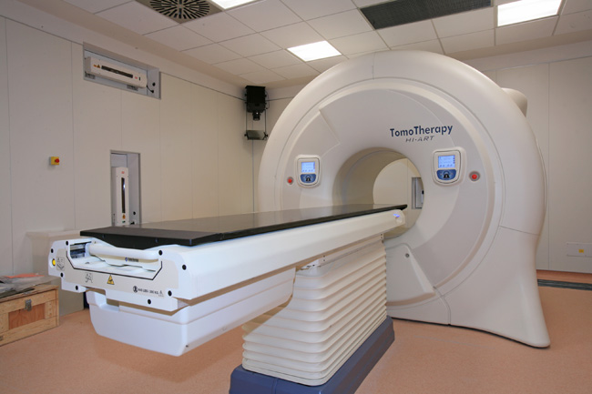
Tomotherapy Hi Art machine
Other namesHelical tomotherapy
SpecialtyOncology

Tomotherapy is a type of radiation therapy treatment machine.[1][2][3] In tomotherapy a thin radiation beam is modulated as it rotates around the patient, while they are moved through the bore of the machine. The name comes from the use of a strip-shaped beam, so that only one "slice" (Greek prefix "tomo-") of the target is exposed at any one time by the radiation. The external appearance of the system and movement of the radiation source and patient can be considered analogous to a CT scanner (computed tomography), which uses lower doses of radiation for imaging. Like a conventional machine used for X-ray external beam radiotherapy (often referred to as a linear accelerator or linac, their main component), it [the tomotherapy machine] generates the radiation beam, but the external appearance of the machine, patient positioning, and treatment delivery differ. Conventional linacs do not work on a slice-by-slice basis but typically have a large area beam which can also be resized and modulated.[4][5][6]

General principles

[edit]

The treatment field's length (the width of the radiation slice) is adjustable using collimator jaws. In static-jaw delivery, the field length remains constant during a treatment; in dynamic-jaw delivery, the field length changes so that it begins and ends at its minimum setting.

A patient undergoing tomotherapy with their face and body covered to prevent movement

Tomotherapy treatment times vary compared to normal radiation therapy treatment times. Tomotherapy treatment times can be as low as 6.5 minutes for common prostate treatment,[7] excluding extra time for imaging. Modern tomotherapy and conventional linac systems incorporate one or both of megavoltage X-ray or kilovoltage X-ray imaging systems, enabling image-guided radiation therapy (IGRT). In tomotherapy, images are acquired in a very similar manner to a CT scanner, thanks to their closely related design.[8][9][10]

There are few head-to-head comparisons of tomotherapy and other IMRT techniques, however there is some evidence that a conventional linac using VMAT can provide faster treatment whereas tomotherapy is better able to spare surrounding healthy tissue while delivering a uniform dose.[11][12][13]

Helical delivery

[edit]

In helical tomotherapy, the linac rotates on its gantry at a constant speed while the beam is delivered; so that from the patient's perspective, the shape traced out by the linac is helical.[14]

While helical tomotherapy can treat very long volumes without a need to abut fields in the longitudinal direction, it does display a distinct artifact due to "thread effect"[15] when treating non-central tumors. Thread effect can be suppressed during planning through good pitch selection.

Fixed-angle delivery

[edit]

Fixed-angle tomotherapy uses multiple tomotherapy beams, each delivered from a separate fixed gantry angle, in which only the couch moves during beam delivery. This is branded as TomoDirect, but has also been called topotherapy.[16]

The technology enables fixed beam treatments by moving the patient through the machine bore while maintaining specified beam angles.

Clinical considerations

[edit]

Lung cancer, head and neck tumors, breast cancer, prostate cancer, stereotactic radiosurgery (SRS) and stereotactic body radiotherapy (SBRT) are some examples of treatments commonly performed using tomotherapy.[17][18][19]

In general, radiation therapy (or radiotherapy) has developed with a strong reliance on homogeneity of dose throughout the tumor. Tomotherapy embodies the sequential delivery of radiation to different parts of the tumor which raises two important issues. First, this method is known as "field matching" and brings with it the possibility of a less-than-perfect match between two adjacent fields with a resultant hot and/or cold spot within the tumor. The second issue is that if the patient or tumor moves during this sequential delivery, then again, a hot or cold spot will result. The first problem is reduced by use of a helical motion, as in spiral computed tomography.[20]

Some research has suggested tomotherapy provides more conformal treatment plans and decreased acute toxicity.[21]

Non-helical static beam techniques such as IMRT and TomoDirect are well suited to whole breast radiation therapy. These treatment modes avoid the low-dose integral splay and long treatment times associated with helical approaches by confining dose delivery to tangential angles.[22][23][24]

This risk is accentuated in younger patients with early-stage breast cancer, where cure rates are high and life expectancy is substantial.[24]

Static beam angle approaches aim to maximize the therapeutic ratio by ensuring that the tumor control probability (TCP) significantly outweighs the associated normal tissue complication probability (NTCP).[25][26][27]

History

[edit]

The tomotherapy technique was developed in the early 1990s at the University of Wisconsin–Madison by Professor Thomas Rockwell Mackie and Paul Reckwerdt.[28] A small megavoltage x-ray source was mounted in a similar fashion to a CT x-ray source, and the geometry provided the opportunity to provide CT images of the body in the treatment setup position. Although original plans were to include kilovoltage CT imaging, current models use megavoltage energies. With this combination, the unit was one of the first devices capable of providing modern image-guided radiation therapy (IGRT).[20]

The first implementation of tomotherapy was the Corvus system developed by Nomos Corporation, with the first patient treated in April 1994.[29][17] This was the first commercial system for planning and delivering intensity modulated radiation therapy (IMRT). The original system, designed solely for use in the brain, incorporated a rigid skull-based fixation system to prevent patient motion between the delivery of each slice of radiation. But some users[30] eschewed the fixation system and applied the technique to tumors in many different parts of the body.

Mobile tomotherapy

[edit]

Due to their internal shielding and small footprint, TomoTherapy Hi-Art and TomoTherapy TomoHD treatment machines were the only high energy radiotherapy treatment machines used in relocatable radiotherapy treatment suites. Two different types of suites were available: TomoMobile developed by TomoTherapy Inc. which was a moveable truck; and Pioneer, developed by UK-based Oncology Systems Limited. The latter was developed to meet the requirements of UK and European transport law requirements and was a contained unit placed on a concrete pad, delivering radiotherapy treatments in less than five weeks.[31][32]

See also

[edit]

References

[edit]
[edit]
Revisions and contributorsEdit on WikipediaRead on Wikipedia
from Grokipedia
Tomotherapy is an advanced form of intensity-modulated radiation therapy (IMRT) that delivers radiation in helical or fixed-angle patterns, utilizing a rotating or static gantry equipped with a linear accelerator to precisely target tumors from multiple angles while minimizing exposure to surrounding healthy tissues.[1] The technique integrates treatment delivery with image-guided capabilities, employing megavoltage computed tomography (MVCT) for daily patient positioning and verification, which enhances accuracy in dose administration.[2] Developed through academic research at the University of Wisconsin-Madison in the early 1990s by Thomas Rockwell Mackie and colleagues, tomotherapy was conceptualized as a method to combine IMRT with volumetric imaging in a single system, with the first prototype implemented in 2001 and initial patient treatments occurring in 2002.[3] The technology was commercialized by TomoTherapy Inc., founded in 1997 as a university spinoff, and later acquired by Accuray Incorporated in 2011, leading to systems like the Hi-Art and subsequent models such as the TomoTherapy H series, including the Radixact system as of 2025.[4] In operation, for helical delivery, the patient lies on a treatment couch that moves continuously through a ring-shaped gantry, where a fan-shaped photon beam—modulated by a binary multileaf collimator with 64 leaves—rotates around the body, delivering radiation slice-by-slice in a spiral trajectory akin to a CT scanner. Fixed-angle delivery uses discrete gantry positions.[5] This approach offers several key advantages over conventional external beam radiotherapy, including superior dose conformity to irregular tumor shapes, reduced radiation to organs at risk (OAR), and the ability to treat both small targets and extended fields in a single session without repositioning.[2] Out-of-field radiation leakage is notably low, typically less than 0.1%, contributing to improved patient safety.[2] Clinically, tomotherapy is applied across a wide spectrum of malignancies, often demonstrating better sparing of critical structures compared to traditional IMRT techniques.[5] Its image-guided features, including MVCT with doses under 3 cGy per scan, enable adaptive radiotherapy adjustments based on daily anatomical changes, further optimizing therapeutic outcomes, with ongoing advancements in adaptive capabilities as of 2025.[2][6]

Principles of Operation

Helical Delivery

Helical tomotherapy employs a dynamic delivery mechanism where the gantry, housing a linear accelerator, performs a continuous 360-degree rotation around the patient at a constant speed, typically completing one full rotation every 6 to 15 seconds in modern systems.[7] Simultaneously, the treatment couch translates the patient linearly through the bore of the ring-shaped gantry, creating a helical trajectory for the radiation beam that envelops the target volume.[8] This synchronized motion ensures comprehensive irradiation from all angles without discrete beam pauses, analogous to the helical scanning path in computed tomography (CT) systems.[9] Beam intensity modulation is achieved using a binary multileaf collimator (MLC) consisting of 64 independent tungsten leaves, each approximately 0.6 cm wide at the isocenter, which rapidly sequence between open and closed states (with transition times of 12-17 ms) to deliver discrete beamlets.[8][10] The beam's fan-shaped profile is defined by adjustable collimator jaws that control the field width, typically ranging from 1 to 5 cm, which determines the slice thickness of the modulated radiation layers.[8] Thousands of such beamlets are superposed across the helical path to sculpt the dose distribution conformally to the tumor while sparing surrounding tissues.[7] The mathematical foundation of dose delivery relies on the superposition of these beamlets, where the total dose at any point is computed as the integral of the beam intensity over the rotation angle θ\theta and the translational coordinate zz:
D(r)=I(θ,z)K(rr)dθdz D(\mathbf{r}) = \iint I(\theta, z) \cdot K(\mathbf{r} - \mathbf{r}') \, d\theta \, dz
Here, I(θ,z)I(\theta, z) represents the modulated intensity function, and KK is the dose kernel accounting for scatter and attenuation; this integral is evaluated using convolution/superposition algorithms during planning and verification.[7] Treatment delivery time is influenced by the pitch ratio, defined as the couch translation distance per gantry rotation divided by the field width (typically 0.2 to 0.5), which balances dose uniformity and efficiency—lower pitches increase overlap for smoother dose gradients but extend treatment duration.[8] This helical delivery integrates seamlessly with onboard megavoltage CT imaging for real-time setup verification, aligning the treatment beam path with anatomical changes.[9]

Fixed-Angle Delivery

Fixed-angle delivery in tomotherapy, known as TomoDirect mode, enables radiation treatment using a static gantry positioned at discrete beam angles, typically ranging from 2 to 5 ports, though up to 12 angles can be employed for specific cases like total body irradiation. In this mode, the gantry pauses at each predefined angle while the treatment couch translates continuously along the craniocaudal axis, allowing sequential delivery of intensity-modulated beams through the system's binary multileaf collimator (MLC). This approach contrasts briefly with the continuous rotational delivery of helical tomotherapy by providing a non-rotating, coplanar beam setup that simplifies treatment for scenarios requiring targeted efficiency.[11][12][13] Modulation in fixed-angle delivery is achieved primarily through MLC leaf sequencing, which adjusts beam intensity across the fan-shaped field geometry without the need for full rotational modulation, thereby reducing planning and delivery complexity compared to helical methods. Dose calculations utilize a convolution/superposition algorithm to compute beamlet contributions from each static port, often incorporating inverse planning with iterative optimization to meet dose-volume histogram (DVH) constraints for targets and organs at risk (OARs), though forward planning is supported for simpler 3D conformal setups. This results in fewer monitor units overall, with delivery times typically shortened to under 15 minutes for most fractions, and as low as 2-5 minutes in stereotactic body radiation therapy (SBRT) applications.[12][11][14] Fixed-angle delivery finds particular utility in SBRT for lung and thoracic wall tumors, as well as palliative treatments for bone metastases and adjuvant breast irradiation using tangential fields (e.g., two lateral ports plus an optional supraclavicular angle). For superficial targets like chest wall lesions, it offers dosimetric advantages such as reduced low-dose exposure to ipsilateral lung (V5Gy of 30% versus 43% in helical mode).[15][16][11] However, conformality is generally lower for targets with curved anatomies, such as prostate or central lung tumors, where conformity indices are higher (indicating poorer dose wrapping) and rectal or lung doses may increase compared to helical delivery, making it less ideal for complex volumetric coverage.[11]

Technical Components

Hardware Systems

The TomoTherapy system, as implemented in devices like the Radixact series, features a linear accelerator (linac) mounted on a rotating ring gantry, designed to deliver intensity-modulated radiation therapy through continuous 360-degree rotations around the patient. This linac produces 6 MV X-rays for treatment, enabling precise beam modulation while the patient couch translates through the gantry bore to support helical delivery patterns.[17][18] The gantry incorporates a helical bore with an approximate diameter of 85 cm, accommodating patient comfort and accessibility similar to a CT scanner, and allows for couch translation speeds corresponding to a pitch factor of up to 0.5 (couch movement per gantry rotation equal to half the field width), ensuring adequate beam overlap in treatments. The collimation system includes adjustable jaws that define the field width, typically ranging from 1 cm to 5 cm or more in dynamic modes, paired with a binary multileaf collimator (MLC) consisting of 64 interlaced leaves, each with a 6.25 mm width at isocenter, providing a maximum field length of 40 cm without the need for tertiary collimators.[17][18][19][17] Integrated imaging capabilities include a megavoltage (MV) CT system for onboard verification, utilizing the treatment linac's X-rays with xenon detectors to generate helical scans for image-guided radiation therapy (IGRT). Newer configurations, such as those in the Radixact, also incorporate a kilovoltage (kV) source and detector array for high-fidelity helical kVCT imaging, such as the ClearRT system, enhancing soft-tissue contrast and setup accuracy. Additionally, the system supports Synchrony real-time motion tracking, utilizing fiducial markers and MVCT/kVCT imaging to adjust beam delivery dynamically for moving tumors, as implemented in 2024.[17][2][17][20] The dose rate is nominally up to 850-1000 monitor units (MU) per minute at a depth of 1.5 cm, calibrated to deliver approximately 1 cGy per MU.[17][2][17] Safety mechanisms are embedded throughout the hardware, including interlocks that monitor gantry rotation speed (typically 15-60 seconds per rotation) and radiation output constancy across angles, ensuring compliance with tolerances such as ±1% variation in beam output relative to baseline. These features, along with real-time couch position verification, prevent delivery errors and protect patient safety during the integrated delivery and imaging processes.[19][21]

Treatment Planning and Imaging

Treatment planning in tomotherapy relies on inverse planning algorithms that optimize beam intensity modulation to achieve desired dose distributions. The core dose calculation employs a convolution/superposition method, which models photon transport using Monte Carlo-derived kernels to account for tissue heterogeneities and scatter contributions, enabling accurate predictions in heterogeneous anatomies.[22] Optimization proceeds iteratively via objective functions that prioritize planning target volume (PTV) coverage while minimizing doses to organs at risk (OARs), such as penalizing hot spots in critical structures through quadratic penalties or dose-volume constraints.[23] This process generates a delivery sinogram—a matrix of multileaf collimator leaf open times across projections—that defines the intensity-modulated beam profile for helical delivery.[24] The TomoTherapy Planning Station serves as the primary software platform, integrating CT import, contouring, and optimization tools to handle complex modulation patterns. It supports up to 51 projections per gantry rotation, allowing for breathing-synchronized sinograms in respiratory-gated treatments to mitigate motion artifacts by aligning beam delivery with patient breathing cycles.[25][26] For instance, in lung or abdominal cases, the software adjusts the sinogram to alternate beam-on periods with exhalation phases, improving conformity without extending treatment duration excessively.[27] Image guidance is integral to tomotherapy, utilizing daily megavoltage computed tomography (MVCT) scans acquired via the system's onboard imaging hardware for setup verification and adaptive replanning. These scans enable automatic rigid registration using bony anatomy or soft-tissue matching, with fusion tolerances typically set at 2-3 mm to ensure positional accuracy before treatment.[28][29] If deviations exceed these thresholds or anatomical changes are detected, adaptive replanning recalculates the dose distribution on the updated MVCT dataset, incorporating deformation models to maintain PTV coverage while sparing OARs.[30] Quality assurance for tomotherapy plans involves patient-specific verification using the Delta4 phantom, a biplanar diode array that measures 3D dose distributions during delivery to confirm agreement with planned sinograms. This phantom captures transmitted dose through its detectors, achieving gamma pass rates above 95% for 3%/3 mm criteria in most cases, thus validating modulation fidelity.[31] Modulation index metrics, such as the modulation factor—defined as the ratio of maximum to average non-zero leaf open times—quantify plan complexity, with values typically ranging from 2 to 4 for clinical cases to balance conformity and delivery efficiency.[32] Computational demands of tomotherapy planning are addressed through GPU-accelerated solvers in modern systems like VoLO technology, which parallelize the superposition/convolution calculations across thousands of beamlets for 3D optimization. This reduces typical planning times to 10-30 minutes per case, compared to hours on traditional CPU-based systems, facilitating rapid iterations for adaptive workflows.[33][34]

Clinical Applications

Indications and Treatment Sites

Tomotherapy is primarily indicated for the treatment of prostate, head and neck, breast, lung, and gynecological cancers, where its intensity-modulated capabilities allow for precise targeting of irregular tumor volumes while minimizing exposure to adjacent organs at risk.[35][36] In prostate cancer, helical tomotherapy is commonly used for whole-pelvis irradiation, delivering 45-50 Gy in 25 fractions to cover nodal regions effectively.[37] For gynecological malignancies such as cervical cancer, it facilitates conformal dosing to the pelvis, supporting postoperative or definitive therapy.[36] Additionally, tomotherapy is applied in pediatric cases, particularly for sarcomas or brain tumors, to spare developing tissues through enhanced normal tissue avoidance.[38] In head and neck cancers, including oropharyngeal carcinoma, tomotherapy demonstrates improved local control rates, with studies reporting up to 77% at 2 years, alongside reduced xerostomia through parotid gland sparing when mean doses are kept below 26 Gy.[39][40] For lung cancer, hypofractionated stereotactic body radiotherapy (SBRT) schemes, such as 50 Gy in 5 fractions, are utilized for nodules smaller than 5 cm, leveraging daily megavoltage imaging for setup accuracy.[41] Breast cancer treatments often involve partial breast or comprehensive nodal irradiation, benefiting from the system's ability to modulate doses around contoured targets.[35] Patient selection for tomotherapy favors cases with irregular or concave target shapes near critical structures, such as tumors abutting the spinal cord or brainstem, where conformality is paramount.[42] It is less suitable for very large treatment fields exceeding 40 cm in the cranio-caudal direction due to hardware limitations in beam coverage.[43] Fixed-angle delivery modes, like TomoDirect, are preferred for stereotactic radiosurgery in brain metastases, enabling single-fraction doses of approximately 20 Gy with submillimeter precision.[44] These adaptations underscore tomotherapy's role in scenarios requiring dosimetric benefits for organ-at-risk sparing without compromising tumor coverage.[40]

Dosimetric Advantages and Limitations

Tomotherapy offers superior target conformity, achieving conformity indices typically in the range of 1.2 to 1.3, which allows for precise radiation delivery that closely matches the shape of the planning target volume (PTV).[45] This is facilitated by the continuous helical beam rotation and binary multileaf collimator modulation, enabling thousands of beamlets to sculpt the dose distribution effectively. In prostate cancer cases, for example, this results in notable organ-at-risk (OAR) sparing, with reductions in rectal dose volumes of approximately 20% at intermediate levels (e.g., V40Gy).[46] Dose homogeneity is another key strength, with low hot spots characterized by a D5% to D95% difference often below 6% of the prescription dose, attributable to the fine granularity of beamlets that minimize dose variations within the PTV.[47] While the integral dose to the body remains a consideration, tomotherapy provides effective control over the low-dose bath through optimized modulation, reducing unnecessary exposure to surrounding healthy tissues compared to less refined delivery methods.[48] Despite these benefits, tomotherapy has dosimetric limitations, particularly the potential for interplay effects in tumors affected by respiratory motion, such as those in the lung, where tumor tracking discrepancies can degrade target coverage.[49] This often necessitates 4D planning to account for motion phases. Treatment durations of 10 to 15 minutes per fraction also heighten the risk of intrafraction motion, potentially affecting dose delivery accuracy.[50] Clinical toxicity profiles reflect these dosimetric qualities, with reduced rates of grade ≥3 dysphagia in head and neck treatments (around 13-15%) compared to historical 3D conformal radiotherapy benchmarks (up to 25%).[51] However, the technique can result in a higher integral dose to non-target tissues, potentially increasing the risk of secondary effects from the extended low-dose region.[52] To mitigate these issues, adaptive replanning based on daily megavoltage CT imaging is employed, allowing adjustments for anatomical changes like tumor shrinkage or weight loss to maintain dosimetric integrity throughout the course.[53]

Comparisons to Other Radiation Therapies

Versus Conventional IMRT

Tomotherapy employs a helical, rotational delivery mechanism with continuous gantry rotation around the patient, in contrast to conventional intensity-modulated radiation therapy (IMRT), which utilizes fixed gantry angles typically limited to 4-7 static beams. This rotational approach in tomotherapy enables more uniform intensity modulation across the target volume by sampling the beam from multiple angles in a single delivery, potentially improving dose conformity for complex geometries. However, it often requires higher monitor units due to the finer modulation and longer beam-on time.[54][55][56] In treatment planning, tomotherapy utilizes a dedicated inverse planning solver optimized for its helical geometry, which streamlines optimization for rotational beams and integrates imaging data more seamlessly than the multi-angle optimization required for conventional IMRT. Both techniques achieve comparable planning target volume (PTV) coverage, with homogeneity indices around 0.95-0.98, but tomotherapy demonstrates superior performance for cylindrical or elongated targets, such as in head-and-neck or spinal cases, due to its ability to avoid junction artifacts inherent in fixed-beam IMRT. Tomotherapy planning can be more computationally intensive due to helical optimization.[23][57][58] Clinical outcomes between tomotherapy and conventional IMRT are largely equivalent in terms of tumor control, with studies reporting high 5-year disease-free survival rates around 90% for prostate cancer with tomotherapy, comparable to IMRT outcomes. Tomotherapy, however, offers advantages in reducing organ-at-risk (OAR) toxicity; for instance, in pelvic irradiations, it may reduce certain toxicities such as bowel and gastrointestinal compared to IMRT, attributed to sharper dose gradients and better small-bowel sparing.[59][54][60] Regarding resource utilization, conventional IMRT allows for higher machine throughput with shorter delivery sessions (4-6 minutes per fraction) versus tomotherapy's 5-8 minutes, enabling more patients per day on linear accelerators. Nonetheless, tomotherapy's integrated image-guided radiation therapy (IGRT) via daily megavoltage CT improves setup accuracy, typically achieving sub-centimeter precision, thereby minimizing geometric misses and replanning needs. This IGRT advantage enhances overall treatment precision without additional hardware.[23][61][62] Meta-analyses of randomized and observational studies confirm no significant differences in overall survival or locoregional control between tomotherapy and conventional IMRT for head-and-neck cancers, with studies indicating comparable survival rates, such as 2-year overall survival around 87-97%. However, tomotherapy is associated with improved quality-of-life scores, particularly in domains of xerostomia and swallowing function, due to enhanced parotid sparing and reduced late dysphagia rates. These benefits stem from tomotherapy's finer modulation without compromising efficacy.[63][64][65]

Versus VMAT and Emerging Techniques

Volumetric modulated arc therapy (VMAT) utilizes continuous gantry rotation around the patient with variable gantry speed and dose rate, enabling dynamic modulation of the beam intensity and shape via a multileaf collimator, in contrast to tomotherapy's fixed-speed helical delivery where the patient couch moves continuously through a rotating fan beam at a constant velocity. This fundamental difference results in VMAT treatments typically lasting 2-4 minutes per fraction, significantly shorter than tomotherapy's delivery times, which often exceed 5-10 minutes due to the sequential helical traversal of the treatment volume. Despite VMAT's efficiency, tomotherapy provides superior longitudinal dose uniformity, particularly for elongated targets such as in craniospinal irradiation or whole-body treatments, achieving a lower homogeneity index (mean HI of 1.07 versus 1.09 for VMAT in sarcoma cases).[66][67][68] Dosimetrically, tomotherapy and VMAT demonstrate equivalence in target conformity, with conformity indices (CI) typically around 1.2 for both in various sites, reflecting comparable ability to sculpt dose to irregular volumes while minimizing spillage. In breast cancer post-mastectomy radiotherapy, tomotherapy can achieve lower mean heart doses compared to VMAT in some studies, attributed to its tighter beam modulation in the helical geometry. VMAT, however, offers advantages for non-coplanar beam needs, where multiple partial arcs from varied angles improve organ-at-risk sparing in complex anatomies like the thorax, outperforming tomotherapy's inherently coplanar helical slices.[69][70][71] Among emerging techniques, tomotherapy contrasts with MR-Linac systems like the Elekta Unity, which incorporate real-time 1.5T MRI for intrafraction motion tracking and adaptive replanning, enabling precise adjustments for soft-tissue changes during delivery; tomotherapy, while lacking onboard MRI, provides dedicated helical megavoltage CT (MVCT) for image-guided radiotherapy (IGRT), ensuring sub-millimeter setup accuracy through daily imaging fused with planning CT. Compared to proton therapy, tomotherapy's photon-based delivery remains more cost-effective, as proton therapy is generally more expensive due to facility and equipment demands, making tomotherapy preferable for widespread photon-accessible indications without the need for specialized proton centers.[72][73] Post-2020 studies underscore tomotherapy's niche in adaptive workflows; a 2023 dosimetric analysis for hippocampal-avoidance whole-brain radiotherapy found tomotherapy superior to VMAT in reducing optic structure doses and improving conformity without compromising target coverage. Tomotherapy's integrated adaptive planning via MVCT supports margin reductions in stereotactic body radiotherapy while maintaining target coverage, as shown in motion management studies.[74][75] Adoption trends reflect tomotherapy's maturing role, with over 500 installations worldwide as of 2015; adoption has continued but at a slower pace relative to VMAT due to the latter's compatibility with versatile conventional linear accelerators and shorter delivery times; tomotherapy persists for specific sites requiring high uniformity, such as head-and-neck or pediatric cancers, where its helical IGRT excels.[76][77]

History and Development

Origins and Early Innovations

Tomotherapy was conceived in the late 1980s by Thomas Rockwell Mackie, Paul Reckwerdt, Simon Swerdloff, and Tim Holmes at the University of Wisconsin–Madison, with the term "tomotherapy" coined by Holmes. Drawing inspiration from the helical scanning mechanics of computed tomography (CT) scanners, the helical delivery concept was adopted in 1991 to enable continuous, rotational radiation delivery. This approach aimed to overcome the static beam limitations of early 3D conformal radiation therapy by integrating imaging and treatment in a unified system.[78] A central innovation was the development of helical intensity-modulated radiation therapy using binary collimation, where a fan-shaped beam is modulated slice-by-slice via a rapidly adjusting multileaf collimator to achieve precise dose sculpting. This concept, which addressed challenges in delivering conformal doses to complex tumor shapes while sparing surrounding tissues, was patented in 1992 as a method and apparatus for radiation therapy.[79] Development of a benchtop prototype began in 1994 in collaboration with GE Medical Systems, leading to further refinements. Phantom studies in the mid-1990s validated the system's dosimetric accuracy, paving the way for further refinement. The first animal treatment occurred in spring 2002. The foundational physics of tomotherapy were detailed in a 1993 publication by Mackie and colleagues, which introduced the sinogram—a projection-space representation of beam intensities—as a key tool for optimizing helical modulation and inverse planning. This work emphasized the synergy between sequential CT-like slices and dynamic collimation for improved therapeutic ratios. Research efforts were supported by National Institutes of Health (NIH) grants, including the programmatic award P01-CA088960, which funded advancements in adaptive helical delivery. These collaborations culminated in the formation of TomoTherapy Incorporated in 1997 by Mackie and Reckwerdt to advance the technology toward clinical application.[78]

Commercialization and Mobile Adaptations

The commercialization of tomotherapy began with the Hi-Art system, which received FDA 510(k) clearance in January 2002 for clinical use as an integrated planning and delivery platform for intensity-modulated radiation therapy.[80] The prototype unit was installed at the University of Wisconsin, where the first MVCT scans occurred in spring 2002 and the first patient treatment took place on August 21, 2002. The first commercial Hi-Art system was installed in July 2003 at the Thompson Cancer Survival Center in Knoxville, Tennessee.[79] In 2011, Accuray Incorporated acquired TomoTherapy Incorporated for approximately $277 million, integrating the technology into its portfolio and rebranding the platform under the TomoTherapy name while expanding its global reach.[81] By 2015, more than 500 TomoTherapy systems had been installed worldwide, reflecting widespread adoption in radiation oncology centers across over 30 countries.[82] Key milestones in technological extensions included the introduction of TomoDirect in 2008, a fixed-angle delivery mode that enabled discrete beam angles for more efficient treatments similar to conventional radiotherapy while maintaining intensity modulation.[83] In 2015, Accuray partnered with MIM Software to develop advanced adaptive therapy software for the TomoTherapy platform, allowing real-time adjustments based on patient anatomy changes during treatment.[84] Regulatory approvals supported international expansion, with CE marking obtained in 2003 to confirm compliance with European standards for medical devices.[85] Tomotherapy reached peak clinical use in the 2010s as a specialized form of IMRT, particularly for complex cases requiring precise dose conformity.[86] Mobile adaptations emerged in the 2010s to address accessibility in remote or underserved areas, including relocatable systems like the TomoMobile, a truck-mounted unit designed for transport between clinics.[87] By 2022, feasibility studies and trials demonstrated the potential of fully mobile radiation oncology units, including tomotherapy-compatible configurations, for delivering treatments in rural settings such as those in Missouri.[88] As of 2025, updates incorporate AI-assisted planning tools to enhance adaptive workflows, improving efficiency in dose optimization for tomotherapy deliveries.[89]

References

User Avatar
No comments yet.