Hubbry Logo
search
logo
2122334

Diplopia

logo
Community Hub0 Subscribers
Read side by side
from Wikipedia
Diplopia
Other namesDouble vision
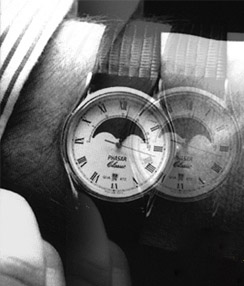
One way a person might experience double vision
SpecialtyNeurology, ophthalmology

Diplopia is the simultaneous perception of two images of a single object that may be displaced in relation to each other.[1] Also called double vision, it is a loss of visual focus under regular conditions, and is often voluntary. However, when occurring involuntarily, it results from impaired function of the extraocular muscles, where both eyes are still functional, but they cannot turn to target the desired object.[2] Problems with these muscles may be due to mechanical problems, disorders of the neuromuscular junction, disorders of the cranial nerves (III, IV, and VI) that innervate the muscles, and occasionally disorders involving the supranuclear oculomotor pathways or ingestion of toxins.[3]

Diplopia can be one of the first signs of a systemic disease, particularly to a muscular or neurological process,[4] and it may disrupt a person's balance, movement, or reading abilities.[2][5]

Presentation

[edit]

Causes

[edit]

Diplopia has a diverse range of ophthalmologic, infectious, autoimmune, neurological, and neoplastic causes:[6]

  • Diagnosis

    [edit]

    Diplopia is diagnosed mainly by information from the patient. Doctors may use blood tests, physical examinations,[14] computed tomography (CT), or magnetic resonance imaging (MRI) to find the underlying cause.[15]

    Classification

    [edit]

    One of the first steps in diagnosing diplopia is often to see whether one of two major classifications may be eliminated. That involves blocking one eye to see which symptoms are evident in each eye alone. Persisting blurry or double vision with one eye closed is classified as monocular diplopia.[16]

    Binocular

    [edit]

    Binocular diplopia is the other one in which the blurring of vision occurs only when the patient looks through both eyes simultaneously. It is common and occurs in approximately 10.0% to 40.0% of zygomatic complex injuries. Furthermore, diplopia may be transient or persistent. Inadequate diagnosis and treatment at improper times and tethering or fibrosis of muscles may lead to persistent diplopia.[17]

    Binocular diplopia is double vision arising as a result of strabismus[18] (in layman's terms "cross-eyed"), the misalignment of the two eyes relative to each other, either esotropia (inward) or exotropia (outward). In such a case while the fovea of one eye is directed at the object of regard, the fovea of the other is directed elsewhere, and the image of the object of regard falls on an extrafoveal area of the retina. Acute diplopia is a diagnostic challenge. The most common cause of acute diplopia are ocular motor nerve palsies (OMP).[19]

    The brain calculates the visual direction of an object based upon the position of its image relative to the fovea. Images falling on the fovea are seen as being directly ahead, while those falling on retina outside the fovea may be seen as above, below, right, or left of straight ahead depending upon the area of retina stimulated. Thus, when the eyes are misaligned, the brain perceives two images of one target object, as the target object simultaneously stimulates different, noncorresponding, retinal areas in either eye, thus producing double vision.[20]

    This correlation of particular areas of the retina in one eye with the same areas in the other is known as retinal correspondence. This relationship also gives rise to an associated phenomenon of binocular diplopia, although one that is rarely noted by those experiencing diplopia. Because the fovea of one eye corresponds to the fovea of the other, images falling on the two foveae are projected to the same point in space. Thus, when the eyes are misaligned, two different objects will be perceived as superimposed in the same space. This phenomenon is known as 'visual confusion'.[21]

    The brain naturally guards against double vision. In an attempt to avoid double vision, the brain can sometimes ignore the image from one eye, a process known as suppression. The ability to suppress is to be found particularly in childhood when the brain is still developing. Thus, those with childhood strabismus almost never complain of diplopia, while adults who develop strabismus almost always do. This ability to suppress might seem an entirely positive adaptation to strabismus, but in the developing child it can prevent the proper development of vision in the affected eye, resulting in amblyopia. Some adults can also suppress their diplopia, but their suppression is rarely as deep or as effective and takes much longer to establish; thus, they are not at risk of permanently compromising their vision. In some cases, diplopia disappears without medical intervention, but in other cases, the cause of the double vision may still be present.

    Certain people with diplopia who cannot achieve fusion and yet do not suppress may display a certain type of spasm-like irregular movement of the eyes in the vicinity of the fixation point (see: Horror fusionis).

    Monocular

    [edit]

    Diplopia can also occur when viewing with only one eye; this is called monocular diplopia, or where the patient perceives more than two images, monocular polyopia. While serious causes rarely may be behind monocular diplopia symptoms, this is much less often the case than with binocular diplopia.[16] The differential diagnosis of multiple image perception includes the consideration of such conditions as corneal surface keratoconus, subluxation of the lens, a structural defect within the eye, a lesion in the anterior visual cortex, or nonorganic conditions, but diffraction-based (rather than geometrical) optical models have shown that common optical conditions, especially astigmatism, can also produce this symptom.[22]

    Temporary

    [edit]

    Temporary binocular diplopia can be caused by alcohol intoxication or head injuries, such as concussion (if temporary double vision does not resolve quickly, one should see an optometrist or ophthalmologist immediately). It can also be a side effect of benzodiazepines or opioids, particularly if used recreationally in larger doses, the antiepileptic drugs phenytoin, zonisamide and lamotrigine, as well as the hypnotic drug zolpidem and the dissociative drugs ketamine and dextromethorphan. Temporary diplopia can also be caused by tired or strained eye muscles.[23] If diplopia appears with other symptoms such as fatigue and acute or chronic pain, the patient should see an ophthalmologist immediately.[24][25]

    Voluntary

    [edit]

    Some people can consciously uncouple their eyes, either by overfocusing closely (i.e., going cross-eyed) or unfocusing. Also, while looking at one object behind another object, the foremost object's image is doubled (for example, placing one's finger in front of one's face while reading text on a computer monitor). In this sense, double vision is neither dangerous nor harmful, and may even be enjoyable. It makes viewing stereograms possible.[26]

    Monocular diplopia may be induced in many individuals, even those with normal eyesight, with simple defocusing experiments involving fine, high-contrast lines.[22]

    Treatment

    [edit]

    The appropriate treatment for binocular diplopia depends upon the cause of the condition producing the symptoms. Efforts must first be made to identify and treat the underlying cause of the problem. Treatment options include eye exercises,[2] wearing an eye patch on alternative eyes,[2][25] prism correction,[27][25][28] and in more extreme situations, surgery[5] or botulinum toxin.[29]

    See also

    [edit]

    References

    [edit]

    Further reading

    [edit]
    [edit]
    Revisions and contributorsEdit on WikipediaRead on Wikipedia
    from Grokipedia
    Diplopia, commonly known as double vision, is a visual disturbance characterized by the perception of two images of a single object, resulting from ocular misalignment, optical aberrations, or neurological issues.[1] It is broadly classified into monocular diplopia, which persists when viewing with one eye alone and typically stems from intraocular problems such as cataracts or corneal irregularities, and binocular diplopia, which occurs only when both eyes are open and disappears upon covering one eye, often due to misalignment from extraocular muscle or nerve dysfunction.[2] This condition can manifest as horizontal (side-by-side images), vertical (one image above the other), or oblique separation and may signal benign refractive errors or serious underlying pathologies requiring prompt evaluation.[3] The causes of diplopia are diverse, encompassing ocular disorders like dry eyes, astigmatism, or lens subluxation for monocular cases, and systemic or neurological conditions such as cranial nerve palsies (III, IV, or VI), myasthenia gravis, thyroid eye disease, stroke, trauma, or orbital tumors for binocular cases.[2] Associated symptoms often include eye strain, headaches, reduced depth perception, or shadows around objects, with acute onset potentially indicating emergencies like brainstem lesions or vascular events.[1] In the United States, diplopia prompts over 800,000 annual visits to eye care specialists and around 50,000 emergency department encounters, predominantly affecting adults over age 60.[1] Diagnosis begins with a thorough history to determine onset, duration, and aggravating factors, followed by ophthalmologic examinations including visual acuity testing, cover-uncover tests for alignment, extraocular motility assessment, and pupillary evaluation.[3] For suspected neurological etiologies, advanced imaging such as MRI or CT angiography is employed, alongside blood tests for conditions like diabetes or thyroid dysfunction.[2] Differential diagnoses include pseudodiplopia from uncorrected refractive errors, transient ischemic attacks, or decompensated strabismus.[3] Management is etiology-specific and often effective; monocular diplopia may resolve with updated refraction, cataract surgery, or treatment of corneal issues, while binocular cases can involve temporary measures like prisms, eye patching, or botulinum toxin injections, with surgical correction of muscle imbalances for persistent misalignment.[1] Microvascular causes, such as those from hypertension or diabetes, frequently improve spontaneously within 3 to 6 months, though urgent intervention is critical for compressive or ischemic threats to prevent vision loss or neurological complications.[2]

    Introduction

    Definition

    Diplopia is the perception of two images of a single object, resulting from either misalignment of the eyes or abnormalities in the ocular media.[2][1] The duplicate images may be displaced horizontally, vertically, or obliquely relative to each other.[2][4] For example, in horizontal diplopia, the second image appears shifted to one side of the primary image, often affecting central vision during fixation, whereas vertical diplopia positions the offset image above or below, which may be more noticeable in peripheral vision depending on head position.[2][5] Diplopia is distinct from polyopia, which involves perceiving multiple (more than two) images of a single object, often in a patterned arrangement.[6][7] It also differs from pseudodiplopia, an illusory double vision arising from refractive errors or opacities in the ocular media that produce ghosting or blurred contours rather than true duplication.[8][9] The term diplopia derives from the Greek words diploos (double) and ops (eye).[10] It is classified as binocular, when present only with both eyes viewing simultaneously, or monocular, when observed in one eye alone.[2]

    Epidemiology

    Diplopia affects a small proportion of the general population, though exact prevalence figures are limited due to its episodic nature and underreporting in non-clinical settings. In the United States, approximately 850,000 diplopia-related medical visits occur annually, with 95% classified as ambulatory outpatient encounters and the remainder in emergency departments; this equates to an estimated incidence of about 257 cases per 100,000 population per year based on national health data from 2003 to 2012.[11] International studies report lower rates in emergency settings, such as 3.6 per 10,000 pediatric emergency visits in Italy and similar figures (4.4–4.6 per 10,000) in South Korea and Taiwan, indicating diplopia as an infrequent chief complaint in acute care.[12] Binocular diplopia predominates, accounting for 88–94% of cases across various cohorts.[13] Prevalence increases notably with age, particularly among older adults where age-related orbital changes contribute significantly. Ambulatory visits in the US show a mean patient age of 62.1 years, with 79% of cases in individuals aged 50 or older, reflecting higher occurrence linked to cranial nerve palsies and conditions like sagging eye syndrome.[11] In patients over 60, sagging eye syndrome emerges as a leading cause, comprising up to 31% of acquired binocular diplopia cases in adults aged 40 and older, with its relative proportion rising to 60.9% in those over 90 among diplopia presentations.[14] Demographic patterns reveal a male predominance in certain etiologies, driven by trauma; a hospital-based study in India found 70% of 160 diplopia patients were male, with trauma as the second most common cause overall (24.7%) and predominant in the 20–40 age group.[13] Comorbidities such as diabetes and thyroid disease elevate risk through microvascular ischemia and ophthalmopathy, respectively; vasculopathy, often tied to these conditions, accounts for 28.7% of binocular diplopia cases, particularly in patients over 40.[13][15] Global variations exist, with higher incidence in regions prone to trauma, including developing countries where road traffic accidents are more frequent and contribute to elevated rates of injury-related diplopia. Peaks in trauma- and stroke-associated diplopia align with these patterns, underscoring the influence of environmental and socioeconomic factors on public health burden.[11]

    Pathophysiology

    Mechanisms of Double Vision

    Binocular vision relies on the precise alignment of the two eyes to fuse images from corresponding retinal points into a single percept; disruption of this alignment causes the brain to perceive two distinct images, resulting in diplopia.[2] The ocular motor system coordinates eye movements through a complex network involving extraocular muscles, cranial nerves, brainstem nuclei, and supranuclear pathways. The extraocular muscles—superior rectus, inferior rectus, superior oblique, inferior oblique, medial rectus, and lateral rectus—control eye position and movement, with balanced innervation ensuring alignment.[16][2] Cranial nerves III (oculomotor), IV (trochlear), and VI (abducens) provide motor innervation to these muscles, while brainstem nuclei such as the oculomotor, trochlear, and abducens nuclei integrate signals for conjugate gaze.[16] Supranuclear pathways, including those from the frontal eye fields and parietal lobe, direct voluntary and reflexive eye movements to maintain fusion.[16] Misalignments leading to diplopia can manifest as specific types, such as convergence or divergence insufficiency, where the eyes fail to adduct or abduct properly for near or distance viewing, respectively.[16] Skew deviation produces vertical misalignment due to imbalance in otolith-ocular reflexes from vestibular or brainstem disruptions.[16] Torsional diplopia arises from cyclovertical imbalances, often linked to vestibular or otolith dysfunction, causing perceived rotation of the visual field.[16] Monocular diplopia occurs within a single eye due to optical irregularities that split the incoming light into multiple images on the retina. Irregular astigmatism, often from corneal surface distortions, diffracts light rays unevenly, producing a primary clear image and secondary ghost images.[17] Lens dislocation or subluxation shifts the lens position, allowing its edge to split the visual axis and induce diffraction-based double images.[18][17] Diplopia from misalignment can stem from neural or mechanical factors, classified as incomitant or comitant based on gaze dependency. Incomitant misalignment varies with gaze direction, typically due to neural disruptions like unequal innervation from cranial nerve issues, such as palsies.[16][2] Comitant misalignment remains constant across gaze positions, often from mechanical restrictions or balanced but aberrant muscle forces.[16]

    Classification of Diplopia

    Diplopia is primarily classified based on whether it occurs in one eye (monocular) or requires both eyes (binocular), as this distinction guides clinical evaluation by indicating potential ocular versus alignment issues.[2] Binocular diplopia arises from misalignment between the eyes, resulting in two distinct images that fuse into one when either eye is covered, as the brain suppresses input from the occluded eye.[16] It is further subdivided by the direction of image displacement: horizontal diplopia features side-by-side images, vertical diplopia shows one image above the other, and torsional or oblique diplopia involves rotated or diagonal separation.[10] These directional subtypes reflect variations in how the images deviate but do not imply specific underlying mechanisms.[16] Monocular diplopia, in contrast, persists when viewing with a single eye open, even after covering the other, and typically produces overlapping or ghost-like images rather than fully separated ones.[15] It may affect one or both eyes and can result in more than two images (e.g., triplopia), with one primary image of normal clarity and secondary images of poorer quality.[16] This type often stems from irregularities within the optical pathway of the affected eye itself.[2] Diplopia can also be categorized by its temporal pattern as temporary or intermittent, where episodes are transient and may resolve spontaneously within minutes to hours, versus persistent forms that endure longer.[10] Temporary diplopia is distinguished from constant diplopia by its waxing and waning nature, often noticeable only under certain conditions.[15] Voluntary or physiologic diplopia refers to the intentional or naturally occurring perception of double images achievable in individuals with normal binocular vision, such as by fixating on a near object while a distant one appears doubled due to uncrossed visual axes.[19] This benign phenomenon is common in children and can be demonstrated voluntarily through eccentric gaze or convergence efforts, without pathological significance.[20] An additional classification distinguishes gaze-evoked diplopia, which worsens or appears only in specific directions of eye movement (e.g., lateral or upward gaze), from non-gaze-evoked diplopia that remains consistent across primary and eccentric gazes.[16] Gaze-evoked forms highlight position-dependent misalignment, while non-gaze-evoked types suggest more uniform disruptions, aiding in localizing the level of dysfunction without delving into specific etiologies.[2]

    Clinical Features

    Symptoms

    Diplopia manifests primarily as the perception of two distinct images of a single object, often described by patients as seeing doubles that may overlap partially or separate completely, creating visual confusion.[2] These duplicate images can appear side by side (horizontal), one above the other (vertical), or at an angle (oblique), depending on the misalignment.[1] In binocular diplopia, this doubling occurs only when both eyes are open and resolves immediately upon covering either eye. In contrast, monocular diplopia, which persists when viewing with one eye, often presents as a ghosting or shadowing effect overlapping the primary image rather than two distinct images.[1][2] The intensity of double vision frequently worsens with fatigue, prolonged near tasks such as reading, or specific directions of gaze, making it more noticeable during extended visual effort or lateral eye movements.[2] It may present as constant or intermittent, with triggers including time of day (e.g., worse in the evening) or distance of focus, alternating between clear single vision and duplication.[1] Associated visual complaints often include blurred vision in one or both images, eye strain from efforts to fuse or suppress the secondary image, and headaches due to sustained visual discomfort.[2] These symptoms arise from the brain's struggle to reconcile conflicting inputs, leading to additional strain.[1] Functionally, diplopia impairs daily activities by reducing depth perception and visual stability; patients report significant difficulties with reading (where even small image separations can slow speed by over 60% and decrease accuracy), driving (due to hazardous misjudgment of distances), and walking (increasing fall risk).[21][1] To cope, individuals may adopt compensatory behaviors such as head tilting to align images, closing one eye for monocular vision, or avoiding tasks requiring precise binocular coordination.[2] On a personal level, sudden-onset diplopia often provokes anxiety and emotional distress, particularly if it affects one eye (monocular) or disrupts routine independence, contributing to feelings of inferiority and social withdrawal.[22] This can profoundly impact quality of life, with affected adults often citing interference in work, social interactions, and activities such as driving (82% reported driving difficulties).[22]

    Associated Signs

    Diplopia is frequently accompanied by observable ocular misalignment, which can be detected through clinical examination techniques such as the cover-uncover test. This test reveals deviations including esotropia (inward deviation of the eye), exotropia (outward deviation), hypertropia (upward deviation), or hypotropia (downward deviation) of the non-fixing eye, indicating strabismus that contributes to the double vision.[23][15] Patients often adopt compensatory head posture abnormalities to alleviate diplopia, such as head tilt, turn, or torticollis, which minimize the angular disparity between the visual axes. For instance, a head turn toward the affected side may be seen in abducens nerve involvement to reduce horizontal misalignment.[24][15] Examination of eye movements may uncover nystagmus or gaze palsies, characterized by involuntary oscillations of the eyes or restricted ductions in specific directions. Gaze-evoked nystagmus can appear during sustained eccentric gaze, while limitations in adduction, abduction, elevation, or depression suggest underlying paresis.[24][3] In cases involving the third cranial nerve, pupillary or lid signs such as ptosis (drooping eyelid) and anisocoria (unequal pupil sizes) are commonly observed, with the affected pupil often dilated and poorly reactive. These findings warrant urgent evaluation due to potential compressive etiologies.[23][15] Fundoscopic examination may reveal abnormalities like papilledema, indicating elevated intracranial pressure, or optic atrophy, suggesting chronic optic nerve damage in intracranial pathologies. These posterior segment signs help localize the diplopia to central nervous system involvement.[23][3]

    Etiology

    Binocular Diplopia Causes

    Binocular diplopia arises from misalignment of the eyes due to dysfunction in the extraocular muscles, their innervation, or orbital structures, distinguishing it from monocular causes that involve optical aberrations within a single eye. The most common etiologies are neurological, particularly involving the oculomotor (third), trochlear (fourth), and abducens (sixth) cranial nerves, which collectively account for up to 70% of cases in clinical series.[13] Neurological causes predominate, with isolated cranial nerve palsies being the leading mechanism. Among these, sixth nerve palsy is the most frequent, comprising approximately 50–53% of cases, followed by third nerve palsy at 17–30%, fourth nerve palsy at 11–25%, and multiple nerve involvement in 10–14%, based on recent clinical series.[13][25] Microvascular ischemia, often linked to diabetes mellitus or hypertension, underlies many of these palsies by causing infarction of the vasa nervorum, leading to acute onset of diplopia without pupillary involvement in third nerve cases.[26] Brainstem lesions, such as those from multiple sclerosis, can also produce binocular diplopia through demyelination affecting internuclear pathways or direct nerve nuclei, resulting in patterns like internuclear ophthalmoplegia with horizontal gaze limitation.[27] Parkinson's disease can cause binocular diplopia through mechanisms such as convergence insufficiency, reduced fusion range, and dysfunction in the oculomotor pathway.[28] Traumatic causes contribute significantly, particularly orbital fractures from blunt injury, which may entrap extraocular muscles like the inferior rectus in blowout fractures of the orbital floor, restricting vertical gaze and causing vertical diplopia.[29] Head trauma can additionally lead to intracranial hematomas compressing cranial nerves or direct muscle contusion, with even minor impacts sufficient for fourth nerve palsy due to its long intracranial course.[30] Myasthenia gravis presents with fatigable weakness of the extraocular muscles due to autoantibodies against acetylcholine receptors at the neuromuscular junction, often manifesting as variable diplopia that worsens with prolonged gaze and improves with rest.[31] Thyroid eye disease, an autoimmune inflammatory condition associated with Graves' disease, induces restrictive myopathy through glycosaminoglycan deposition and fibrosis in the extraocular muscles, particularly the inferior rectus and medial rectus, leading to proptosis and incomitant strabismus with diplopia on upward or lateral gaze.[32] Infectious and inflammatory processes, such as orbital cellulitis from bacterial spread via sinuses, can cause diplopia through inflammatory edema or abscess formation restricting muscle movement, while cavernous sinus thrombosis—often complicating facial infections—results in painful ophthalmoplegia and multiple cranial nerve deficits due to venous congestion and ischemia in the cavernous sinus.[33][34] Emerging data since 2020 highlight post-COVID-19 cranial neuropathies as a novel etiology, with cases of isolated third, fourth, or sixth nerve palsies attributed to viral-induced inflammation or immune-mediated damage, often resolving spontaneously but warranting neuroimaging to exclude compressive lesions.[35]

    Monocular Diplopia Causes

    Monocular diplopia arises from abnormalities within a single eye that distort or split the incoming light rays, leading to the perception of multiple images when viewing with that eye alone. Unlike binocular diplopia, which involves misalignment between the eyes, monocular diplopia typically stems from refractive, corneal, lenticular, retinal, or ocular surface irregularities. These causes are often identifiable through targeted ophthalmic examination and can frequently be managed by addressing the underlying optical defect.[23][17] Refractive errors, particularly irregular astigmatism, are among the most common causes of monocular diplopia. Corneal scars, such as those resulting from post-keratitis complications or trauma, can induce irregular astigmatism by altering the corneal surface's smoothness, causing light to scatter and produce ghost images. Similarly, early-stage keratoconus, a progressive corneal ectasia, leads to irregular astigmatism through corneal thinning and protrusion, resulting in distorted central vision and monocular double vision in affected individuals. These refractive anomalies often manifest as overlapping or shadowed images that improve with pinhole testing.[17][36][37] Lens-related issues also frequently contribute to monocular diplopia. Cataracts can cause image splitting due to opacification and irregular refractive indices within the lens, leading to diffraction of light rays and the formation of multiple foci. Lens subluxation, or ectopia lentis, particularly in systemic conditions like Marfan syndrome, displaces the lens from its normal position, inducing astigmatism and fluctuating double vision as the lens shifts with eye movement. In Marfan syndrome, ectopia lentis affects 50–80% of patients, often bilaterally, and is a hallmark ocular feature, often presenting with monocular diplopia alongside blurred vision.[23][37][38][39] Retinal disorders involving the macula can distort the retinal image, producing monocular diplopia through metamorphopsia or irregular light projection. Macular edema, characterized by fluid accumulation in the macular region, warps central vision and can cause perceived image duplication due to retinal surface irregularity. Epiretinal membrane (ERM), a fibrocellular proliferation on the retinal surface, similarly induces image distortion by contracting and displacing the macula, leading to diplopia in cases where the membrane causes significant foveal drag or rivalry between central and peripheral retinal inputs. These conditions often coexist with decreased visual acuity and are more prevalent in older adults or those with prior retinal pathology.[40][41][42] Ocular surface abnormalities further account for many instances of monocular diplopia by disrupting the tear film or corneal integrity. Dry eye syndrome destabilizes the precorneal tear film, creating transient refractive irregularities that result in blurred or doubled vision, particularly during prolonged visual tasks. Pterygium, a fibrovascular growth encroaching on the cornea, induces astigmatism through mechanical distortion of the corneal curvature, occasionally causing monocular diplopia in advanced cases. Post-surgical irregularities, such as those following LASIK, can lead to corneal surface unevenness or persistent dry eye, both of which contribute to ghosting or double images in the operated eye.[23][43][44] Rare causes of monocular diplopia include iris defects and vitreous opacities, which alter light transmission or create interfering shadows. Iris lesions or colobomas can cause light scatter through irregular pupillary apertures, producing superimposed images. Vitreous opacities, such as floaters from posterior vitreous detachment, may project multiple shadows onto the retina, mimicking diplopia in low-light conditions. Additionally, rising cases among young adults have been linked to contact lens overuse, which exacerbates dry eye and corneal epithelial irregularities, leading to increased reports of monocular diplopia in this demographic.[36][45][46]

    Diagnosis

    History and Physical Examination

    The evaluation of diplopia begins with a detailed patient history to characterize the symptom and guide localization of the underlying pathology. Key elements include the onset, which is classified as acute (sudden, often within hours to days) or gradual (progressing over weeks to months), as acute onset raises concern for vascular events such as stroke or aneurysm. Progression should be assessed, noting whether the double vision is constant, intermittent, or worsening in specific gazes or distances (e.g., worse at near suggesting convergence insufficiency or at distance indicating abducens nerve involvement). Associated symptoms are crucial, including neurological features like vertigo, weakness, headache, or dysarthria that may point to brainstem involvement, as well as fatiguability suggesting myasthenia gravis or symptoms of increased intracranial pressure. Risk factors such as recent trauma, vascular disease (e.g., hypertension, diabetes), medications (e.g., those causing neuromuscular blockade), or systemic conditions like giant cell arteritis in patients over 50 years should be elicited to narrow the differential.[2][23][47] The physical examination starts with an assessment of visual acuity in each eye separately, using Snellen charts or equivalent, to identify any underlying refractive errors or optic nerve involvement that could contribute to perceived double vision. Pupillary responses are evaluated for size, reactivity, and relative afferent pupillary defect (RAPD), as anisocoria or non-reactive pupils may indicate third nerve palsy or other urgent conditions. Extraocular motility testing is performed by observing eye movements: ductions assess monocular movements in cardinal directions to detect isolated muscle or nerve weaknesses; versions evaluate binocular gaze in all fields to identify underactions or overactions; and vergences test convergence and divergence for near tasks, revealing issues like insufficiency of convergence. These tests help localize lesions to specific cranial nerves (III, IV, VI) or supranuclear pathways.[2][23][47] Alignment and misalignment are quantified using targeted tests to measure the degree and type of deviation. The Hirschberg test estimates corneal light reflex alignment for gross tropia detection; the alternate cover test distinguishes manifest (tropia) from latent (phoria) deviations by measuring refixation movements in prism diopters; and the Maddox rod quantifies horizontal, vertical, or torsional misalignment by dissociating images for patient-reported alignment. A systemic neurological review follows, screening for deficits such as limb weakness, sensory changes, or other cranial nerve involvement (e.g., facial asymmetry), alongside vascular history to localize supranuclear or infranuclear lesions. Red flags warranting immediate intervention include sudden onset diplopia, especially with pupillary involvement or multiple cranial neuropathies, suggesting potentially life-threatening causes like aneurysm rupture or stroke.[2][23][47]

    Diagnostic Tests

    Diagnostic tests for diplopia encompass a range of ancillary investigations aimed at confirming the underlying etiology and localizing pathology within the ocular, orbital, or neurological structures. These tests are selected based on clinical suspicion from history and examination, targeting specific causes such as compressive lesions, neuromuscular disorders, or vascular abnormalities. Imaging modalities, serological assays, electrophysiological studies, and advanced optical techniques form the cornerstone of this diagnostic approach. For cases with suspected neurological etiologies, consultation with a neuro-ophthalmologist is often indicated to provide specialized evaluation and management of diplopia.[23] Neuroimaging plays a pivotal role in evaluating diplopia, particularly when central or orbital pathology is suspected. Magnetic resonance imaging (MRI) of the brain and orbits is the preferred modality for assessing ocular motor nerve palsies, as it offers superior soft tissue resolution to detect lesions along the neural pathways governing eye movements, such as tumors, infarcts, or inflammatory processes.[2] Computed tomography (CT) is indicated for acute trauma or when bony orbital involvement is suspected, utilizing thin-slice helical protocols (1-2 mm) to identify fractures or foreign bodies that may cause restrictive diplopia.[48] For vascular etiologies, such as aneurysms or carotid-cavernous fistulas, CT or MR angiography is employed to visualize arterial abnormalities, often following initial non-contrast imaging in emergent cases.[49] Blood tests are essential for identifying systemic autoimmune or inflammatory conditions contributing to diplopia. Testing for acetylcholine receptor (AChR) antibodies supports the diagnosis of myasthenia gravis, a common neuromuscular cause of variable diplopia, with seropositivity present in approximately 85% of generalized cases and 50% of ocular-only presentations.[50] Thyroid function tests, including thyroid-stimulating hormone (TSH) and free thyroxine levels, are crucial for detecting Graves' disease-associated ophthalmopathy, which manifests as restrictive diplopia due to extraocular muscle enlargement.[51] Inflammatory markers such as erythrocyte sedimentation rate, C-reactive protein, and complete blood count help evaluate for orbital inflammatory syndromes like idiopathic orbital inflammation or Tolosa-Hunt syndrome.[52] Electrophysiological assessments aid in differentiating paretic from restrictive mechanisms and confirming neuromuscular disorders. Electromyography (EMG), particularly single-fiber EMG, demonstrates jitter or blocking in myasthenia gravis, enhancing diagnostic sensitivity when antibody tests are negative. The forced duction test, performed under topical anesthesia, mechanically rotates the globe to assess passive restriction; resistance indicates mechanical limitation from orbital pathology, such as fibrosis or entrapment, distinguishing it from neurogenic paresis where free movement occurs.[53] In monocular diplopia, optical coherence tomography (OCT) provides high-resolution cross-sectional imaging of the retina and anterior segment to identify structural anomalies causing image duplication. OCT is particularly useful for detecting subtle macular pathologies like epiretinal membranes or cystoid macular edema, which can lead to central-peripheral rivalry and monocular ghosting, as well as lens irregularities such as early cataracts.[54] Emerging AI-assisted tools in the 2020s have introduced precise quantification of eye motility for diplopia evaluation. Cloud-based AI systems, such as Eyeturn Cloud, analyze smartphone-captured videos to measure strabismus angles with high accuracy, aiding in the objective assessment of misalignment in both static and dynamic conditions.[55] Similarly, deep learning models applied to eye movement videos can diagnose ocular motility disorders, identifying affected muscles and quantifying deviations to support strabismus and diplopia classification.[56] These technologies enhance diagnostic precision, especially in resource-limited settings, by automating traditional motility assessments.[57]

    Management

    Non-Surgical Treatments

    Non-surgical treatments for diplopia primarily aim to alleviate symptoms through reversible interventions while addressing underlying etiologies where possible. These approaches include optical aids, pharmacological options, and targeted therapies for specific causes such as cranial nerve palsies or neuromuscular disorders. Selection depends on the type (binocular or monocular), acuity, and etiology of the diplopia, with the goal of improving visual comfort without invasive procedures.[49] Occlusion therapy involves blocking vision in one eye to eliminate double images, particularly useful in acute or intractable cases. This can be achieved with an eye patch, frosted lens, semi-opaque tape, or Bangerter occlusion foils applied to spectacles, which provide partial or total occlusion of the affected eye. It is often employed temporarily for ischemic cranial nerve palsies until spontaneous resolution occurs, offering immediate relief without altering eye alignment.[49][54] Prism glasses represent a non-invasive optical correction to realign visual images and reduce or eliminate diplopia. Fresnel press-on prisms, lightweight and temporary, can be applied directly to existing lenses to compensate for horizontal, vertical, or oblique deviations, making them suitable for trial periods or fluctuating misalignments. For stable cases, ground-in prisms incorporated into prescription eyewear provide a more permanent solution, though they are limited in cases of torsional diplopia or rapidly changing angles. Prism adaptation has been shown to improve alignment in up to 70% of suitable patients with small-angle strabismus.[49][58][59] Pharmacotherapy targets muscle dysfunction or inflammation contributing to diplopia. Botulinum toxin injections temporarily paralyze overactive extraocular muscles by inhibiting acetylcholine release at neuromuscular junctions, with effects onset in 2-4 days and lasting 5-8 weeks, allowing antagonist muscles to strengthen and potentially improve alignment. This is indicated for acute paralytic strabismus, such as sixth nerve palsy, or restrictive conditions like thyroid eye disease, providing symptomatic relief without surgery. Corticosteroids, such as intravenous methylprednisolone (500 mg weekly for 6 weeks), are used for inflammatory causes like active thyroid eye disease, achieving diplopia improvement in approximately 50% of moderate-to-severe cases.[60][61][62] For active thyroid eye disease, teprotumumab, an insulin-like growth factor-1 receptor (IGF-1R) inhibitor, is an FDA-approved infusion therapy (as of 2020) that reduces inflammation and orbital congestion, leading to diplopia improvement in 68-83% of patients after 8 infusions over 24 weeks, offering superior efficacy over steroids in clinical trials.[63] Treatment of underlying conditions is essential for etiology-specific diplopia. In myasthenia gravis, where diplopia arises from neuromuscular transmission defects, corticosteroids like prednisone combined with immunosuppressants such as azathioprine (2-3 mg/kg daily) reduce symptoms and prevent progression to generalized disease, with early initiation reducing generalization risk by approximately 81%. For diabetic cranial neuropathy causing third or sixth nerve palsy, optimizing glycemic control through oral hypoglycemics or insulin—targeting HbA1c below 7%—promotes nerve recovery and resolves diplopia in most cases within 3-6 months, as microvascular ischemia improves with normalized blood glucose.[64][65][66] Vision therapy, particularly orthoptic exercises, addresses convergence insufficiency, a common binocular diplopia cause involving poor eye teaming at near. Home-based or office-supervised programs, including pencil push-ups, stereograms, or computer orthoptics, strengthen fusional vergence and alleviate symptoms like double vision in about 70% of patients after 12 weeks of consistent practice. These exercises focus on improving near point of convergence and reducing accommodative demand, with long-term efficacy maintained through periodic reinforcement.[67][68]

    Surgical Interventions

    Surgical interventions for diplopia are typically reserved for cases where non-surgical measures fail to address persistent structural or restrictive causes, such as muscle misalignment, orbital compression, or optical irregularities. These procedures aim to restore binocular alignment or correct monocular visual distortions, improving single vision fields.[69][70] Strabismus surgery addresses binocular diplopia resulting from extraocular muscle misalignment by adjusting muscle tension through recession or resection techniques. In recession, the muscle is detached and reattached farther from its original insertion point to weaken its pull, while resection involves shortening the muscle by removing a segment and reattaching it closer to the cornea to strengthen it. These adjustments realign the eyes, reducing or eliminating double vision in primary or specific gazes.[69][71] Orbital decompression surgery is indicated for diplopia associated with thyroid eye disease, where inflamed tissues compress extraocular muscles, restricting motility. The procedure involves removing portions of the orbital walls—typically the medial and/or inferior walls—to expand the orbital space, thereby relieving pressure on the muscles and improving eye movement. This can resolve restrictive diplopia by allowing freer globe rotation.[72][73] For diplopia stemming from traumatic orbital entrapments, surgical exploration and neurolysis release trapped tissues or nerves within fracture sites, such as blowout fractures of the orbital floor. Exploration identifies and frees inferior rectus muscle or surrounding soft tissue adhesions, restoring normal motility and alleviating induced misalignment. In cases involving tumors, similar orbital exploration may be performed to excise or debulk masses impinging on neural or muscular structures, thereby mitigating compressive diplopia.[74][75][76] Monocular diplopia due to lens opacities or corneal irregularities may require cataract extraction or topography-guided LASIK. Cataract surgery removes the clouded lens and implants an intraocular lens, eliminating ghosting or doubling from light scatter within the lens. Topography-guided LASIK reshapes the corneal surface to correct irregular astigmatism, smoothing aberrations that cause monocular image duplication.[77][78] Timing of surgery is crucial, particularly for diplopia from cranial nerve palsies, where observation for spontaneous recovery is recommended for 6-12 months before proceeding to operative correction. This period allows potential resolution of temporary paresis, avoiding unnecessary intervention. Preoperative trials with prisms can help predict surgical outcomes by simulating alignment effects.[79][80][81]

    Prognosis and Complications

    Outcomes

    The prognosis for diplopia varies significantly depending on its underlying etiology and type, with many cases achieving favorable outcomes through spontaneous resolution or targeted interventions. In microvascular cranial nerve palsies, a common cause of binocular diplopia, spontaneous resolution occurs in 70-80% of cases within 3-6 months, often without residual deficits.[82][83] For instance, third nerve palsies due to microvascular ischemia show complete recovery in approximately 82% of patients by 3 months and 91% by 12 months.[82] Similarly, sixth nerve palsies from microvascular causes resolve in 70-90% of cases.[82] Treatment outcomes are generally positive for comitant strabismus, where prisms or surgery yield improvement in around 85% of patients, enabling better alignment and reduced double vision.[84] In contrast, success rates for incomitant strabismus are variable, often 60-80% depending on criteria and method, due to the variable misalignment that complicates full correction with prisms or surgical adjustment.[85] Long-term visual function often improves with these approaches, particularly when binocular vision is restored, leading to enhanced depth perception and overall stability.[86] Several factors influence recovery rates and long-term prognosis. Younger age is associated with better outcomes, as decompensating heterophorias in patients under 20 years resolve more readily than in older adults.[13] Etiology plays a key role, with ischemic or microvascular causes faring better than traumatic ones, where persistent deficits are more common.[13][87] Duration of symptoms also matters, as early intervention within the first few months promotes higher resolution rates, especially in microvascular cases that naturally improve within 5 months if uncomplicated.[87] Restoration of binocular vision significantly enhances quality of life, particularly by mitigating fall risks in the elderly, where diplopia increases the likelihood of musculoskeletal injuries and fractures by about 36% compared to those with normal binocular function.[88][89] In the 2020s, telemedicine follow-up has improved outcomes in remote areas by enabling timely monitoring of strabismus and diplopia post-treatment, with high patient satisfaction.[90][91] This approach ensures consistent care, reducing delays that could worsen visual function.[92]

    Potential Complications

    Untreated diplopia can lead to chronic suppression of the affected eye's image, particularly in children, resulting in amblyopia or "lazy eye" with potential permanent vision loss if not addressed during critical developmental periods.[93][94] Additionally, persistent diplopia increases the risk of accidents, such as motor vehicle crashes, due to impaired depth perception and visual confusion; regulatory bodies like the DVLA prohibit driving with unresolved double vision for safety reasons.[95] Treatment for diplopia carries specific risks, including prism-induced visual distortions such as blurring, dazzle, headaches, dizziness, and altered depth perception, which may affect up to 22% of users and limit long-term tolerability.[96][97] Surgical interventions for underlying strabismus can result in overcorrection, inducing new or intractable diplopia in adults who previously lacked it.[98][99] Botulinum toxin injections, used to manage misalignment, may cause ptosis in about 5% of cases, superior oblique weakness, or rare infections like orbital cellulitis.[100][101] When diplopia stems from systemic conditions like myasthenia gravis, failure to treat the underlying disease can progress to severe complications, including myasthenic crisis with respiratory failure requiring mechanical ventilation.[102][103] The visual disability from diplopia often contributes to psychological distress, including depression and reduced quality of life, as patients experience isolation, anxiety, and diminished independence.[104][105] Rarely, vestibular involvement in diplopia cases may lead to decompensation, resulting in persistent oscillopsia—an illusory movement of the visual field during head motion—that exacerbates imbalance and disorientation.[106][107]

    References

    User Avatar
    No comments yet.