Hubbry Logo
Transcription factor II HTranscription factor II HMain
Open search
Transcription factor II H
Community hub
Transcription factor II H
logo
7 pages, 0 posts
0 subscribers
Be the first to start a discussion here.
Transcription factor II H
from Wikipedia

general transcription factor IIH, polypeptide 1, 62kDa
Identifiers
SymbolGTF2H1
Alt. symbolsBTF2
NCBI gene2965
HGNC4655
OMIM189972
RefSeqNM_005316
UniProtP32780
Other data
LocusChr. 11 p15.1-p14
Search for
StructuresSwiss-model
DomainsInterPro
general transcription factor IIH, polypeptide 2, 44kDa
Identifiers
SymbolGTF2H2
Alt. symbolsBTF2, TFIIH, BTF2P44, T-BTF2P44
NCBI gene2966
HGNC4656
OMIM601748
RefSeqNM_001515
UniProtQ13888
Other data
LocusChr. 5 q12.2-13.3
Search for
StructuresSwiss-model
DomainsInterPro
general transcription factor IIH, polypeptide 3, 34kDa
Identifiers
SymbolGTF2H3
Alt. symbolsBTF2, TFIIH
NCBI gene2967
HGNC4657
OMIM601750
RefSeqNM_001516
UniProtQ13889
Other data
LocusChr. 12 q24.31
Search for
StructuresSwiss-model
DomainsInterPro

Transcription factor II H (TFIIH) is a multi-subunit protein complex involved in both the transcription of protein-coding genes and the nucleotide excision repair (NER) pathway. TFIIH was first identified in 1989 as general transcription factor-δ or basic transcription factor 2, an essential factor for transcription in vitro. It was subsequently isolated from yeast and officially named TFIIH in 1992.[1][2]

TFIIH is composed of ten subunits. Seven of these—ERCC2/XPD, ERCC3/XPB, GTF2H1/p62, GTF2H4/p52, GTF2H2/p44, GTF2H3/p34, and GTF2H5/TTDA—constitute the core complex. The remaining three subunits—CDK7, MAT1, and cyclin H—form the cyclin-activating kinase (CAK) subcomplex, which is tethered to the core via the XPD protein.[3] Among the core subunits, ERCC2/XPD and ERCC3/XPB possess helicase and ATPase activities and are essential for unwinding DNA to form the transcription bubble. These activities are necessary during transcription in vitro only when the DNA template is not already denatured or is supercoiled.

The CAK subunits, CDK7 and cyclin H, are responsible for the phosphorylation of serine residues in the C-terminal domain of RNA polymerase II, as well as potentially other targets involved in the cell cycle. In addition to its essential role in transcription initiation, TFIIH also plays a critical part in nucleotide excision repair.

History

[edit]

Before being designated as TFIIH, the complex was known by several names. It was first isolated in 1989 from rat liver and referred to as transcription factor δ. When identified in cancer cells, it was called basic transcription factor 2, and when isolated from yeast, it was known as transcription factor B. The complex was officially named TFIIH in 1992.[4]

Structure

[edit]

TFIIH is a ten‐subunit complex; seven of these subunits comprise the "core" whereas three comprise the dissociable "CAK" (CDK-activating Kinase) module.[5] The core consists of subunits XPB, XPD, p62, p52, p44, p34 and p8 while CAK is composed of CDK7, cyclin H, and MAT1.[5]

Function

[edit]

General functions of TFIIH include:

  1. Initiating transcription of protein-coding genes[6]
  2. Repairing DNA[6]

Gene transcription

[edit]

TFIIH is a general transcription factor that helps recruit RNA polymerase II (Pol II) to gene promoters. It acts as a DNA translocase, sliding along the DNA while feeding it into the RNA polymerase II cleft, thereby generating torsional strain that facilitates local DNA unwinding.[7] TFIIH also plays a critical role in nucleotide excision repair (NER), where it unwinds DNA at sites of damage following lesion recognition by either the global genome repair (GGR) or transcription-coupled repair (TCR) pathway.[8][9]

DNA repair

[edit]
Mechanism of TFIIH repairing DNA damaged sequence

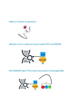
TFIIH participates in nucleotide excision repair (NER) by opening the DNA double helix after damage is initially recognized. NER is a multi-step pathway that removes a wide range of different types of damage that distort normal base pairing, including bulky chemical damage and UV-induced damage. Individuals with mutational defects in genes specifying protein components that catalyze the NER pathway, including the TFIIH components, often display features of premature aging.[10][11]

Clinical significance

[edit]

Trichothiodystrophy

[edit]

Mutation in genes ERCC3 (XPB), ERCC2 (XPD) or GTF2H5 (TTDA) cause trichothiodystrophy, a condition characterized by photosensitivity, ichthyosis, brittle hair and nails, intellectual impairment, decreased fertility and/or short stature.[10]

Cancer

[edit]

Genetic polymorphisms of genes that encode subunits of TFIIH are known to be associated with increased cancer susceptibility in many tissues, e.g. skin tissue, breast tissue and lung tissue. Mutations in the subunits (such as XPD and XPB) can lead to a variety of diseases, including xeroderma pigmentosum (XP) or XP combined with Cockayne syndrome.[12]

Viral infection

[edit]

Virus-encoded proteins target TFIIH.[13]

Inhibitors

[edit]

Potent, bioactive natural products such as triptolide, which inhibit mammalian transcription by targeting the XPB subunit of the general transcription factor TFIIH, have recently been developed as glucose conjugates to selectively target hypoxic cancer cells with elevated glucose transporter expression.[14]

References

[edit]
[edit]
Revisions and contributorsEdit on WikipediaRead on Wikipedia
from Grokipedia
Transcription factor II H (TFIIH) is a multi-subunit eukaryotic essential for the initiation of II-dependent transcription and (NER) of DNA damage. Composed of ten subunits, TFIIH adopts a modular with a seven-subunit core (XPB, XPD, p62, p52, p44, p34, and p8) and a three-subunit cyclin-dependent -activating (CAK) module (CDK7, H, and MAT1), enabling dynamic conformational changes that support its multifunctional roles. This complex plays a pivotal role in promoter DNA unwinding during transcription initiation and in verifying and excising DNA lesions during NER, thereby maintaining genome integrity and regulating . In transcription, TFIIH integrates into the pre-initiation complex at gene promoters, where the XPB subunit acts as a 3'-5' and 5'-3' to melt DNA and form the transcription bubble, while the CDK7 subunit of the CAK module phosphorylates the C-terminal domain (CTD) of at serine residues 5 and 7, facilitating promoter clearance and elongation. The core subunits, such as p62 and p44, provide structural stability and modulate activities, ensuring precise coordination with other general transcription factors. Beyond basal transcription, TFIIH influences progression through CAK-mediated activation of cyclin-dependent kinases and participates in the of nuclear receptors like , linking it to broader regulatory networks. In NER, TFIIH is recruited to sites of DNA damage, such as UV-induced lesions, where XPB initiates limited DNA unwinding and XPD scans single-stranded DNA for distortions using its 5'-3' activity, enabling the assembly of repair factors for oligonucleotide excision. Conformational switching between transcription and repair modes—such as repositioning of p62 to activate XPD in NER—underlies TFIIH's ability to prioritize repair without disrupting transcription globally. Mutations in core subunits like XPB, XPD, and p8 disrupt these processes, leading to rare genetic disorders including (characterized by extreme UV sensitivity and predisposition), (with neurological and developmental abnormalities), and (featuring brittle hair and ). TFIIH's central involvement in both transcription and has positioned it as a promising therapeutic target in cancer, where inhibitors of its subunits (e.g., XPB or CDK7) exploit dependencies in highly proliferative tumor cells while sparing normal tissues with lower transcription rates. Structural studies using cryo-electron microscopy have revealed the intricate dynamics of TFIIH assembly and function, highlighting how disease-associated mutations impair subunit interfaces and overall complex stability. More recent studies (2024-2025) have further elucidated TFIIH's recruitment in transcription-coupled repair, including the role of STK19 in positioning the complex.

Introduction

Overview

Transcription factor II H (TFIIH) is a heterodecameric that functions as a essential for the initiation of (Pol II)-dependent transcription in eukaryotes. It plays critical roles not only in basal transcription but also in (NER) of DNA damage and cell cycle regulation through its (CDK)-activating kinase (CAK) module. In transcription, TFIIH unwinds promoter DNA using its helicase subunits XPB and XPD, facilitating open complex formation and promoter clearance by Pol II. The complex comprises 10 subunits organized into a core subcomplex of seven subunits (XPB, XPD, p62, p52, p44, p34, and p8) and a CAK subcomplex consisting of three subunits (CDK7, H, and MAT1), with the CAK loosely associated and sometimes dissociable. This modular architecture enables TFIIH's multifunctional capabilities, allowing it to integrate signaling from transcription, repair, and pathways. Through its involvement in Pol II transcription, TFIIH is responsible for synthesizing all protein-coding messenger RNAs (mRNAs) and many noncoding RNAs in eukaryotic cells, accounting for the vast majority of . TFIIH is evolutionarily conserved across eukaryotes, from to humans, with its core subunits showing high sequence and functional similarity that underscores its fundamental role in cellular . In humans, mutations in TFIIH subunits are linked to genetic disorders such as and , highlighting its indispensability. Studies on human TFIIH have revealed its dynamic conformational changes that coordinate these diverse activities.

Nomenclature

Transcription factor II H (TFIIH) is the established nomenclature for this eukaryotic multi-subunit essential for -mediated transcription initiation. The full designation reflects its role as a specific to , with the "H" indicating its classification within the general transcription factor series (TFIIA through TFIIJ). The (HGNC) recognizes TFIIH as comprising subunits collectively termed IIH complex subunits, emphasizing their coordinated function in basal transcription machinery. Synonyms for TFIIH include 2H, reflecting its basal role in promoter recognition and pre-initiation complex assembly. These alternative terms appear frequently in to denote the complex's multifunctional nature, though TFIIH remains the primary identifier in modern . Basal transcription factor is another occasional synonym, underscoring its necessity for RNA polymerase II-dependent across eukaryotes. Subunit nomenclature follows HGNC-approved conventions, combining functional descriptors, molecular weights, and disease associations. For instance, the core subunits are designated XPB (Xeroderma pigmentosum group B protein, encoded by ERCC3) and XPD ( group D protein, encoded by ERCC2), reflecting their identification through complementation studies in disorders. Structural subunits include p62 (encoded by GTF2H1, previously BTF2), p44 (GTF2H2), p34 (GTF2H3), p52 (GTF2H4), and p8 (GTF2H5, also known as TTDA for complementation group A). The cyclin-activating kinase (CAK) subcomplex subunits are named cyclin H (CCNH), 7 (CDK7), and MNAT1 (MAT1). These names often derive from biochemical purification (e.g., "p" for apparent molecular weight in kilodaltons) or orthologs (e.g., BTF2 from basic transcription factor 2). Historically, TFIIH was first purified and named transcription factor δ (delta) in the late 1980s from liver extracts, where it was identified as a multifunctional with and activities required for promoter opening. This early designation shifted to the current TFIIH nomenclature in the early 1990s as its full composition and roles in both transcription and were elucidated through genetic and biochemical studies in and systems. The transition aligned with standardized naming for general transcription factors, distinguishing TFIIH from other complexes like TFIIF or TFIIE.

History

Discovery

The discovery of Transcription factor II H (TFIIH) stemmed from pioneering biochemical studies on eukaryotic mRNA synthesis in the 1980s, particularly those conducted in Robert G. Roeder's laboratory at The Rockefeller University. In 1980, Roeder and colleagues developed an in vitro transcription system using soluble extracts from human cells and purified , demonstrating that multiple accessory protein factors were essential for accurate initiation at the promoter of the adenovirus major late gene. This seminal work revealed the complexity of the basal transcription apparatus and set the stage for systematic fractionation to identify individual components, shifting the field from crude extracts to defined systems. Between 1983 and 1985, Roeder's group and others performed extensive chromatographic fractionation of and liver nuclear extracts, successfully resolving the general transcription factors required for II-mediated basal transcription into distinct activities labeled TFIIA through TFIIF, with an additional heat-labile fraction initially termed factor G or δ. This fraction was indispensable for reconstituting accurate transcription but resisted further purification due to its multisubunit nature and instability. By 1989, Ron C. Conaway and Joan W. Conaway, working in Roeder's lab, achieved over 3,000-fold purification of this factor (named δ) from liver nuclear extracts, identifying it as a ~500 kDa complex essential for open complex formation and promoter opening in assays. Notably, the purified factor exhibited DNA-dependent activity strongly stimulated by TATA box-containing promoters, providing early evidence of its capacity for DNA unwinding during transcription initiation. Independently, Jean-Marc Egly's group at the Institut de Génétique et de Biologie Moléculaire purified a functionally equivalent multisubunit complex from cells, termed basic transcription factor 2 (BTF2), which complemented the rat δ activity and confirmed its universal role in human basal transcription. These parallel efforts established TFIIH (unifying the nomenclature for δ and BTF2) as a core . Subsequent investigations in the early refined TFIIH's mechanistic contributions. In , Hiroyuki Serizawa, along with the Conaways in Roeder's laboratory, demonstrated through kinetic analyses in reconstituted systems that TFIIH (factor δ) facilitates promoter clearance—the ATP-dependent transition from to processive elongation—via its / activities, independent of its associated function for C-terminal domain . This finding underscored TFIIH's active role beyond preinitiation complex assembly, linking its enzymatic properties directly to promoter escape and early elongation. These discoveries, grounded in rigorous biochemical and functional assays, positioned TFIIH as a pivotal multifunctional complex in transcription.

Subunit identification

The identification of TFIIH subunits began in the early 1990s through genetic complementation studies in (XP) cell lines, which revealed the core subunits XPB (encoded by ERCC3) and XPD (encoded by ERCC2). The ERCC3 was cloned in by complementing the repair defect in an XP patient-derived cell line, identifying XPB as an ATP-dependent DNA essential for (NER). Subsequent work in 1993 demonstrated that XPB is a component of the basal BTF2, later renamed TFIIH, establishing its dual role in transcription and repair. Similarly, the ERCC2 was cloned around the same period via complementation of XP group D cells, with XPD identified as the second subunit of TFIIH in 1993, confirming its integration into the complex for both processes. Sequential cloning efforts followed, elucidating the remaining core subunits. The p62 subunit (encoded by GTF2H1) was cloned in 1992 as the 62-kDa component of BTF2/TFIIH through biochemical purification and cDNA isolation from cell extracts, revealing its role in stabilizing the complex. The p44 subunit (GTF2H2) and p34 subunit (GTF2H3) were identified in 1994 via cDNA and , with p44 showing homology to yeast Ssl1 and p34 exhibiting homology to a domain of Ssl1, confirming their presence in the TFIIH core through studies. The p52 subunit (GTF2H4) was cloned in 1997 as the fifth core subunit, with homology to Tfb2, and its integration into TFIIH verified by co-immunoprecipitation with other subunits. The p8 subunit (also known as TTDA or GTF2H5) was discovered in 2004 through genetic analysis of (TTD) patients, where s in this gene were linked to reduced TFIIH stability; early hints from TTD patient cell lines in the late 1990s suggested a small stabilizing factor, but formal identification came via complementation and mutation screening. This subunit acts as a repair-specific stabilizer, with patient-derived mutations causing TFIIH instability without affecting transcription as severely as repair. Parallel identification of the CAK subcomplex occurred in the mid-1990s. The kinase subunit CDK7 (previously MO15) was characterized in 1994 as part of TFIIH through co-purification with the core complex from cells, demonstrating its role in phosphorylating the C-terminal domain. Cyclin H was cloned and associated with CDK7 in the same year, forming the core kinase module essential for TFIIH's transcriptional activity. MAT1 was identified in 1995 as the third CAK component, stabilizing the CDK7-Cyclin H assembly and bridging it to the TFIIH core via interactions with XPD. A key milestone came in the mid-2000s with biochemical and genetic studies confirming TFIIH's heterodecameric composition, integrating the seven core subunits (XPB, XPD, p62, p44, p52, p34, p8) and the three CAK subunits (CDK7, Cyclin H, MAT1) into a stable 500-kDa assembly, resolving earlier uncertainties about subcomplex stoichiometry.

Composition

Core subunits

The core of Transcription factor II H (TFIIH) consists of seven stably associated subunits that form the minimal complex required for nucleotide excision repair (NER) and basal transcription activities, excluding the detachable CAK subcomplex. These subunits are XPB (encoded by ERCC3, 782 amino acids, ~89 kDa), an ATP-dependent DNA helicase with 3'→5' polarity that unwinds DNA to promote promoter opening during transcription initiation; XPD (ERCC2, 760 amino acids, ~87 kDa), a 5'→3' DNA helicase essential for damage verification in NER; p62 (GTF2H1, 548 amino acids, ~62 kDa), a scaffold protein that mediates interactions with TFIIE and stabilizes the core architecture; p52 (GTF2H4, 464 amino acids, ~52 kDa), which binds and stabilizes XPB; p44 (GTF2H2, 395 amino acids, ~44 kDa), containing an iron-sulfur cluster that regulates XPD helicase activity; p34 (GTF2H3, 294 amino acids, ~34 kDa), involved in DNA binding and core structural integrity; and p8 (GTF2H5, 71 amino acids, ~8 kDa), a stabilizer that bridges XPB and XPD interactions within the complex.
SubunitGeneSize (aa, kDa)Key Properties
XPBERCC3782, ~89/helicase (3'→5'), promoter unwinding
XPDERCC2760, ~87 (5'→3'), damage sensing in NER
p62GTF2H1548, ~62Scaffold, TFIIE interaction
p52GTF2H4464, ~52XPB stabilizer and recruiter
p44GTF2H2395, ~44Fe-S cluster, XPD regulator
p34GTF2H3294, ~34DNA binding, structural support
p8GTF2H571, ~8Core stabilizer, XPB/XPD bridging
The assembly of the TFIIH core proceeds through modular, sequential incorporation of subcomplexes. It begins with the formation of a p34-p44-XPD trimer, where p44's von Willebrand factor A (vWFA) domain binds XPD, and p34 provides structural support via its eZnF domain interaction with p44. This trimer then associates with the XPB-p52-p8 module, in which p52 recruits XPB through a pseudo-symmetric "" interaction involving XPB's N-terminal domain, while p8 dimerizes with p52's to cradle XPB's RecA2 domain and enhance stability. Finally, p62 integrates as a central scaffold, encircling the emerging complex and linking all modules through multiple interfaces with p34, p44, XPD, and p52, thereby completing the ring-like core architecture. All seven core subunits are essential for TFIIH function, as evidenced by studies of their yeast homologs—Ssl2 (XPB), Rad3 (XPD), Ssl1 (p44), Tfb1 (p62), Tfb2 (p52), Tfb4 (p34), and Tfb5 (p8)—where deletion or depletion of any leads to lethality due to defects in both transcription and . In humans, mutations in these subunits, particularly XPB, XPD, and p8, underlie severe disorders such as and , underscoring their indispensability for cellular viability.

CAK subcomplex

The CAK subcomplex of TFIIH consists of three subunits: CDK7, the catalytic subunit; Cyclin H (encoded by CCNH), the regulatory subunit that confers cyclin-dependent -activating kinase (CAK) activity; and MAT1 (encoded by MNAT1), an assembly factor featuring a that stabilizes the complex. CDK7, a 39 kDa protein, primarily phosphorylates serine 5 of the C-terminal domain (CTD) when within TFIIH, facilitating promoter clearance during transcription initiation. Cyclin H, at 38 kDa, binds CDK7 to form the core module, while MAT1, comprising 309 , enhances activity and complex assembly through its N-terminal interactions. The CAK subcomplex associates transiently with the TFIIH core via an interaction between MAT1 and the core subunit p62, allowing dissociation under conditions such as high salt concentration, which separates free CAK from the core. This detachable nature enables CAK to function independently in regulation, where the free form predominates and activates cyclin-dependent kinases. Regarding kinase specificity, the free CAK complex preferentially phosphorylates the T-loop of regulators such as CDK1, CDK2, CDK4, and CDK6, promoting G1/S phase progression, whereas the TFIIH-associated CAK exhibits altered substrate preference, favoring of the Pol II CTD over these CDKs. This switch in specificity is mediated by MAT1 and core TFIIH components, ensuring context-dependent roles in both transcription and control.

Structure

Overall architecture

Transcription factor II H (TFIIH) is a heterodecameric consisting of seven core subunits (XPB, XPD, p62, p52, p44, p34, and p8) and three subunits from the cyclin-dependent kinase-activating kinase (CAK) subcomplex (CDK7, H, and MAT1), with a total of approximately 460 kDa. The core subcomplex forms the structural foundation, while the CAK module associates peripherally, often flexibly, enabling TFIIH's dual roles in transcription and . High-resolution cryo-electron microscopy (cryo-EM) structures have revealed the overall architecture of the TFIIH core as a ring-like assembly adopting a horseshoe shape, approximately 150 Å in height and 120 Å in width, with the XPB and XPD helicases positioned at the open end to facilitate DNA clamping. The central scaffold is built by intertwined interactions among p34, p44, p52, and p8, creating a modular framework that encircles and orients the ATPase domains of XPB and XPD. The p62 subunit caps the top of this structure like a crown, extending α-helices that contact XPD and mediate interactions with transcriptional mediators such as the Mediator complex. Key structural interfaces within the core include the XPB-p52-p8 module, where p52 and p8 form a collar around XPB's N-terminal domain to support ATP-dependent DNA unwinding and bubble opening during transcription initiation. Similarly, the XPD-p44 interface positions XPD's iron-sulfur cluster domain for damage verification in nucleotide excision repair, with p44 stabilizing XPD's helicase motifs through extensive β-sheet and helical contacts. Initial cryo-EM models of the TFIIH core were determined at 4.4 Å resolution in 2017, including partial CAK via MAT1 density, highlighting the flexible attachment of the full CAK subcomplex. This was refined in 2019 to 3.7 Å resolution for the complete core, enabling de novo atomic modeling of all subunits and revealing precise interdomain contacts. Updates in 2023 incorporated simulations and refined models to depict baseline compact and extended conformations of the holo-complex, confirming the conserved horseshoe motif across functional states.

Conformational dynamics

Transcription factor II H (TFIIH) exhibits dynamic conformational changes that enable it to alternate between transcription initiation and nucleotide excision repair (NER) functions. In transcription, ATP-dependent remodeling by the XPB helicase subunit drives structural rearrangements within the preinitiation complex. XPB's RecA1 and RecA2 domains rotate in opposite directions, translocating double-stranded DNA approximately 3 Å per ATP hydrolysis cycle, which progressively opens a promoter DNA bubble of about 15-20 base pairs to facilitate RNA polymerase II promoter escape. In NER, TFIIH adopts a distinct conformation where the XPD helicase subunit becomes active for damage-specific DNA processing. XPD translocates along single-stranded DNA, causing an approximately 5 Å opening between its Arch and iron-sulfur (Fe-S) domains to separate DNA strands and verify lesions. This repair-specific opening contrasts with the transcription mode, where XPB activity predominates, highlighting the mutually exclusive helicase functions within the same complex. A key regulatory mechanism involves conformational switching where the XPD subunit's Fe-S cluster senses DNA damage via constriction between its Arch and Fe-S domains. Upon lesion detection, XPA binding displaces the CAK subcomplex (comprising CDK7, cyclin H, and MAT1), redirecting TFIIH toward NER by repositioning p62 to activate XPD translocation while modulating XPB activity and enhancing . This damage-responsive switching ensures efficient mode transition without compromising the core scaffold's stability. Allosteric regulation further modulates these dynamics, with the p8 subunit stabilizing XPB in a closed, inactive state during transcription to prevent premature unwinding. p8 forms part of a collar with p52 that latches XPB's N-terminal extension and recognition domain, suppressing translocation until appropriate signaling. Mutations disrupting these dynamics, such as those in XPB-p44 or XPD-p62 interfaces (e.g., XP-associated R683W in XPD or TTD-linked variants in p44), impair switching and contribute to genetic disorders by altering exclusivity and complex stability.

Functions

Transcription initiation

Transcription factor II H (TFIIH) is recruited to the pre-initiation complex (PIC) at eukaryotic promoters following the assembly of RNA polymerase II (Pol II) with TFIIF, through direct interactions mediated by TFIIE and the p62 subunit of TFIIH. Specifically, the α-subunit of TFIIE binds to the pleckstrin homology (PH) domain of p62 with high affinity (K_d ≈ 45 nM), anchoring the TFIIH core complex to the PIC, while TFIIEβ further stabilizes the association of the CAK kinase module. This stepwise recruitment ensures that TFIIH integrates into the PIC only after TFIIA, TFIIB, Pol II/TFIIF, and TFIIE are positioned, enabling coordinated assembly at the promoter. Once incorporated, TFIIH facilitates promoter opening through the ATPase activity of its XPB subunit, which hydrolyzes ATP to drive DNA unwinding and form the transcription bubble. XPB acts as a 3'–5' DNA translocase, engaging promoter DNA approximately 25–30 base pairs downstream of the transcription start site (TSS) and progressively separating the DNA strands from position -9 to +2 relative to the TSS, creating an initial open complex of about 11–13 base pairs. This melting is essential for Pol II to access the template strand and initiate RNA synthesis, with XPB's helicase-like motion reeling downstream DNA into the PIC cleft to stabilize the open promoter conformation. Concomitant with promoter melting, the CAK subcomplex of TFIIH, comprising cyclin-dependent kinase 7 (CDK7), cyclin H, and MAT1, the C-terminal domain (CTD) of Pol II's largest subunit (RPB1) at serine 5 (Ser5) of the heptapeptide repeat (YSPTSPS). This , occurring primarily during early transcription, disrupts interactions between the unphosphorylated CTD and PIC components, such as and factors, thereby promoting promoter clearance and the transition to productive elongation. Ser5-phosphorylated CTD also recruits the capping enzyme (guanylyltransferase and RNA triphosphatase), which adds the 7-methylguanosine cap to the nascent transcript within the first 20–30 , protecting the mRNA and facilitating further processing. The activity follows Michaelis-Menten kinetics, with the phosphorylation rate approximated as v = k [CDK7][CTD], where k reflects the catalytic efficiency enhanced by cyclin H binding. Following promoter clearance, TFIIH dissociates from the elongating Pol II complex after synthesis of approximately 30–40 of , allowing the transition to a paused early elongation complex stabilized by DSIF and NELF. This release is triggered by CTD Ser5 and the displacement of TFIIE and TFIIH by incoming elongation factors, ensuring efficient recycling of TFIIH for subsequent events without hindering processive transcription.

Nucleotide excision repair

TFIIH plays a central role in (NER), a pathway that removes bulky DNA lesions such as ultraviolet-induced cyclobutane (CPDs) and chemical adducts by excising a short containing the damage. NER operates through two subpathways: global genome repair (GGR), which scans the entire genome for lesions, and transcription-coupled repair (TCR), which prioritizes damage in actively transcribed strands. In GGR, the XPC-RAD23B-CETN2 complex initially recognizes helix-distorting lesions and recruits TFIIH via direct interaction with the pleckstrin homology domain of its p62 subunit. In TCR, stalling of (Pol II) at a lesion triggers recruitment of CSB (ERCC6), which in turn brings in the CSA-containing CRL4 complex and UVSSA, facilitating TFIIH loading to the stalled polymerase. Upon recruitment, TFIIH verifies the lesion and unwinds the DNA duplex to form a repair bubble. The XPD helicase subunit threads along the damaged single-stranded DNA to confirm the lesion's presence, while the XPB translocase subunit initiates duplex opening, generating an approximately 25-30 nucleotide bubble asymmetrically around the damage site. This unwinding, aided by the release of TFIIH's CAK subcomplex, recruits XPA and replication protein A (RPA) to stabilize the bubble and further verify the damage. Subsequently, TFIIH coordinates the endonucleases XPG and ERCC1-XPF, which make incisions 24-32 nucleotides 3' and 5' to the lesion, respectively, excising the damaged oligonucleotide for gap-filling by DNA polymerase and ligation. A 2025 study elucidated the molecular details of TFIIH recruitment in TCR, demonstrating that the ELOF1 works with the CSA-CRL4 complex to ate Pol II at RPB1-K1268 and UVSSA at K414 following Pol II stalling. These modifications stabilize the TCR complex and enable specific tethering of TFIIH via interactions between UVSSA's TIR domain and TFIIH's p62 pleckstrin homology domain, ensuring efficient handover from transcription to repair. The efficiency of NER varies between subpathways, with TCR generally outpacing GGR to protect transcribed genes. In cells, GGR repairs CPDs with a of ~10-24 hours in non-transcribed DNA, while TCR achieves faster removal with a of ~1-4 hours in the transcribed strand of active genes.

Cell cycle regulation

Transcription factor II H (TFIIH) contributes to cell cycle regulation primarily through its CAK subcomplex, which consists of the kinase CDK7, H, and the assembly factor MAT1. This module functions independently when dissociated from the TFIIH core, serving as a CDK-activating kinase that phosphorylates the loop threonine residues on cell cycle-dependent to enable phase transitions. Specifically, free CAK phosphorylates CDK4 and CDK6 at Thr172 to promote G1 progression, CDK2 at Thr160 to facilitate the , and CDK1 at Thr161 to drive G2/ entry. In cellular contexts, only about 20% of the total CAK pool is associated with TFIIH, leaving the majority as free CAK to dynamically respond to cell cycle demands and checkpoint signals. This partitioning allows the tethered CAK within TFIIH to balance transcription and repair functions while the unbound fraction prioritizes CDK activation for timely progression through the . The limited availability of free CAK thus modulates checkpoint enforcement, ensuring DNA integrity before advancing phases. UV-induced DNA integrates TFIIH into checkpoint responses by recruiting the complex to lesion sites for , which triggers CAK dissociation from via XPA-mediated mechanisms. This sequestration of TFIIH reduces the immediate pool of intact holoenzyme, delaying CAK re-association and thereby limiting free CAK availability for CDK , which contributes to prolonged G1/S until repair completion. and cellular studies demonstrate that such dynamics enhance repair efficiency while enforcing checkpoints, with CAK release peaking at ~70-85% within minutes of . Inhibition of CDK7 kinase activity, as achieved with selective inhibitors like BS-181 or YKL-5-124, disrupts CAK function and halts progression, leading to accumulation of cells in ; for instance, treatment induces G1 arrest in a substantial fraction of cells (often exceeding 50% in responsive lines like ), underscoring CAK's essential role in .

Regulation and interactions

Post-translational modifications

Post-translational modifications play a critical role in regulating the assembly, activity, and localization of transcription factor II H (TFIIH), enabling it to switch between transcription initiation and (NER) functions. Phosphorylation is a key modification affecting TFIIH's enzymatic subunits. The XPB subunit is phosphorylated at serine 751 (Ser751) by CK2, which inhibits TFIIH's 5'-excision activity in NER without impacting XPB's intrinsic function. This fine-tunes NER efficiency by modulating the complex's repair-specific conformation, sparing transcription. In the DNA damage response, and ATR kinases contribute to events that influence TFIIH recruitment; related signaling through XPB regulates ATR activation in non-replicating cells following damage. Ubiquitination targets TFIIH components during transcription-coupled repair (TCR), a subpathway of NER. The CSA-DDB1-CUL4 ubiquitin ligase complex ubiquitinates CSB to facilitate TFIIH's involvement in recognition and repair initiation in TCR. This modification promotes TFIIH dissociation from stalled , allowing repair factor handover. Deubiquitination by USP7 counteracts this process, recycling TFIIH by stabilizing associated proteins like CSB and preventing degradation, thereby maintaining the pool of active complexes for repeated repair cycles. USP7's activity ensures TFIIH localization and reuse post-repair, highlighting the dynamic cycle in NER regulation.

Inhibitors

Inhibitors of the TFIIH complex primarily target its enzymatic subunits, such as the and XPB/XPD helicases, to disrupt transcription initiation, (NER), and related processes for therapeutic applications in cancer and genetic disorders. These small molecules and compounds often exploit covalent or degradative mechanisms to impair TFIIH function, leading to selective in diseased cells while sparing normal ones. CDK7, the subunit of TFIIH, phosphorylates the C-terminal domain (CTD) of to facilitate transcription elongation, making it a key target for halting oncogenic transcription. THZ1 is a selective covalent inhibitor of CDK7 with an IC50 of 3.2 nM, binding irreversibly to a residue (Cys312) outside the kinase domain to block CTD and super-enhancer-driven . This inhibition increases pausing and downregulates oncogenes like , inducing in various cancer cells, including those from and small-cell . The XPB helicase subunit of TFIIH unwinds DNA during transcription and NER, and its inhibition sensitizes cells to DNA-damaging agents. Spironolactone, an FDA-approved aldosterone antagonist, acts off-target as an XPB inhibitor at concentrations around 5 μM, inducing rapid proteasomal degradation of XPB via ubiquitin-dependent pathways without affecting other TFIIH subunits like XPD. This degradation disrupts XPB's and activities, impairing NER and enhancing UV-induced DNA damage accumulation, which chemosensitizes tumor cells to platinum-based therapies and UV radiation. Modulators of the XPD helicase subunit influence TFIIH's in transcription-coupled NER (TC-NER), a subpathway of NER that repairs lesions on transcribed strands. Neddylation inhibitors like MLN4924 target the NEDD8-activating to prevent activation of cullin-RING E3 ligases, including CRL4CSA, which ubiquitinates to recruit TFIIH for TC-NER. At 200 μM, MLN4924 blocks CRL4 neddylation, halting TFIIH recruitment via XPD and suppressing repair of UV-induced lesions in cell-free assays, thereby potentiating DNA damage in repair-deficient cancers. This mechanism indirectly modulates XPD function by disrupting the TC-NER scaffold, with implications for sensitizing cells to genotoxic therapies. Clinical development of TFIIH-targeted inhibitors focuses on CDK7 antagonists for cancer and indirect modulators for TFIIH-related disorders like (TTD). Samuraciclib, an oral CDK7 inhibitor, in the phase II SUMIT-BC trial (NCT05963984) with , demonstrated benefit in hormone receptor-positive, HER2-negative advanced patients without TP53 or liver metastases, as of May 2025. For TTD, which involves TFIIH causing brittle hair and skin defects, elevated JAK/STAT signaling drives inflammation; Opzelura (ruxolitinib cream), a topical JAK1/2 inhibitor, shows indirect potential via this pathway to alleviate symptoms like itch and barrier dysfunction, though dedicated trials are lacking.

Clinical significance

Genetic disorders

Mutations in subunits of transcription factor II H (TFIIH), particularly XPB (ERCC3), XPD (ERCC2), and p8 (GTF2H5/TTDA), underlie several rare autosomal recessive genetic disorders characterized by defects in (NER) and basal transcription. These disorders arise from the dual roles of TFIIH in and RNA polymerase II-mediated transcription, with specific mutations disrupting one or both functions to varying degrees. The incidence of these conditions is low, with (XP) occurring at approximately 1–2.3 per million live births in . Xeroderma pigmentosum (XP) results primarily from mutations in XPB or XPD, corresponding to complementation groups B and D, respectively. These mutations severely impair NER, reducing repair efficiency to as low as 10% of normal levels in affected cells, which prevents the removal of ultraviolet (UV)-induced DNA lesions such as cyclobutane pyrimidine dimers. This deficiency leads to extreme UV hypersensitivity, manifesting as acute sunburns after minimal sun exposure, freckling, and a dramatically elevated risk of skin cancers, including basal cell carcinoma and melanoma, with incidence up to 2,000-fold higher than in the general population. Neurological abnormalities are rare in pure XP but can occur in overlap forms. Trichothiodystrophy (TTD) is caused by mutations in p8, XPB, or XPD, with the latter two genes accounting for most cases. Unlike XP, TTD mutations preferentially disrupt TFIIH's transcriptional activity over NER, although repair capacity is reduced to 15–25% of normal. This selective impairment results in multisystem developmental defects, including sulfur-deficient brittle with a characteristic "tiger-tail" banding pattern under polarized microscopy, , nail dystrophy, and , but notably without increased cancer predisposition. affects about 50% of patients, linked to partial NER defects. Cockayne syndrome (CS) arises from mutations in XPD or p8 that specifically block transcription-coupled NER (TCR), a subpathway prioritizing repair of actively transcribed genes. This leads to accumulation of DNA damage in neuronal cells, causing progressive neurodegeneration, , retinal degeneration, and with . Patients exhibit premature aging features, such as wrinkled skin, sunken eyes, and , with death often occurring in the second decade due to neurological complications. XPB mutations can also contribute to CS or hybrid phenotypes. Overlap syndromes, such as XP/CS or XP/TTD, occur when mutations in XPB, XPD, or p8 disrupt both NER and transcription functions simultaneously, leading to combined phenotypes like UV sensitivity with neurological degeneration or brittle hair. For instance, certain XPD variants cause XP/CS, featuring skin cancers alongside and premature aging. These overlaps highlight the nuanced impact of TFIIH's dual roles, with disease severity depending on the mutation's position and effect on protein stability or interactions.

Cancer

Somatic mutations in the XPD subunit of TFIIH, often resulting in loss-of-function, have been identified at low frequencies (1-4%) in non-small cell lung cancers and skin cancers such as squamous cell carcinoma, leading to impaired nucleotide excision repair (NER) and heightened mutagenesis from unrepaired DNA lesions. These mutations disrupt TFIIH's helicase activity, promoting accumulation of UV- or tobacco-induced DNA adducts, which contributes to tumor initiation and progression by elevating the overall somatic mutation burden. Overexpression of TFIIH components, including the CDK7 kinase subunit, is observed in breast and prostate cancers, where it drives hyper-transcription of oncogenes and correlates with aggressive disease and poor prognosis. In breast cancer, elevated levels of CDK7, cyclin H, and MAT1 enhance RNA polymerase II phosphorylation, supporting rapid cell proliferation and resistance to endocrine therapies. Similarly, in prostate cancer, TFIIH upregulation facilitates androgen receptor-dependent transcription, exacerbating tumor growth and . Therapeutic strategies targeting TFIIH, particularly its CDK7 subunit, are under investigation for cancers with MYC amplification, where inhibitors like SY-1365 reduce RNA polymerase II pause-release and suppress hyper-transcription of MYC-driven genes. SY-1365, a selective covalent CDK7 inhibitor, was investigated in Phase 1 clinical trials for advanced solid tumors including and ovarian cancers, demonstrating tolerability and preliminary antitumor activity by inducing cell cycle arrest and in preclinical models of MYC-overexpressing tumors, but development was discontinued as of 2021. A 2023 review highlights that TFIIH , akin to reduced activity in certain mutants, fosters genomic instability in cancer cells by compromising NER efficiency, resulting in a 2-5-fold elevation in rates compared to wild-type cells. This instability amplifies oncogenic transformations through persistent DNA damage accumulation, underscoring TFIIH's role in maintaining genome integrity during tumorigenesis.

Viral infections

In human immunodeficiency virus () infection, the Tat transactivator protein directly binds to the p62 subunit of TFIIH, leading to hyperactivation of the CDK7 within the complex. This interaction boosts (Pol II) processivity and elongation on the HIV (LTR) promoter, overcoming early transcriptional pausing and enabling high-level viral gene production. Tat's association with TFIIH also facilitates of the Pol II C-terminal domain, essential for productive viral transcription during latency reactivation. Additionally, Tat interacts with other TFIIH components like cyclin H, further amplifying these effects to support in infected cells.

References

Add your contribution
Related Hubs
User Avatar
No comments yet.