Hubbry Logo
VampyrellidaVampyrellidaMain
Open search
Vampyrellida
Community hub
Vampyrellida
logo
8 pages, 0 posts
0 subscribers
Be the first to start a discussion here.
Vampyrellida
Vampyrellida
from Wikipedia

Vampyrellida
"Vampyrella lateritia"
Vampyrella lateritia
Scientific classification Edit this classification
Domain: Eukaryota
Clade: Sar
Clade: Rhizaria
Phylum: Endomyxa
Superclass: Proteomyxia
Class: Vampyrellidea
Cavalier-Smith 2018[2]
Order: Vampyrellida
West 1901, emend. Hess et al. 2012[1]
Clades[3]
Diversity[3]
48 species
Synonyms[4]

Aconchulinida De Saedeleer 1934

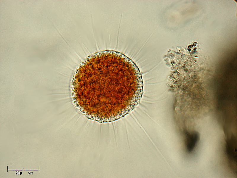
The vampyrellids (order Vampyrellida, class Vampyrellidea), colloquially known as vampire amoebae, are a group of free-living predatory amoebae classified as part of the lineage Endomyxa. They are distinguished from other groups of amoebae by their irregular cell shape with propensity to fuse and split like plasmodial organisms, and their life cycle with a digestive cyst stage that digests the gathered food. They appear worldwide in marine, brackish, freshwater and soil habitats. They are important predators of an enormous variety of microscopic organisms, from algae to fungi and animals.[3] They are also known as aconchulinid amoebae (order Aconchulinida).[4]

Cell morphology and movement

[edit]

Vampyrellids are traditionally considered filose amoebae, i.e. they generate slender pseudopodia (filopodia). They are naked, devoid of external structures such as scales, cell coats or a glycocalyx, although there may be a temporary mucilage coat in the trophozoite stage. The trophozoites vary greatly in shape, size and color between species, but can be grouped into three cell states or 'morphotypes': isodiametric, expanded, and 'filoflabellate'.[1][3]

  • Isodiametric (spherical) morphotype, common in algivorous Vampyrella and Lateromyxa, with radiating filopodia. Some species float in the water column, resembling heliozoa in shape. Others crawl on the surface by concentrating stiff filopodia at the anterior region of the cell, attaching them to the surface, retracting and moving them towards the posterior region.[3]
  • Expanded morphotype, the most common, bound to the surface, with a variety of shapes (for example, either fan-shaped or branched in Leptophrys; with large, hyaline lamellae with thread-like filopodia in Sericomyxa; highly branched or reticulate, in Platyreta and Thalassomyxa).[3]
  • Filoflabellate morphotype, only found in Placopus, with flattened elliptical, spherical or fan-shaped cells that exhibit a clear separation between the granuloplasmic cell hump and the hyaloplasmic lamellae, sometimes called 'lamellipodia'. There are numerous filopodia on the ventral side of the cell. Some of these trophozoites resemble amoebozoans such as vannellids, except for the presence of filopodia. They move by rolling over the filopodia that are anchored to the substrate.[3]
The three distinct morphotypes of vampyrellid amoebae

Life cycle

[edit]
Motile cell after feeding (J), early (K) and mature (L) digestive cysts, cyst with daughter cells after internal plasmotomy (M)
Large, bulky plasmodium of Vampyrella lateritia
Vampyrella lateritia resting cyst with four envelopes

Nutrition stages

[edit]

All known vampyrellids are heterotrophic amoebae with a free-living (non-parasitic) life cycle that lacks flagellate stages, except for Lateromyxa gallica, and is characterized by an alternation between mobile and immobile cellular stages:[3]

  • The mobile, amoeboid cells, called 'trophozoites' or 'swarmers'[a] in old literature. Their main activity is to disperse, search and gather food through phagocytosis.[3]
  • The immobile but highly metabolically active 'digestive cyst' stage, that appears after the feeding. In some species it is called a 'resting phase', but it is different from a true resting cyst (or spore) that is metabollically inactive to survive adverse conditions. To reach this stage, the trophozoite retracts its filopodia, secretes a layered cell wall, and strongly attaches itself to the substrate or floats freely. Either a central main vacuole or multiple separate vacuoles appear to digest the food. The cytoplasm color may change to a bright red, orange or yellow color, or remain colorless. When the digestive phase is finished, one or multiple trophozoites hatch from the cyst through holes in the cell wall.[3]

Reproduction

[edit]

In some species, near the end of the digestive cyst stage, asexual reproduction takes place inside the cyst through a cell division (called 'internal plasmotomy'), resulting in 2–4 daughter cells. These cells are released as young trophozoites through the holes. Other species do not divide inside the closed cyst, and instead divide during or after the hatching process ('external plasmotomy'). Lateromyxa gallica shows an unusual mode of reproduction: while feeding on the inside of algal cells, the plasmodia shed and develop into digestive cysts.[3]

There is a lack of evidence for sexual reproduction in vampyrellids, except for some meiotic stages in resting cysts revealed in Lateromyxa gallica through ultrastructural studies.[5]

Plasmodial behavior

[edit]

Many vampyrellid species have more than one nucleus and behave like plasmodia. They can fuse their cells upon contact, and split apart when moving in opposite directions. Some species readily grow plasmodia as large as a Petri dish under laboratory conditions, while others only fuse when the cell density is high and the food availability is low. It is uncertain to what extend this can happen in the natural environment. In contrast, Placopus species are rarely ever seen with more than two nuclei.[3]

Resting stages

[edit]

Under adverse environmental conditions, vampyrellids can transform into several types of resting stages:[3]

  • Hypnocysts, thin-walled and devoid of food content, formed when the trophozoite is disturbed by external stress.[3]
  • Secondary cysts, thin-walled and devoid of food content, formed as a result of starvation.[3]
  • True resting cysts, also called 'sporocysts' or 'spores' in old literature. They form in natural samples and old cultures, when there is no food or the conditions are unfavorable. They build several cyst walls and condense their cellular contents. They can survive events of desiccation or freezing, up to at least three years.[3]

Ecology

[edit]

Distribution

[edit]

Vampyrellids have a cosmopolitan distribution: they appear in all continents except Antarctica and all marine ecosystems. They inhabit a wide range of marine, brackish and freshwater habitats, and are frequently isolated from soil samples.[3] Marine ecosystems hold a surprisingly high diversity,[6] and they are found mostly in benthic habitats (e.g. tidal pools, diatom lawns, associated with red algae...). There is a significant positive correlation between the diversity of Vampyrellida and the nutrient availability in the sediment.[7] According to environmental sequencing vampyrellids colonize neotropical soil,[8] glacial cryoconite systems,[9] Brassicaceae leaves,[10] Sphagnum-inhabited peat bogs,[11] hydrothermal sediments[6] and the deep sea.[12]

Trophic diversity

[edit]
Trophozoite (left) and digestive cyst (right) of Vampyrella pendula on a Oedogonium filament

Vampyrellids display a great trophic diversity. They are predators of a long list of organisms of diverse evolutionary affinities, structures and sizes, including chlorophyte and streptophyte green algae, diatoms, chrysophytes, cryptophytes, euglenids, heterotrophic flagellates, ciliate cysts, fungal hyphae and spores, yeasts, and even micrometazoa such as nematodes and rotifer eggs. Bacterivory is rare and mostly involves filamentous cyanobacteria. Though there are generalist omnivorous predators such as Leptophrys, some vampyrellid species are specialized predators; for example, the algivorous Vampyrella and Placopus are restricted to few species of hard-walled green algae, while Arachnomyxa and Planctomyxa prefer Volvocales and euglenids.[3]

Feeding strategies

[edit]
Vampyrella lateritia extracting algal cell content with a pseudopodium (arrow)

Vampyrellids have evolved strategies to deal with relatively large bulky prey that are difficult to consume. They display at least four different feeding strategies to engulf entire prey or to devour the contents of other eukaryotic cells. These feeding strategies are not mutually exclusive, and the same species can display each with a different type of prey.[3]

  • Free capture. Similarly to amoebae of other supergroups, they catch and enclose their prey within a food vacuole by usual phagocytosis. Some can paralyse their prey before the enclosement. The size of the enveloping is widely varied, from numerous small cells at the same time to entire nematodes or colonial green algae.[3]
  • Colony invasion. They attach to the colonies of volvocalean algae, dissolve and penetrate the extracellular gelatinous mucilage matrix, and phagocytose individual cells inside the colony. Possibly for protection against predators, they transform into digestive cysts inside of the colony.[3]
  • Protoplast extraction, the most famous strategy. They specifically remove, ingest and digest the cellular contents of their prey, always by dissolving the prey's organic cell wall or simply displacing the prey's siliceous wall, and invading through pseudopodia (called 'calyculopodia') to remove the cell contents . Some species eject the prey cytoplasm by applying pressure, a process known as 'plasmoptysis', which is followed by a rapid formation of a large vacuole. This process resembles a sucking motion, and is likely the reason for their comparison to vampires. In marine species no plasmoptysis is observed, which suggests that the osmotic pressure given by salinity is important for plasmoptysis.[3]
  • Prey infiltration. Similarly to protoplast extractors, they perforate the cell wall of an algal prey, but invade the cell itself and completes the cycle within it. Some are able to move laterally from one cell to the next in filamentous prey. They divide into smaller portions that turn into digestive cysts.[3]

History of research

[edit]
1899 illustration of Vampyrella invading Spirogyra

Vampyrellids have a long history of research. They are known for the vampire-like feeding habit of several vampyrellid amoebae, which pierce the cell walls of other eukaryotic cells to feed specifically on the cell contents, a feeding mechanism known as protoplast extraction. This similarity lead to the origin of the name for their most popular genus, Vampyrella, and their colloquial name 'vampire amoebae'.[3]

One of the earliest unambiguous reports of a vampyrellid is the mid-19th century description of Amoeba lateritia (now known as Vampyrella lateritia) by the German botanist Georg Fresenius.[13] The first extensive documentation of their life history and feeding behavior was provided in 1865 by the Polish protozoologist Leon Cienkowski, who created the genus Vampyrella and classified it in a subgroup of the 'monads',[14] a polyphyletic assemblage of parasitoid protists. Posterior works and monographs described numerous aquatic vampyrellid species, with important observations of their behaviour and ecology. In 1885, the German mycologist Wilhelm Zopf demonstrated the presence of nuclei in vampyrellids and erected the first family, Vampyrellidae.[15][16][3]

In the mid-20th century the first discoveries of soil-dwelling Vampyrellida were made. The first vampyrellid laboratory culture was established, containing the soil amoeba Theratomyxa weberi that fed on nematodes. Similar soil amoebae were isolated later, and studied as possible pest control against plant-pathogenic nematodes.[17] Other studies identified a giant soil vampyrellid as the organism responsible for perforations found in fungal spores.[18][3]

In the early 1980s the feeding process and life cycle of the algivorous freshwater Vampyrella lateritia was filmed in unsurpassed detail.[19][20] At the same time, the genus of large, plasmodial amoebae Thalassomyxa, was discovered in marine waters from remote parts of the world.[21]

Before genetic analyses, the taxonomic placement of vampyrellids was difficult: they were regarded as relatives of myxomycete slime moulds,[16] heliozoa,[22] proteomyxids,[23] filose rhizopods[24] and even monera.[25] In 2009 the mystery was solved through phylogenies of 18S ribosomal RNA genes, which placed vampyrellids as part of Rhizaria.[26] A revised taxonomy in 2012 reconstituted the order Vampyrellida.[1] In 2013, a huge unexpected diversity of marine vampyrellids was detected.[6][3]

Evolution and systematics

[edit]

External relationships

[edit]

Vampyrellida represents one of the major groups of free-living amoebae, phylogenetically separate from other groups of amoebae such as Amoebozoa, Heterolobosea and Nucleariidae. Instead, Vampyrellida is an isolated clade within the Rhizaria supergroup.[26] They are the closest relatives of the Phytomyxea, parasites of plants and algae that, unlike vampyrellids, disperse through flagellated stages during their life cycle and spend most of their active life within host cells.[3] Current classifications place both Vampyrellida and Phytomyxea, along with other small groups of Rhizaria, within the phylum Endomyxa.[4] Several phylogenetic analyses have recovered a sister group relationship between Vampyrellida and Phytomyxea and have named their clade Proteomyxia[2] or Phytorhiza.[27]

Internal classification

[edit]
Vampyrellid phylogeny
Phylogeny of Vampyrellida published in 2023, inferred from SSU rRNA gene sequences.[28] The lineages B1, B2 and B4 are clades that contain only environmental DNA sequences, with no described species.[3]

There are currently 48 credible vampyrellid species distributed in 10 genera, scattered across five well-established clades found through genetic data, four of which are families. Despite the advances, most of the vampyrellid diversity is still unknown or undescribed.[3]

The following taxa have been associated with Vampyrellida, but their placement is uncertain or might not belong to the group.[3]

Notes

[edit]

References

[edit]
[edit]
Revisions and contributorsEdit on WikipediaRead on Wikipedia
from Grokipedia
Vampyrellida is an order of free-living, predatory naked amoebae belonging to the infraphylum Endomyxa within the eukaryotic supergroup . These filose amoebae are distinguished by their thin, filamentous and a biphasic life cycle featuring mobile trophozoites for locomotion and feeding, alternating with immobile digestive cysts where prey is broken down. Primarily heterotrophic, they employ phagocytic feeding strategies, often penetrating the cell walls of eukaryotic prey to access protoplasts or engulfing entire organisms, targeting a range of microbes such as , fungi, and micrometazoa. Taxonomically, Vampyrellida encompasses approximately 55 described distributed across 16 genera and four families: Vampyrellidae, Leptophryidae, Placopodidae, and Sericomyxidae (as of 2025). The order is monophyletic, as confirmed by small subunit () phylogenies, and forms a to the Phytomyxea (including plasmodiophorids) within Endomyxa. Key genera include Vampyrella (algivorous like V. lateritia and V. pendula), Leptophrys (omnivorous, such as L. vorax), Theratromyxa, Platyreta, and Hyalodiscus, with evolutionary divergence evident in feeding modes: wall-perforating in Vampyrellidae versus whole-prey engulfment in Leptophryidae. Recent additions include the genera Pseudovampyrella (2023) and Strigomyxa (2024), and such as Vampyrella crystallifera (2025), further illustrating the expanding diversity. Only about 17 have been molecularly characterized as of 2025, highlighting gaps in linking morphology to amid broader undescribed diversity. Ecologically, vampyrellids are globally distributed in limnetic (freshwater), terrestrial (), and marine environments, functioning as key microbial predators that regulate populations of and fungi. Some , like those in Vampyrella, specialize in freshwater algae such as Zygnematales, potentially influencing algal blooms, while terrestrial forms like Theratromyxa weberi and Platyreta germanica prey on fungi in rhizospheres, contributing to nutrient cycling in soils. Their predatory role extends to parasitoid-like behaviors in some cases, and they serve as prey for larger organisms or hosts in complex food webs, underscoring their ecological significance despite limited study of their , , and . Recent molecular surveys suggest high cryptic diversity, with improved primers aiding detection in environmental samples.

Description

Morphology

Vampyrellida are naked filose amoebae characterized by slender, branching that lack scales or other external ornamentations, distinguishing them from scaled cercozoans within . These pseudopodia are typically rigid and supported by bundled filaments, as observed in like Vampyrella lateritia and Lateromyxa gallica, though some genera such as Thalassomyxa incorporate , vesicles, and mitochondria for structural reinforcement. The cell body exhibits granular, opaque , often divided into a central granuloplasm rich in organelles and a peripheral hyaloplasm extending into the filopodia; in pigmented like V. lateritia, the cytoplasm acquires reddish hues from compounds derived from algal prey. Vampyrellids are multinucleate, with vesicular nuclei and mitochondria featuring tubular cristae, and they notably lack centrioles and flagella, undergoing closed orthomitosis supported by intranuclear . Three primary morphotypes define the structural diversity of vampyrellid cells: isodiametric, expanded, and filoflabellate. Isodiametric forms, exemplified by V. lateritia and L. gallica, present a compact, nearly spherical body from which radiating extend, creating a heliozoan-like appearance in floating states. Expanded morphotypes, such as Leptophrys vorax and Sericomyxa perlucida, feature a flattened, fan-shaped or irregularly branched body that adheres to substrates, allowing for broad surface coverage during feeding. Filoflabellate types, represented by Placopus species, display a distinctly flattened profile with marginal lamellipodia surrounding a central granuloplasmic hump, facilitating substrate interaction. Overall, cell sizes range from 10 to 100 μm in diameter, with V. lateritia trophozoites measuring 25–70 μm, though can extend well beyond the body length. A key morphological is the formation of digestive cysts, which are immobile, walled structures enabling after prey engulfment. These cysts vary by species but typically consist of a roundish or elongate enclosed in an organic envelope of one or two layers; for instance, V. lateritia produces spherical cysts (35–100 μm) with a double-layered wall, from which undigested remnants are expelled upon . In contrast, L. vorax forms larger, variably shaped cysts (up to several hundred μm) with a single envelope, reflecting adaptations to different prey sizes. in these cysts are retracted, emphasizing the static nature of this phase.

Locomotion

Vampyrellids primarily achieve locomotion through the extension, adhesion, and retraction of , slender that enable substrate crawling and slow progression across surfaces. These , supported by filament bundles, anchor to the substrate while the cell body advances via coordinated retraction, often resulting in rolling or rowing motions characteristic of isodiametric forms. In Vampyrella lateritia, for instance, extend beneath the globular cell body to facilitate a slow rolling movement, with internal particle streaming aiding the process. within the filopodia propels granules and membranosomes bidirectionally, enhancing motility without reliance on flagella or cilia. Expanded morphotypes, such as those in Leptophrys vorax and Sericomyxa perlucida, employ broad lamellipodia—hyaline, sheet-like extensions—for gliding over substrates, allowing smoother and more directional travel compared to filopodial rolling. These lamellipodia form frontal or lateral fringes that spread and contract, supporting creeping locomotion in fan-shaped or branched cells. Filoflabellate forms, exemplified by Placopus species, utilize marginal ruffling along pseudopodial lamellae to generate rapid directional movement through a distinctive rolling mechanism, where the cell body rotates over anchored . Environmental factors, including substrate texture and food availability, influence locomotion by affecting filopodial attachment and morphotype transitions; rougher surfaces enhance for stable crawling, while nutrient scarcity prompts shifts to floating or expanded forms with altered patterns. All vampyrellid locomotion depends exclusively on pseudopodial dynamics, as no flagellar or ciliary structures are present.

Life cycle

Trophic stages

The trophic stages of Vampyrellida constitute the core feeding cycle in their life history, characterized by an alternation between a mobile phase and an immobile digestive phase. In the mobile stage, vampyrellids exist as free-living, amoeboid cells that actively search for and capture prey using or other . These typically exhibit compact, spherical to irregular shapes, ranging from 20–70 µm in diameter, with a central granular body often colored orange or greenish due to ingested material. For instance, in Vampyrella lateritia, the advances via radiating , allowing it to stalk or float toward suitable prey such as filamentous . The engulfment process begins with the trophozoite attaching to the prey and employing to ingest , either partially or wholly, depending on the and prey type. Many vampyrellids, particularly in the genus Vampyrella, specialize in protoplast extraction, where perforate the prey's —such as that of algal cells—and the trophozoite injects or sucks the into a while leaving the wall intact. In contrast, more omnivorous forms like Leptophrys vorax engulf entire prey items, including or cells, using expanded to surround and incorporate them. This targeted ingestion enables vampyrellids to feed on a range of eukaryotes, from to fungi, with the process often completing in minutes. Following engulfment, the transitions to the immobile digestive stage by retracting its and secreting a protective wall, forming a sessile for . These , typically 25–100 µm in size, feature one to three envelopes and exhibit color changes as progresses; for example, in V. lateritia, the content shifts from greenish to orange-red over the course of enzymatic breakdown. occurs within the , where high metabolic demands drive the breakdown of prey nutrients via lysosomal enzymes, lasting from hours to several days—such as 1–2 days in V. lateritia. The wall, often stalked or attached to the prey remnant, isolates the process, allowing absorption of digested materials into the vampyrellid's . Upon completion of digestion, a new emerges from the by dissolving a circular in the wall, exiting while often expelling undigested remnants through . In V. pendula, for instance, the emerging leaves behind brown conglomerates of waste material attached to the original prey filament. This exit marks the return to mobility, restarting the feeding cycle. Energy allocation during these stages is uneven, with the rapid phase prioritizing locomotion and capture, while the prolonged phase imposes high metabolic costs for enzymatic and assimilation, reflecting the obligatory nature of encystment in vampyrellid trophic ecology.

Reproduction

Vampyrellida primarily reproduce asexually through plasmotomy, in which a multinucleate divides its to produce multiple uninucleate daughter cells. This process is particularly evident in infiltrating species such as Lateromyxa gallica, where the becomes multinucleate while feeding on algal cell contents and then undergoes division into several uninucleate individuals. In genera like Sericomyxa and Vampyrella, is linked to the digestive stage, where internal plasmotomy occurs after feeding, yielding up to eight daughter amoebae that excyst as new . The form serves as the main reproductive unit, with divisions typically triggered by availability following successful feeding. Simpler vampyrellids, such as species in the Vampyrella, exhibit resembling binary fission during the free-living phase or within cysts, producing two daughter cells from a . Nutrient-rich conditions post-ingestion promote these divisions, ensuring in favorable environments. remains poorly documented across Vampyrellida, with limited evidence suggesting it occurs in certain species. In Lateromyxa gallica, syngamy involving fusion has been inferred from observations of meiotic divisions in resting cysts, indicating a potential sexual phase that restores after polyploid stages. Nuclear behavior during reproduction involves acentriolar , lacking centrioles or microtubule-organizing center plaques, as observed in all mitotic stages of Lateromyxa gallica. This process features synchronous karyokinesis with an intranuclear spindle and intact until , often leading to polyploid nuclei in digestive and reproductive cysts before reduction via .

Plasmodial phase

The plasmodial phase represents a distinctive syncytial stage in the life cycle of certain vampyrellid amoebae, characterized by the fusion of multiple trophozoites into a multinucleate plasmodium that facilitates enhanced nutrient acquisition and growth. In genera such as Theratromyxa and Leptophrys, this phase initiates through the coalescence of individual trophozoites, often observed in high-density cultures or under conditions of limited food availability, allowing the organism to form a larger, collective structure for more efficient predation on prey like or fungi. During this phase, the undergoes extensive growth primarily through repeated nuclear divisions (karyokinesis) without accompanying (), resulting in a highly multinucleate . These structures can attain considerable sizes, reaching up to several millimeters in —for instance, plasmodia of Leptophrys vorax have been documented exceeding 1 mm, while in soil-dwelling species like Theratromyxa weberi, they may expand to cover significant substrate areas. Internally, the exhibits dynamic organization, with streaming cytoplasm that facilitates the distribution of nuclei, organelles, and ingested nutrients throughout the structure, often supported by anastomosing that aid in locomotion and feeding. Upon achieving sufficient growth and resource accumulation, the transitions by fragmenting into numerous individual trophozoites, enabling the dispersal and resumption of the standard trophic cycle. This phase is not universal across Vampyrellida but is predominantly restricted to specific clades, such as the Leptophryidae family, where it enhances survival in nutrient-scarce environments.

Resting stages

Resting stages in Vampyrellida represent a dormant phase that enables survival under unfavorable environmental conditions, distinct from active trophic or digestive forms. These stages typically form as thick-walled cysts in response to stressors such as , low temperatures, or prolonged , allowing the amoebae to enter a state of reduced metabolic activity. Formation is triggered when trophozoites or plasmodia detect adverse cues, leading to encystment where the cell retracts and secretes multiple protective envelopes. This process is particularly vital in fluctuating habitats like and freshwater, where Vampyrellida species endure periodic drying or cooling. The structure of these resting cysts features dehydrated, condensed cytoplasmic contents enclosed by several robust walls, often 1-2 μm thick, which confer resistance to environmental extremes. is greatly diminished, with the cysts acting as resilient spores capable of viability for extended periods, up to at least three years in species like Vampyrella lateritia. For instance, in soil-dwelling Vampyrellida such as those related to Arachnomyxa, s facilitate survival during drying events, maintaining integrity amid stress common in terrestrial ecosystems. The walls, composed of layered organic materials similar to those in digestive cysts, provide mechanical and osmotic protection without active metabolic investment. Excystation occurs when conditions improve, such as rehydration or warming, prompting the cyst to rupture and release a motile that resumes feeding and locomotion. This transition can happen rapidly, within hours, upon exposure to and suitable temperatures. These resting cysts also play a key role in dispersal, as their hardy nature allows via wind or water currents, enabling Vampyrellida to colonize new habitats beyond the range of active stages.

Ecology

Habitat and distribution

Vampyrellida exhibit a , occurring across six continents including , , , , and North and , as well as in marine ecosystems worldwide. They inhabit diverse environments such as marine benthic sediments, freshwater bodies like and lakes, brackish waters, and terrestrial s including agricultural, , and areas. While less common in running waters, they are frequently isolated from soil samples and have been recorded in specific locales such as Lac Pavin and Lac Chauvet in , Loch Leven in , Lake Oshimma-ohnumma in , Lake Sidney Lanier in the , and Rodrigo de in . Microhabitat preferences center on organic-rich environments that support their predatory lifestyle, including sediments, algal mats, detritus layers, and associations with or lawns. In freshwater settings, they often occur among plants and submerged vegetation in nutrient-poor, acidic . Marine populations favor benthic zones, such as tidal pools and coastal areas, with records extending to deep-sea and hydrothermal sediments. The order encompasses approximately 50 credibly described across 14 genera, with particularly high diversity reported in temperate marine benthic habitats, where molecular surveys have revealed unexpectedly rich assemblages. Zonation patterns include prevalence in intertidal zones and profundal-like deep benthic areas. Recent post-2020 surveys have documented new populations in Arctic cyanobacterial mats and soils, such as in , highlighting their adaptability to extreme cold environments through forms like resting cysts.

Trophic diversity

Vampyrellida exhibit remarkable trophic diversity, preying on a broad spectrum of microbial organisms, including eukaryotes and some prokaryotes such as , across aquatic, terrestrial, and marine ecosystems. They primarily target algae, including chlorophyte and streptophyte such as Zygnema spp. and Oedogonium spp., diatoms like Stephanodiscus rotula and Chaetoceros minimus, and such as Dolichospermum planctonicum. Additionally, vampyrellids consume fungi, including hyphae, spores, and yeasts like Saccharomyces cerevisiae, as well as such as euglenids, heterotrophic flagellates, and . Their diet extends to small metazoa, encompassing nematodes and rotifer eggs, showcasing a range from specialized algivory to generalist predation. In microbial food webs, vampyrellids function predominantly as primary consumers, preying on primary producers like microalgae and decomposers such as fungi, though some species occupy higher trophic levels by consuming protozoa. Some species display omnivory, incorporating diverse prey like algae, fungi, and protozoa, which enhances their adaptability across ecosystems. They play key roles as regulators of microalgal blooms, for instance, by contributing to the decline of diatom populations in freshwater lakes and modifying plankton dynamics in marine environments. For example, the recently described Vampyrella crystallifera (2025) engulfs entire cells of the green alga Closterium in Sphagnum habitats, representing a rapid whole-cell predation mode. As fungal predators, they aid in decomposition processes, potentially supporting nutrient cycling and biological control of soil pathogens. Their global distribution further amplifies these trophic interactions in varied habitats. Vampyrellids exert significant community effects by reducing prey populations, which influences microbial diversity and stability. For example, predation by like Asterocaelum algophilum on diatoms leads to species replacement and bloom crashes, altering composition. In and aquatic systems, their suppresses algal overgrowth and fungal proliferation, promoting balanced microbial communities. Through these dynamics, vampyrellids facilitate carbon transfer from algal and fungal sources to higher trophic levels, integrating into broader energy flows, though specific isotopic tracing remains underexplored in dedicated studies.

Feeding strategies

Vampyrellids exhibit a range of specialized feeding strategies that enable them to exploit diverse prey, primarily through heterotrophic phagocytosis of live organisms such as algae, fungi, and protozoa. The most characteristic method is protoplast extraction, where amoebae penetrate the prey's cell wall using pseudopodia to extract and ingest the internal contents, leaving behind the emptied cell wall. This process is exemplified by Vampyrella lateritia, which targets filamentous green algae like Oedogonium, piercing the wall and sucking out the protoplast via a feeding tube-like structure. Penetration involves localized dissolution of the cell wall, facilitated by extracellular lytic enzymes that weaken structural components such as cellulose. In addition to protoplast extraction, vampyrellids employ whole-cell engulfment, or free capture, to phagocytose smaller or softer prey items that lack robust walls. Species such as Leptophrys vorax extend filose pseudopodia to enclose and internalize entire cells, including ciliates and small algae, forming a food vacuole for digestion. This strategy is particularly effective against motile protozoans, where the amoeba adheres to or immobilizes the prey before complete enclosure. For colonial prey, vampyrellids use coordinated colony invasion, as seen in Arachnomyxa cryptophaga, which attaches to gelatinous matrices of algal colonies like Eudorina (Volvocales), dissolves the mucilage with enzymes, and penetrates to phagocytose individual cells within the structure. Enzymatic digestion plays a crucial role across these strategies, with extracellular carbohydrate-active enzymes (e.g., glycoside hydrolases like GH5_5 endocellulase) secreted at the attack site to perforate walls and facilitate entry. Experimental inhibition of these enzymes in related protoplast feeders reduces feeding success to approximately 5%, underscoring their essential function in prey access. Feeding efficiency varies by prey type but can be high on soft-bodied algae; for instance, Kinopus chlorellivorus achieves grazing rates of up to 131 Chlorella cells per individual per day under laboratory conditions, demonstrating rapid predation on unicellular green algae. These tactics highlight vampyrellids' adaptability, linking their predatory behaviors to broader trophic roles in microbial ecosystems dominated by algal prey.

Taxonomy and evolution

Phylogenetic relationships

Vampyrellida are classified within the eukaryotic supergroup , specifically in the subclade Endomyxa, where they form a to Phytomyxea, encompassing plasmodiophorids and phagomyxids. This positioning was established through molecular phylogenetic analyses of small subunit () genes, with initial confirmation of their rhizarian affinity emerging from studies in 2009 that analyzed environmental sequences and cultured strains like Platyreta germanica. Subsequent SSU rRNA-based phylogenies have consistently supported this placement, using methods such as maximum likelihood and to resolve deep eukaryotic relationships. Within , Vampyrellida exhibit distant relations to the shell-bearing and the silica-skeletoned , which represent distinct lineages adapted to marine environments. In contrast, they share a closer phylogenetic proximity to Gromiida, another endomyxan group characterized by , reflecting shared traits in their non-flagellate, amoeboid morphology within the broader Endomyxa clade. These external relationships highlight Vampyrellida's position as free-living predators in a supergroup otherwise dominated by parasitic or sediment-dwelling forms. The evolutionary innovation of filopodial predation in Vampyrellida— involving thin, branching pseudopods for penetrating and digesting prey—likely originated from cercozoan-like ancestors in the early diversification of . This strategy distinguishes them from the more generalized feeding in related groups and underscores their adaptation for exploiting fungal and algal hosts. A recent 2023 SSU rRNA phylogeny has further refined this framework by integrating the genus Pseudovampyrella into the family Leptophryidae, supported by high bootstrap values and Bayesian posterior probabilities, thus updating the order's internal diversification without altering its broader rhizarian context.

Internal classification

The order Vampyrellida comprises approximately 52 credibly described distributed across 16 and organized into four encompassing eight major genetic subclades. A pivotal revision occurred in 2012, when Hess, Sausen, and Melkonian emended the family Vampyrellidae to include only the genus Vampyrella and established the new Leptophryidae based on gene phylogenies that resolved two robust clades within the order. Subsequent updates have expanded the , including the addition of Placopodidae and Sericomyxidae as distinct families, while further genera have been described in existing families. In 2023, the genus Pseudovampyrella was established within Leptophryidae. In 2024, the genus Strigomyxa (with species S. ruptor) was erected within Leptophryidae, representing a novel lineage characterized by multinucleate trophozoites and a unique feeding mechanism involving internal extraction from algal prey. In 2025, the species Vampyrella crystallifera was described within Vampyrellidae, notable for engulfing and dissolving entire algal cells rather than protoplast extraction. Key families include Vampyrellidae, typified by Vampyrella species (e.g., V. lateritia) with isodiametric, filose that perforate prey cell walls; and Leptophryidae, encompassing genera such as Leptophrys, Gobiella, and Theratromyxa that often display expanded, plasmodial-like morphotypes capable of engulfing entire food items. Genus delimitation relies primarily on morphotype (e.g., isodiametric vs. expanded) and wall structure, supplemented by molecular data. The former taxon Aconchulinida, once used for filose amoebae with similar feeding habits, is now synonymized with Vampyrellida following phylogenetic integration.

Undescribed diversity

Despite the recognition of approximately 52 described vampyrellid species, environmental DNA surveys indicate a substantially larger undescribed diversity, with hundreds of phylotypes detected across various habitats, suggesting over 100 potential undescribed species. For instance, marine sediment analyses have revealed novel clades, such as those within the Endomyxa incertae sedis and previously understudied lineages like Novel Clade 12, highlighting the incompleteness of current taxonomic inventories. These findings underscore the limitations of traditional morphology-based descriptions, as only about 20 of the known species have been integrated into molecular phylogenies. A primary challenge in documenting this hidden diversity stems from cryptic species complexes, often obscured by morphological convergence among vampyrellids, where similar appearances mask genetic distinctions. This convergence arises from the group's morphological plasticity, with trophozoites exhibiting variable shapes—ranging from isodiametric to filoflabellate forms—that shift under environmental conditions, complicating identification without integrative approaches combining , morphology, and . Integrative is thus essential to resolve these ambiguities and formally describe novel lineages. Recent metagenomic studies from 2022 to 2024 have further illuminated undescribed vampyrellid lineages in underrepresented environments, including deep-sea sediments and microbiomes. For example, targeted 18S rRNA sequencing in coastal marine habitats has uncovered over 200 operational taxonomic units (OTUs) attributable to Vampyrellida, the majority representing novel deep-branching clades in anoxic sediments. Similarly, and surveys have detected uncharacterized sequences affiliated with vampyrellid families, expanding known distributions into terrestrial extremes. These discoveries point to potential new ecological roles, such as predation in deep-ocean microbial loops or nutrient cycling in oligotrophic , where vampyrellids may influence carbon flux in ways not yet observed in described taxa. Barriers to formal description persist, including the scarcity of axenic cultures for experimental validation and the inherent morphological plasticity that hinders consistent characterization from environmental samples alone. Without advances in culturing techniques or high-throughput phenotyping, many of these lineages may remain as sequences in databases, limiting insights into vampyrellid evolution and function.

Research history

Early discoveries

The earliest documented observation of a vampyrellid occurred in 1856, when Georg Fresenius described Amoeba lateritia as a parasitic on , characterizing it as a spherical, reddish form adhering to algal surfaces without detailing its full life cycle. This description, published in Beiträge zur Kenntniss der Rhizopoden, marked the initial recognition of vampyrellids as algal associates, though Fresenius interpreted it primarily through a parasitic lens rather than as a free-living predator. The species was later transferred to the genus Vampyrella by Joseph Leidy in 1879, establishing Vampyrella lateritia as a foundational in the group. In 1865, Leon Cienkowski provided the first comprehensive account of vampyrellid biology, erecting the genus Vampyrella and describing such as V. spirogyrae, V. pendula, and V. vorax. Using light microscopy, Cienkowski revealed key features including the extension of slender to perforate algal cell walls for intracellular feeding, as well as the formation of digestive cysts where engulfed algal contents were processed. These observations shifted perceptions from mere to active predation, though early workers like Cienkowski still grappled with misconceptions, such as affiliating vampyrellids with fungi or acellular due to their apparent lack of visible nuclei under basic staining techniques. Wilhelm Zopf's 1885 studies further clarified nuclear presence and cyst stages, formalizing the family Vampyrellidae while emphasizing their ecological role as algal consumers. Throughout the late 19th and early 20th centuries, researchers like Édouard Penard expanded the known diversity in the 1900s, describing genera such as Leptophrys (initially noted by Hertwig and Lesser in 1874 but detailed by Penard in works like his 1904 faunal surveys) and numerous based on morphological variations in shape and pseudopodial patterns observed via improved light microscopy. Penard's contributions highlighted expanded, net-like forms in some vampyrellids, contrasting with the compact bodies of Vampyrella. However, reliance on morphology alone led to taxonomic fragmentation, with often reassigned across groups like heliozoans or myxomycetes due to superficial similarities in cyst walls or filose extensions. Henri de Saedeleer's 1934 monograph, Beitrag zur Kenntnis der Rhizopoda, synthesized these efforts into a systematic framework, proposing the order Aconchulinida for shell-less filose amoebae including vampyrellids, though ambiguities persisted without ultrastructural or genetic data. By the mid-20th century, approximately 40 had been described, underscoring the challenges of pre-molecular classification.

Modern molecular insights

Molecular analyses of () genes have revolutionized the understanding of Vampyrellida phylogeny, firmly establishing their position within the supergroup. In 2009, Bass et al. analyzed the sequence of the soil-dwelling, mycophagous vampyrellid Platyreta germanica, demonstrating its placement in a distinct of the Endomyxa, a of closely related to Phytomyxea. This study provided the first molecular evidence linking vampyrellids to cercozoans, overturning prior morphological classifications that had ambiguously allied them with lobose amoebae or heliozoans. Building on this foundation, Hess et al. in 2012 employed an integrative taxonomic approach, combining SSU rRNA sequencing from eight clonal cultures of algivorous vampyrellids with morphological data to revise internal clades. Their phylogenetic analyses, using maximum likelihood and Bayesian methods, resolved Vampyrellida into two monophyletic families: Vampyrellidae (encompassing Vampyrella species) and the newly erected Leptophryidae (including Leptophrys, Theratromyxa, and Platyreta). This work highlighted cryptic diversity within genera, such as distinct lineages among Leptophrys vorax strains, and emphasized the limitations of morphology alone for delimiting species. Post-2020 studies have further refined Vampyrellida through expanded phylogenomic sampling. A comprehensive 2022 review in Protist discussed more than 40 credibly described species based on available data from 12 molecularly characterized taxa, identifying eight major subclades and underscoring the ecological implications of their predatory roles across aquatic and terrestrial habitats. In 2023, Suthaus et al. described Pseudovampyrella gen. nov. in the Journal of Eukaryotic Microbiology, reclassifying Vampyrella closterii and introducing P. minor sp. nov. based on phylogenies that positioned the genus within Leptophryidae, revealing underestimated in -feeding lineages. Similarly, the 2024 description of Strigomyxa ruptor gen. et sp. nov. in Ecology and Evolution integrated sequences (94.46% identity to Pseudovampyrella) with ultrastructural observations, assigning it to Leptophryidae and documenting a internal extraction feeding strategy. In 2025, Suthaus and Hess described Vampyrella crystallifera sp. nov., a species that engulfs and rapidly dissolves entire algal cells, further expanding the known diversity within Vampyrellidae. Metagenomic approaches have illuminated the uncultured diversity of Vampyrellida, particularly in underrepresented environments. Environmental DNA sequencing from marine coastal sites has uncovered hundreds of phylotypes, including novel lineages (e.g., B1, B2, B4) absent from culture-based studies, expanding known diversity by over tenfold in oceanic sediments. These sequences, often retrieved via pan-eukaryote V4 region primers, highlight vampyrellids' global distribution and trophic roles in microbial food webs, though phenotypic linkages remain challenging. Looking ahead, single-cell emerges as a promising avenue to resolve cryptic species complexes and link genotypes to ecophysiological traits in Vampyrellida. By enabling whole-genome amplification from individual cells, this method could address cultivation biases and elucidate evolutionary innovations in feeding and cyst formation, as advocated in recent syntheses.
Add your contribution
Related Hubs
User Avatar
No comments yet.