Hubbry Logo
Lateral geniculate nucleusLateral geniculate nucleusMain
Open search
Lateral geniculate nucleus
Community hub
Lateral geniculate nucleus
logo
8 pages, 0 posts
0 subscribers
Be the first to start a discussion here.
Lateral geniculate nucleus
from Wikipedia
Lateral geniculate nucleus
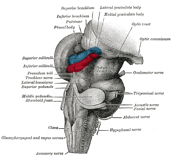
Hind- and mid-brains; postero-lateral view. (Lateral geniculate body visible near top.)
Details
Part ofThalamus
SystemVisual
ArteryAnterior choroidal and Posterior cerebral
VeinTerminal vein
Identifiers
Latincorpus geniculatum laterale
AcronymLGN
NeuroNames352
NeuroLex IDbirnlex_1662
TA98A14.1.08.302
TA25666
FMA62209
Anatomical terms of neuroanatomy

In neuroanatomy, the lateral geniculate nucleus (LGN; also called the lateral geniculate body or lateral geniculate complex) is a structure in the thalamus and a key component of the mammalian visual pathway. It is a small, ovoid, ventral projection of the thalamus where the thalamus connects with the optic nerve. There are two LGNs, one on the left and another on the right side of the thalamus. In humans, both LGNs have six layers of neurons (grey matter) alternating with optic fibers (white matter).

The LGN receives information directly from the ascending retinal ganglion cells via the optic tract and from the reticular activating system. Neurons of the LGN send their axons through the optic radiation, a direct pathway to the primary visual cortex. In addition, the LGN receives many strong feedback connections from the primary visual cortex.[1] In humans as well as other mammals, the two strongest pathways linking the eye to the brain are those projecting to the dorsal part of the LGN in the thalamus, and to the superior colliculus.[2]

Structure

[edit]
Nuclei of the Thalamus

Both the left and right hemispheres of the brain have a lateral geniculate nucleus, named after its resemblance to a bent knee (genu is Latin for "knee"). In humans as well as in many other primates, the LGN has layers of magnocellular cells and parvocellular cells that are interleaved with layers of koniocellular cells.

In humans the LGN is normally described as having six distinctive layers. The inner two layers, (1 and 2) are magnocellular layers, while the outer four layers, (3, 4, 5 and 6), are parvocellular layers. An additional set of neurons, known as the koniocellular layers, are found ventral to each of the magnocellular and parvocellular layers.[3]: 227ff [4] This layering is variable between primate species, and extra leafleting is variable within species.

The average volume of each LGN in an adult human is about 118mm. (This is the same volume as a 4.9mm-sided cube.) A study of 24 hemispheres from 15 normal individuals with average age 59 years at autopsy found variation from about 91 to 157mm.[5] The same study found that in each LGN, the magnocellular layers comprised about 28mm in total, and the parvocellular layers comprised about 90mm in total.

M, P, K cells

[edit]
Relative locations of the M-, P-, and K-layers (macaque monkey)
Type Size* RGC Source Type of Information Location Response Number
M: Magnocellular cells Large Parasol cells perception of movement, depth, and small differences in brightness Layers 1 and 2 rapid and transient ?
P: Parvocellular cells (or "parvicellular") Small Midget cells perception of color (red-green)[6] and form (fine details) Layers 3, 4, 5 and 6 slow and sustained ?
K: Koniocellular cells (or "interlaminar") Very small cell bodies Bistratified cells color (yellow-blue)[6] Between each of the M and P layers

*Size describes the cell body and dendritic tree, though also can describe the receptive field

The magnocellular, parvocellular, and koniocellular layers of the LGN correspond with the similarly named types of retinal ganglion cells. Retinal P ganglion cells send axons to a parvocellular layer, M ganglion cells send axons to a magnocellular layer, and K ganglion cells send axons to a koniocellular layer.[7]: 269 

Koniocellular cells are functionally and neurochemically distinct from M and P cells and provide a third channel to the visual cortex. They project their axons between the layers of the lateral geniculate nucleus where M and P cells project. Their role in visual perception is presently unclear; however, the koniocellular system has been linked with the integration of somatosensory system-proprioceptive information with visual perception[citation needed], and it may also be involved in color perception.[8]

The parvo- and magnocellular fibers were previously thought to dominate the Ungerleider–Mishkin ventral stream and dorsal stream, respectively. However, new evidence has accumulated showing that the two streams appear to feed on a more even mixture of different types of nerve fibers.[9]

The other major retino–cortical visual pathway is the tectopulvinar pathway, routing primarily through the superior colliculus and thalamic pulvinar nucleus onto posterior parietal cortex and visual area MT.

Non-primates

[edit]

Other mammals such as cats generally only have L and M cones, hence no red-green differentiation. They also have three cell types denoted as X (magno), Y (parvo), and W (konio). The W type is beyond most doubt homologous to the primate K type. There are some subtle differences between the M and X types as well as the Y and P types to make the correspondence unclear.[6] There is additional evidence of a yellow-blue opponent process in the LGN of mice.[10]

Ipsilateral and contralateral layers

[edit]

Both the LGN in the right hemisphere and the LGN in the left hemisphere receive input from each eye. However, each LGN only receives information from one half of the visual field. Retinal ganglion cells (RGCs) from the inner halves of each retina (the nasal sides) decussate (cross to the other side of the brain) through the optic chiasma (khiasma means "cross-shaped"). RGCs from the outer half of each retina (the temporal sides) remain on the same side of the brain. Therefore, the right LGN receives visual information from the left visual field, and the left LGN receives visual information from the right visual field. Within one LGN, the visual information is divided among the various layers as follows:[11]

  • the eye on the same side (the ipsilateral eye) sends information to layers 2, 3 and 5
  • the eye on the opposite side (the contralateral eye) sends information to layers 1, 4 and 6.

This description applies to the LGN of many primates, but not all. The sequence of layers receiving information from the ipsilateral and contralateral (opposite side of the head) eyes is different in the tarsier.[12] Some neuroscientists suggested that "this apparent difference distinguishes tarsiers from all other primates, reinforcing the view that they arose in an early, independent line of primate evolution".[13]

Input

[edit]

The LGN receives input from the retina and many other brain structures, especially visual cortex.

The principal neurons in the LGN receive strong inputs from the retina. However, the retina only accounts for a small percentage of LGN input. As much as 95% of input in the LGN comes from the visual cortex, superior colliculus, pretectum, thalamic reticular nuclei, and local LGN interneurons. Regions in the brainstem that are not involved in visual perception also project to the LGN, such as the mesencephalic reticular formation, dorsal raphe nucleus, periaqueuctal grey matter, and the locus coeruleus.[14] The LGN also receives some inputs from the optic tectum (known as the superior colliculus in mammals).[15] These non-retinal inputs can be excitatory, inhibitory, or modulatory.[14]

Output

[edit]

Information leaving the LGN travels out on the optic radiations, which form part of the retrolenticular portion of the internal capsule.

The axons that leave the LGN go to V1 visual cortex. Both the magnocellular layers 1–2 and the parvocellular layers 3–6 send their axons to layer 4 in V1. Within layer 4 of V1, layer 4cβ receives parvocellular input, and layer 4cα receives magnocellular input. However, the koniocellular layers, intercalated between LGN layers 1–6 send their axons primarily to the cytochrome-oxidase rich blobs of layers 2 and 3 in V1.[16] Axons from layer 6 of visual cortex send information back to the LGN.

Studies involving blindsight have suggested that projections from the LGN travel not only to the primary visual cortex but also to higher cortical areas V2 and V3. Patients with blindsight are phenomenally blind in certain areas of the visual field corresponding to a contralateral lesion in the primary visual cortex; however, these patients are able to perform certain motor tasks accurately in their blind field, such as grasping. This suggests that neurons travel from the LGN to both the primary visual cortex and higher cortex regions.[17]

Function in visual perception

[edit]

The output of the LGN serves several functions.

Temporal and spatial processing

[edit]

Computations are achieved to determine the position of every major element in object space relative to the principal plane. Through subsequent motion of the eyes, a larger stereoscopic mapping of the visual field is achieved.[18]

It has been shown that while the retina accomplishes spatial decorrelation through center-surround inhibition, the LGN accomplishes temporal decorrelation.[19] This spatial–temporal decorrelation makes for much more efficient coding. However, there is almost certainly much more going on.

Like other areas of the thalamus, particularly other relay nuclei, the LGN likely helps the visual system focus its attention on the most important information. That is, if you hear a sound slightly to your left, the auditory system likely "tells" the visual system, through the LGN via its surrounding peri-reticular nucleus, to direct visual attention to that part of space.[20] The LGN is also a station that refines certain receptive fields.[21]

Axiomatically determined functional models of LGN cells have been determined by Lindeberg [22][23] in terms of Laplacian of Gaussian kernels over the spatial domain in combination with temporal derivatives of either non-causal or time-causal scale-space kernels over the temporal domain. It has been shown that this theory both leads to predictions about receptive fields with good qualitative agreement with the biological receptive field measurements performed by DeAngelis et al.[24][25] and guarantees good theoretical properties of the mathematical receptive field model, including covariance and invariance properties under natural image transformations.[26][27] Specifically according to this theory, non-lagged LGN cells correspond to first-order temporal derivatives, whereas lagged LGN cells correspond to second-order temporal derivatives.

Color processing

[edit]

The LGN is also integral in the early steps of color processing, where opponent channels are created that compare signals between the different photoreceptor cell types: a type of inter-channel decorrelation. The output of P-cells comprises red-green opponent signals. The output of M-cells does not include much color opponency, rather a sum of the red-green signal that evokes luminance. The output of K-cells comprises mostly blue-yellow opponent signals.[6]

Rodents

[edit]

In rodents, the lateral geniculate nucleus contains the dorsal lateral geniculate nucleus (dLGN), the ventral lateral geniculate nucleus (vLGN), and the region in between called the intergeniculate leaflet (IGL). These are distinct subcortical nuclei with differences in function.

dLGN

[edit]

The dorsolateral geniculate nucleus is the main division of the lateral geniculate body. In the mouse, the area of the dLGN is about 0.48mm. The majority of input to the dLGN comes from the retina. It is laminated and shows retinotopic organization.[28]

vLGN

[edit]

The ventrolateral geniculate nucleus has been found to be relatively large in several species such as lizards, rodents, cows, cats, and primates.[29] An initial cytoarchitectural scheme, which has been confirmed in several studies, suggests that the vLGN is divided into two parts. The external and internal divisions are separated by a group of fine fibers and a zone of thinly dispersed neurons. Additionally, several studies have suggested further subdivisions of the vLGN in other species.[30] For example, studies indicate that the cytoarchitecture of the vLGN in the cat differs from rodents. Although five subdivisions of the vLGN in the cat have been identified by some,[31] the scheme that divides the vLGN into three regions (medial, intermediate, and lateral) has been more widely accepted.

IGL

[edit]

The intergeniculate leaflet is a relatively small area found dorsal to the vLGN. Earlier studies had referred to the IGL as the internal dorsal division of the vLGN. Several studies have described homologous regions in several species, including humans.[32]

The vLGN and IGL appear to be closely related based on similarities in neurochemicals, inputs and outputs, and physiological properties.

The vLGN and IGL have been reported to share many neurochemicals that are found concentrated in the cells, including neuropeptide Y, GABA, encephalin, and nitric oxide synthase. The neurochemicals serotonin, acetylcholine, histamine, dopamine, and noradrenaline have been found in the fibers of these nuclei.

Both the vLGN and IGL receive input from the retina, locus coreuleus, and raphe. Other connections that have been found to be reciprocal include the superior colliculus, pretectum, and hypothalamus, as well as other thalamic nuclei.

Physiological and behavioral studies have shown spectral-sensitive and motion-sensitive responses that vary with species. The vLGN and IGL seem to play an important role in mediating phases of the circadian rhythms that are not involved with light, as well as phase shifts that are light-dependent.[30]

Additional images

[edit]

See also

[edit]

References

[edit]
[edit]
Revisions and contributorsEdit on WikipediaRead on Wikipedia
from Grokipedia
The lateral geniculate nucleus (LGN) is a sensory nucleus within the that serves as the primary gateway for visual information from the to the , processing and segregating signals related to color, motion, form, and . Located in the posteroventral portion of the , adjacent to the pulvinar nucleus and posterior to the inferior choroidal point, the LGN is anatomically divided into six layered sheets in : two ventral magnocellular layers, four dorsal parvocellular layers, and interspersed koniocellular layers. The magnocellular layers contain large cells that handle motion-sensitive (Y-type) inputs with broad receptive fields and low color acuity, while the parvocellular layers feature smaller cells processing color-sensitive (X-type) signals for fine detail and form detection; koniocellular cells, positioned ventrally, contribute additional color-opponent and blue-yellow processing. axons from the optic tract monosynaptically onto LGN cells in a retinotopic manner, with contralateral eye inputs terminating in layers 1, 4, and 6, and ipsilateral inputs in layers 2, 3, and 5, enabling binocular integration and maintaining spatial organization. Beyond relaying signals, the LGN modulates visual processing through extensive feedback and lateral connections, receiving inputs from the primary (V1) layer 6, the for attentional gating, brainstem nuclei like the and pedunculopontine tegmental area for arousal-related noradrenergic and modulation, and the for serotonergic influences. Its outputs project via the optic radiations directly to V1, preserving parallel magnocellular, parvocellular, and koniocellular pathways that underpin distinct visual functions. The intergeniculate leaflet (IGL), an accessory structure, links the LGN to circadian regulation by connecting to the and influencing release in the . Embryologically, LGN layers form through retinogeniculate beginning around 13 weeks , with full laminar organization emerging by 22–25 weeks, though plasticity persists into early development. Physiologic variants include reduced gray matter volume in conditions like strabismic and atrophy in , while lesions often result in homonymous hemianopia or due to disrupted representation. Blood supply derives from the anterior and lateral choroidal branches of the posterior cerebral and internal carotid arteries, making it vulnerable to ischemic events.

Anatomy

Location and gross structure

The lateral geniculate nucleus (LGN) is situated in the posteroventral region of the , immediately adjacent to the pulvinar nucleus and forming part of the visual thalamus alongside it. It lies posterior to the inferior choroidal point and is enclosed within the internal medullary lamina, a thin sheet of that separates nuclei. This positioning places the LGN in the lateral posterior , where it serves as a key relay in the visual pathway. In , the human LGN presents as a small, ovoid measuring approximately 4-6 mm in and 10-12 mm in anteroposterior length, with a typical volume ranging from 70 to 100 mm³. It is subdivided into a dorsal LGN (dLGN), which functions as the primary relay for visual information, and a smaller ventral LGN (vLGN), considered accessory and less prominent in including humans. An intergeniculate leaflet separates these dorsal and ventral regions. The overall shape resembles an asymmetric cone on the dorsolateral aspect of the . The LGN displays a retinotopic topographic organization, mirroring the contralateral visual field such that the lower is represented dorsally and the upper ventrally, with the horizontal meridian oriented medially and the vertical meridian laterally. The blood supply to the LGN is dual, primarily from the (a branch of the ) and the thalamogeniculate arteries (lateral posterior choroidal branches of the ). This vascular arrangement ensures robust perfusion to support its role in visual processing, though disruptions can lead to specific visual field defects.

Layered organization

The lateral geniculate nucleus (LGN) in is characterized by a distinct laminar that segregates visual information based on origin and processing pathways. This structure consists of six primary layers, numbered from ventral to dorsal, with layers 1 and 2 comprising the magnocellular division and layers 3 through 6 forming the parvocellular division. Koniocellular layers are interposed between these main layers, particularly between layers 1 and 2, 2 and 3, 5 and 6, as well as in the anterior and medial regions known as the koniocellular "hilum." Eye-specific segregation is a hallmark of this organization, ensuring that inputs from each retina remain separate to maintain binocular correspondence. Layers 1, 4, and 6 receive afferents exclusively from the contralateral eye, while layers 2, 3, and 5 receive inputs from the ipsilateral eye. This pattern arises from the orderly projection of retinal ganglion cells, with temporal retina projecting ipsilaterally and nasal retina contralaterally, preventing overlap within individual layers. Within each layer, the neuronal arrangement follows a precise retinotopic that mirrors the topographic organization of the , allowing spatial relationships from the to be preserved through the LGN relay. This mapping is consistent across layers, with the central represented in the posterior pole of the LGN and the peripheral field toward the anterior end. Interlaminar zones between the primary layers contain scattered koniocellular neurons and dense , which support local interconnections and modulate signals across layers without disrupting the main laminar segregation. These zones are particularly prominent in New World primates like , where koniocellular elements are more diffuse, but the overall layered framework remains conserved across species.

Ipsilateral and contralateral layers

The lateral geniculate nucleus (LGN) organizes visual inputs from the two eyes into strictly segregated layers, with the contralateral eye projecting primarily to layers 1, 4, and 6, and the ipsilateral eye to layers 2, 3, and 5. This eye-specific laminar arrangement preserves monocular signals through the thalamic relay, preventing premature mixing of inputs from the left and right visual fields until they reach the primary visual cortex (V1). By maintaining this separation, the LGN enables precise binocular integration at the cortical level, where aligned inputs from corresponding retinal locations can be combined to support depth perception and stereopsis. The development of this segregation begins with overlapping (RGC) axons from both eyes innervating the immature LGN during embryonic stages, around 7-11 weeks gestation in . Activity-dependent mechanisms, driven by spontaneous waves of correlated firing in the mediated by transmission, refine these projections postnatally, typically completing eye-specific layering by 15-20 weeks gestation. This process involves competitive interactions where correlated activity strengthens intra-eye connections and weakens inter-eye overlaps, ensuring robust territorial segregation without requiring visual experience. This layered organization directly influences cortical processing, contributing to the emergence of columns in V1, where alternating bands preferentially respond to inputs from one eye or the other, shaping the retinotopic map of the . Disruptions in this segregation can manifest in binocular rivalry, a perceptual where incompatible stimuli alternate in awareness; studies show modulated responses in eye-specific LGN layers during rivalry, highlighting the nucleus's role in early competitive dynamics before full cortical arbitration. Exceptions to normal segregation occur in conditions like , where deficiency at the causes aberrant , directing more temporal retinal fibers contralaterally and reducing ipsilateral projections to LGN layers 2, 3, and 5, often resulting in fused or displaced laminae. In , misalignment of the eyes during critical developmental periods can lead to anomalous ipsilateral projections and weakened segregation, impairing of inputs across layers and contributing to through reduced binocular competition.

Cell Types

Magnocellular cells

Magnocellular (M) cells occupy the two ventralmost layers (layers 1 and 2) of the lateral geniculate nucleus (LGN), where they form distinct magnocellular laminae characterized by large, darkly stained neurons. These cells account for approximately 10% of the total LGN neuronal population, a proportion that reflects their specialized role within the visual system. M cells possess notably large somata, with diameters typically ranging from 30 to 50 μm, and extensive dendritic arbors that facilitate broad integration of incoming signals across the layers. These neurons receive direct afferent inputs from parasol retinal ganglion cells, which convey signals along the Y-like pathway from the to the LGN. This connectivity endows M cells with heightened sensitivity to low-contrast, high-luminance modulations in the visual scene, supported by rapid conduction velocities that enable swift transmission of luminance-based information. Unlike other pathways, the Y-like route emphasizes achromatic processing and fast signaling, allowing M cells to prioritize dynamic changes over fine details. Receptive fields of M cells demonstrate nonlinear spatial summation, organized in a center-surround antagonistic configuration that enhances detection of contrasts. The central regions of these fields are comparatively broad, extending up to 0.5° in near the fovea, which permits summation over larger retinal areas and contributes to reduced acuity but increased robustness to noise. This organization underpins their key functional contributions to motion detection and binocular , where they excel at signaling rapid environmental changes. M cells further exhibit a phasic, profile, with optimal activation at temporal frequencies of 10-20 Hz, aligning with their emphasis on high-speed visual events.

Parvocellular cells

Parvocellular cells, or P cells, are the predominant neuronal population in the lateral geniculate nucleus (LGN), comprising approximately 80% of its neurons and occupying layers 3 through 6. These layers are organized to receive segregated inputs from the ipsilateral and contralateral eyes, with layers 3 and 5 processing ipsilateral visual fields and layers 4 and 6 handling contralateral inputs. P cells are characterized by their small soma sizes, typically ranging from 10 to 20 μm in diameter, which contrasts with the larger somata of other LGN cell types and supports their role in detailed visual processing. They exhibit linear spatial summation within their receptive fields, allowing for precise integration of retinal signals without nonlinear distortion. P cells receive primary afferent inputs from midget cells, which convey signals predominantly from individual photoreceptors in the . Their receptive fields are notably narrow, often around 0.1° in near the fovea, enabling sustained responses to fine spatial details and high spatial frequencies up to 40 cycles per degree. This high-resolution tuning is particularly dense in the foveal representation of the LGN, where P cell populations are concentrated to facilitate and the detection of subtle patterns. Many P cells display opponent color responses, such as red-green opponency, arising from differential inputs from medium- and long-wavelength , which enhances chromatic discrimination. Compared to other LGN pathways, P cells have slower al conduction velocities, typically around 5-10 m/s, reflecting their smaller diameters and emphasis on sustained rather than transient signaling. This property aligns with their specialization for processing stable, detailed visual information over rapid changes.

Koniocellular cells

Koniocellular (K) cells in the lateral geniculate nucleus (LGN) of are located primarily in the interlaminar zones and thin koniocellular layers, such as those situated between magnocellular layer 1 and parvocellular layer 2, as well as between parvocellular layers 5 and 6. These cells constitute approximately 10% of the total relay neurons in the LGN, numbering around 100,000 per hemisphere in the rhesus monkey. Morphologically, K cells feature small somata and sparse, elongated dendrites that extend across broader regions compared to those of parvocellular or magnocellular cells, facilitating diffuse integration of inputs. K cells receive direct afferent inputs from specific retinal ganglion cell types, including small bistratified cells that convey blue-ON/yellow-OFF signals and melanopsin-expressing intrinsically photosensitive s (ipRGCs). These inputs contribute to large receptive fields, typically measuring 0.5–1° in diameter at eccentricities of 2–30° from the fovea, which exhibit poor relative to parvocellular pathways due to their broader center-surround organization. The primary functional specialization of K cells involves sensitivity to blue-yellow color opponency, particularly short-wavelength (S-cone) light increments and decrements, enabling detection of chromatic contrasts in low-luminance environments. Physiologically, K cells display slow, sustained response profiles with low spontaneous firing rates and prolonged latency to visual stimuli, contrasting with the transient dynamics of other LGN cell types. This sustained activity supports processing of stable chromatic and signals rather than rapid changes. Additionally, projections from melanopsin-containing ipRGCs to K layers link these cells to non-image-forming visual functions, such as circadian photoentrainment and modulation, by conveying absolute light intensity information independent of pattern vision.

Neural Connections

Afferent inputs

The primary afferent inputs to the lateral geniculate nucleus (LGN) originate from ganglion cells (RGCs), which convey visual information via the optic tract and account for approximately 5-10% of the total synapses onto LGN relay cells, despite being the dominant driver of visual signaling. These inputs are topographically organized, preserving such that axons from the nasal project to the contralateral LGN and those from the temporal to the ipsilateral LGN, resulting in a representation of the contralateral visual hemifield across the nucleus's layers. The inputs are segregated by RGC type into parallel pathways: large alpha-like (parasol) RGCs target the magnocellular layers, smaller beta-like (midget) RGCs innervate the parvocellular layers, and small bistratified RGCs project to the koniocellular layers (or interlaminar zones). Additional afferents include feedback projections from the primary (V1), primarily from pyramidal neurons in layer 6, which form about 20-30% of synapses on LGN relay cells and contribute to gain control, sharpening, and attentional modulation of visual responses. These corticogeniculate inputs are also retinotopically aligned with retinal projections, exciting the same LGN neurons that drive cortical activity. Cholinergic inputs from the , particularly from nuclei such as the pedunculopontine tegmental nucleus and parabigeminal nucleus, provide another major source of afferents, comprising roughly 20-30% of synapses and modulating LGN excitability to influence states and attention-dependent visual processing. Noradrenergic inputs from the modulate -related visual processing, while serotonergic projections from the influence attention and response properties. Inhibitory projections from the (TRN), which envelops the , target LGN relay cells and , contributing to the remaining synaptic inputs (along with local LGN ) to regulate burst firing, surround inhibition, and overall thalamic rhythmicity.

Efferent outputs

The principal efferent projections from the lateral geniculate nucleus (LGN) target the primary (V1) via the optic radiations, forming the main pathway for visual information to the . These axons originate from cells in the LGN's magnocellular, parvocellular, and koniocellular layers and maintain parallel processing streams to specific V1 sublayers. Magnocellular cells in the ventral LGN layers project primarily to layer 4Cα of V1, supporting processing of low-spatial-frequency, motion-related signals. Parvocellular cells from the dorsal layers target layer 4Cβ, contributing to high-acuity form and color discrimination. Koniocellular cells, located interlaminarly, send outputs to the oxidase-rich blobs in layers 2 and 3 of V1, with some extensions to layer 1. These projections exhibit precise topographic organization, with ipsilateral and contralateral LGN layers mapping retinotopically to corresponding columns in V1, preserving eye-specific segregation from the . Minor efferent projections from the LGN include connections to the pulvinar nucleus for integration with higher-order visual pathways. Additionally, geniculocortical axons emit collaterals to the , where they onto inhibitory neurons to modulate surround inhibition and attentional gating of LGN activity.

Functions

Relay and integration

The lateral geniculate nucleus (LGN) serves as an obligatory station in the geniculostriate pathway, transmitting visual information from the to the primary (V1) while performing initial filtering and sharpening of signals. Retinal ganglion cell axons form precise retinotopic maps in the LGN layers, where nearly all visual input to the cortex passes through this structure, ensuring organized representation of the . This function is essential, as lesions or disruptions in the LGN severely impair conscious vision, underscoring its gatekeeping in the visual pathway. Beyond passive transmission, the LGN integrates signals through intrinsic circuits that enhance properties, particularly via intralaminar processing involving excitatory inputs from retinal afferents and inhibitory . These , which constitute about 25% of LGN neurons, provide and feedback inhibition that sharpens center-surround s, amplifying contrast at edges while suppressing uniform illumination. This push-pull mechanism—excitatory drive to relay cells paired with inhibitory surround—extends the of responses and refines spatial selectivity beyond inputs. Intrinsic circuits further improve contrast sensitivity, with LGN relay cells exhibiting nonlinear gain control that enhances detection of low-contrast stimuli compared to retinal responses. Gating mechanisms in the LGN, influenced by cortical feedback from V1, modulate relay cell gain to prioritize relevant signals, particularly during attentional tasks. Layer 6 corticogeniculate projections, comprising up to 30% of synaptic inputs to the LGN, adjust excitability through both direct excitation and indirect inhibition via the , suppressing irrelevant or distracting inputs. This top-down modulation stabilizes response precision and enhances signal-to-noise ratios, allowing the LGN to dynamically filter visual information based on behavioral context without altering basic retinotopic organization.

Spatial and temporal processing

The lateral geniculate nucleus (LGN) contributes to spatial processing in vision primarily through the parallel magnocellular () and parvocellular () pathways, which segregate information based on . cells are preferentially tuned to low spatial frequencies, typically in the range of 1–5 cycles per degree, facilitating the detection of coarse visual structures such as overall and global motion cues. In contrast, cells respond robustly to high spatial frequencies, often 10–40 cycles per degree, enabling detailed analysis of edges, textures, and fine patterns essential for . These tuning differences arise from the larger centers of cells compared to the smaller, more precise fields of cells, as characterized in recordings. Temporal processing in the LGN is also segregated along these pathways, with M cells displaying profiles characterized by phasic bursts at stimulus onset and offset, which support motion detection across temporal frequencies of 10–50 Hz. P cells, conversely, exhibit sustained responses that maintain activity throughout stimulus presentation, ideal for processing stable or slowly changing scenes at lower temporal frequencies around 10 Hz. This enhances the visual system's ability to handle dynamic environments, where rapid transients signal change and sustained signals provide continuity. Receptive field organization further refines spatial and temporal analysis: P cells show largely linear spatial summation within their center-surround structure, while M cells exhibit nonlinear summation, contributing to their broader tuning. Surround suppression is prominent in both but stronger in M cells, reducing response overlap and enhancing contrast sensitivity by inhibiting activity from adjacent regions.

Color processing

The lateral geniculate nucleus (LGN) plays a key role in color vision by maintaining opponent-process mechanisms that segregate chromatic information along red-green and blue-yellow axes. Parvocellular (P) cells, located in layers 3–6, primarily mediate red-green color opponency through L-M cone differences, where long-wavelength-sensitive (L) cone excitation is opposed by medium-wavelength-sensitive (M) cone excitation (+L − M). In contrast, koniocellular (K) cells, situated in the interlaminar zones between the main layers, handle blue-yellow opponency via short-wavelength-sensitive (S) cone signals opposed by a combination of L and M cone inputs (+S − (L + M)). These segregated pathways ensure that color signals are relayed without initial mixing, preserving the retinal origins of chromatic selectivity. Opponent color signals in the LGN are largely preserved from the , with minimal transformation occurring until they reach the primary (V1). cells transmit these single-opponent responses directly to LGN neurons, which exhibit nearly identical chromatic properties, such as center-surround tuned to specific color contrasts. This fidelity allows the LGN to act as a station that organizes but does not substantially alter the basic opponent structure, enabling V1 to perform more complex integrations like double opponency. The lack of full in the LGN underscores its role in maintaining distinct chromatic channels for efficient transmission. Magnocellular (M) cells, found in layers 1 and 2, contribute achromatically to color processing by detecting luminance-based boundaries within chromatic scenes, facilitating the segmentation of objects even when color cues are ambiguous. These cells respond primarily to overall light intensity changes, providing essential context for interpreting color edges against luminance gradients. Wavelength-specific tuning in the LGN aligns with sensitivities, with red-green opponent cells peaking around 570 nm (reflecting L-M differences) and blue-yellow cells at 440 nm (S-cone driven). This tuning supports the perceptual stability of opponent colors across varying illuminations.

Comparative Anatomy

In primates

In primates, the lateral geniculate nucleus (LGN) displays a highly laminated structure with distinct segregation of magnocellular (M), parvocellular (P), and koniocellular (K) pathways, tailored to support trichromatic vision and elevated foveal acuity. Catarrhine primates, encompassing monkeys, apes, and humans, feature six well-defined layers in the dorsal LGN, where layers 1 and 2 house large M cells for processing low-spatial-frequency and achromatic information, layers 3 through 6 contain smaller P cells optimized for high-spatial-frequency and color signals, and K cells reside in the interlaminar regions for additional chromatic and achromatic contributions. This layered organization, with eye-specific inputs alternating between layers, enhances parallel processing efficiency for complex visual scenes characteristic of diurnal lifestyles. Relative to other mammals, the LGN, particularly in humans, occupies a larger proportion of volume, estimated at approximately 0.03% based on total LGN volumes of around 407 mm³ and average volumes of 1.3 million mm³, reflecting correlated expansion with the to accommodate advanced visual demands. This scaling pattern shows positive in , where LGN volume increases disproportionately with brain size compared to non-primate mammals like , underscoring evolutionary pressures for enhanced visual relay capacity. Evolutionary adaptations in primates emphasize parvocellular dominance, with the P pathway comprising over 80% of LGN neurons in species like macaques, enabling refined color discrimination integral to trichromatic vision arising from opsin gene duplications. This shift from the more balanced M-P ratios in primates highlights adaptations for fruit detection and social signaling in arboreal environments. Slight sexual dimorphisms in LGN volume have been observed, with some studies reporting marginally larger volumes in males compared to females, though not always statistically significant and with recent data showing volumes around 175 mm³ per hemisphere and no significant sex differences. These differences, if present, align with broader sex-based variations in and color perception modulated by and testosterone.

In non-primates

In non-primate mammals, the lateral geniculate nucleus (LGN) displays a generally simpler with reduced lamination compared to , often consisting of fewer distinct layers and exhibiting greater topographic scatter in projections. For instance, in carnivores such as cats and dogs, the dorsal LGN is divided into six principal layers—A (contralateral input), A1 (ipsilateral input), and the C complex (C, C1, C2, C3)—but these layers lack the sharp segregation of functional cell classes observed in LGN. This reduced lamination facilitates a less compartmentalized of visual information, with afferents showing broader overlap across layers. Cell types in the non-primate LGN are analogous to the magnocellular () and parvocellular () pathways of primates but are less strictly segregated, resulting in a more integrated processing of spatial and temporal features. In cats, for example, X-cells (sustained, parvo-like responses with small receptive fields) and Y-cells (transient, magno-like responses with larger fields) are distributed across the A and C layers without the dedicated layering seen in primates, while W-cells handle low-acuity inputs. Koniocellular-like small cells, which in primates form distinct intercalated zones for color and high-frequency information, appear more diffusely scattered throughout non-primate layers rather than being zonally organized, contributing to a blended rather than parallel stream architecture. The non-primate LGN plays a larger role in reflexive behaviors, with stronger projections to the that support rapid orienting responses to visual stimuli. In cats, W-cells in the C layers provide a dedicated pathway to the , enabling quick subcortical processing for eye and head movements, which is more prominent than the cortex-dominant projections in . This emphasis on tectal outputs underscores the LGN's function in survival-oriented visuomotor integration rather than higher perceptual analysis. Evolutionarily, the LGN in non-primate mammals represents a simpler station, particularly in basal groups like monotremes and early placental mammals, where it lacks the specialized subdivisions for color processing that emerged in alongside trichromatic vision. In monotremes such as the , the LGN is a relatively undifferentiated structure focused on basic retinocortical without layered complexity, reflecting an ancestral mammalian design adapted for nocturnal or low-light environments. Across placental non-, this trend persists with alpha and beta cells (analogous to Y and X types) handling motion and form without dedicated color-opponent mechanisms, highlighting a conservative evolutionary trajectory prioritizing efficiency over perceptual specialization.

In rodents

In rodents, the lateral geniculate nucleus (LGN) is subdivided into the dorsal lateral geniculate nucleus (dLGN), ventral lateral geniculate nucleus (vLGN), and intergeniculate leaflet (IGL), each with distinct structural and functional specializations adapted to nocturnal lifestyles and behavioral needs such as prey detection and circadian regulation. Unlike in primates, the rodent LGN lacks prominent laminar organization overall, reflecting broader non-primate traits with more integrated retinotopic mapping across subdivisions. The dLGN serves as the primary visual nucleus, characterized by partial into a ventromedial core and dorsolateral shell, though without the distinct cellular layers seen in other mammals. It maintains precise retinotopic organization, where axons from neighboring positions project to adjacent dLGN regions, preserving spatial visual maps that are refined postnatally through Eph/ signaling and activity-dependent mechanisms. The core receives inputs from diverse (e.g., ON/OFF sustained and transient types), while the shell targets direction-selective cells; neurons—classified as X-like (ventral, ), Y-like (central, binocular), and W-like (shell)—project primarily to layer 4 of the primary (V1), with shell neurons additionally innervating layers 1 and 2. These cells are smaller than their magnocellular/parvocellular counterparts in primates, comprising about 90% of dLGN neurons alongside 10% . The vLGN functions as an accessory structure for non-image-forming visual processing, particularly wide-field motion detection and behaviors like prey capture in nocturnal environments. It is organized into an external lamina with large cells and an internal lamina with minimal input, dominated by neurons and few ones, and lacks projections to the cortex. inputs, including from intrinsically photosensitive ganglion cells (ipRGCs), arrive alongside superior colliculus afferents, enabling integration of visual and visuomotor signals; outputs target the , , and indirectly the (SCN) to support reflexive responses and orientation. In mice, the vLGN volume is comparable to that of the dLGN, underscoring its significant role in visual . The IGL, a thin leaflet embedded between the dLGN and vLGN, specializes in circadian entrainment and modulation of nonphotic cues, integrating retinal signals from ipRGCs via the . It contains mostly neurons, including (NPY)-expressing cells that convey photic and behavioral information to the SCN through the geniculohypothalamic tract, influencing phase shifts and rhythm stability without topographic mapping. Lesions of the IGL disrupt entrainment to skeleton photoperiods (brief light pulses at transitions) but spare standard light-dark cycles, highlighting its role in fine-tuning circadian responses to environmental light patterns. Outputs also extend to the , linking visual processing to visuomotor and vestibular functions. Overall, the LGN is compact, with the dLGN volume approximately 0.25 mm³ in adults, adapted for efficient processing in small brains suited to low-light and rapid threat detection.

References

Add your contribution
Related Hubs
User Avatar
No comments yet.