Hubbry Logo
TrismusTrismusMain
Open search
Trismus
Community hub
Trismus
logo
8 pages, 0 posts
0 subscribers
Be the first to start a discussion here.
Trismus
Trismus
from Wikipedia
Trismus
Other namesLockjaw
Trismus caused due to muscle rigidity. Diagram of jaw muscles are shown here.

Trismus is a condition of restricted opening of the mouth.[1][2] The term was initially used in the setting of tetanus.[2] Trismus may be caused by spasm of the muscles of mastication or a variety of other causes.[3] Temporary trismus occurs much more frequently than permanent trismus.[4] It is known to interfere with eating, speaking, and maintaining proper oral hygiene. This interference, specifically with an inability to swallow properly, results in an increased risk of aspiration. In some instances, trismus presents with altered facial appearance. The condition may be distressing and painful. Examination and treatments requiring access to the oral cavity can be limited, or in some cases impossible, due to the nature of the condition itself.

Definition

[edit]

Trismus is defined as painful restriction in opening the mouth due to a muscle spasm,[5] however it can also refer to limited mouth opening of any cause.[6] Another definition of trismus is simply a limitation of movement.[4] Historically and commonly, the term lockjaw was sometimes used as a synonym for both trismus[2] and tetanus.[7]

Normal mouth-opening ranges from 35 to 45 mm.[6] Males usually have slightly greater mouth opening than females. (40–60 mm, average of 50 mm). The normal lateral movement is 8–12 mm,[8] and normal protrusive movement is approximately 10 mm.[medical citation needed] Some have distinguished mild trismus as 20–30 mm interincisal opening, moderate as 10–20 mm and severe as less than 10 mm.[9]

Trismus is derived from the Greek word trigmos/trismos meaning "a scream; a grinding, rasping or gnashing".[10]

Differential diagnosis

[edit]

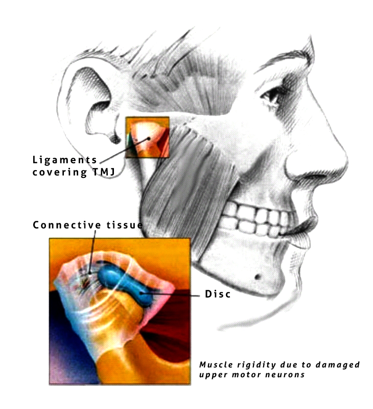
Traditionally causes of trismus are divided into intra-articular (factors within the temporomandibular joint [TMJ]) and extra-articular (factors outside the joint, see table).[4]

Commonly listed causes of trismus

Intra-articular:

  • Internal derangement of TMJ / meniscus displacement.[3][6]
  • Fractured mandibular condyle or intracapsular fracture.[3]
  • TMJ dislocation.[4]
  • Traumatic synovitis.[3]
  • Septic arthritis.[6]
  • Osteoarthritis.[3]
  • Inflammatory arthritis (e.g. rheumatoid or psoriatic).[3]
  • Ankylosis.[3]
  • Osteophyte formation.[6]

Extra-articular:

  • Trauma not involving the mandibular condyle (e.g. a fracture of another part of the mandible, fractures of the middle third of the facial skeleton, fractures of the zygoma or zygomatic arch).[3]
  • Post surgical edema, e.g. removal of impacted lower wisdom teeth,[3] or other dentoalveolar surgery.[6]
  • Recent prolonged dental treatment (e.g. root canal therapy).[3]
  • Following administration of inferior alveolar nerve block with local anesthetic (medial pterygoid).[6]
  • Hematoma of medial pterygoid.[4]
  • Acute infections of the oral tissues, especially involving the buccal space or muscles of mastication.[3]
    • Odontogenic infection.[3][6]
    • Peritonsillar abscess.[6]
    • Acute parotitis, e.g. mumps.[4]
    • Pericoronitis.[4]
    • Submasseteric abscess.[4]
  • Tetanus.[4][6]
  • Tetany.[4]
  • Local malignancy.[6]
  • Myofascial pain / temporomandibular joint dysfunction.[6]
  • Radiation fibrosis.[6]
  • Fibrosis from burns.[4]
  • Submucous fibrosis.[6]
  • Systemic sclerosis.[11]
  • Myositis ossificans.[11]
  • Coronoid hyperplasia.[6]
  • Malignant hyperpyrexia.[6]
  • Epidermolysis bullosa.[8]
  • Drug associated dyskinesia.[4]
  • Psychotic disturbances, hysteria.[4]

Joint problems

[edit]

Ankylosis

[edit]
  • True bony ankylosis: can result from trauma to chin, infections and from prolonged immobilization following condylar fracture
    • Treatment – several surgical procedures are used to treat bony ankylosis, e.g.: Gap arthroplasty using interpositional materials between the cut segments.
  • Fibrous ankylosis: usually results due to trauma and infection
    • Treatment – trismus appliances in conjunction with physical therapy.

Arthritis synovitis

[edit]

Meniscus pathology

[edit]

Extra-articular causes

[edit]

Infection

[edit]
  • Odontogenic- Pulpal
    • Periodontal
    • Pericoronal
  • Non-odontogenic- Peritonsillar abscess
    • Tetanus
    • Meningitis
    • Brain abscess
    • Parotid abscess
  • The hallmark of a masticatory space infection is trismus or infection in anterior compartment of lateral pharyngeal space results in trismus. If these infections are unchecked, can spread to various facial spaces of the head and neck and lead to serious complications such as cervical cellulitis or mediastinitis.
    • Treatment: Elimination of etiologic agent along with antibiotic coverage
  • Trismus or lock jaw due to masseter muscle spasm, can be a primary presenting symptom in tetanus, Caused by Clostridium tetani, where tetanospasmin (toxin) is responsible for muscle spasms.
    • Prevention: primary immunization (DPT)

Dental treatment

[edit]
  • Dental trismus is defined by difficulty in opening the jaw. It is a temporary condition that usually lasts no more than two weeks. Dental trismus is caused by an injury to the masticatory muscles, such as opening the jaw for an extended period of time or having a needle pass through a muscle. Typical dental anesthesia for the lower jaw involves inserting a needle into or through a muscle. In these cases it is usually the medial pterygoid or the buccinator muscles.
  • Oral surgery procedures, as in the extraction of lower molar teeth, may cause trismus as a result either of inflammation to the muscles of mastication or direct trauma to the TMJ.
  • Barbing of needles at the time of injection followed by tissue damage on withdrawal of the barbed needle causes post-injection persistent paresthesia, trismus and paresis.
    • Treatment: in acute phase:
      • Heat therapy
      • Analgesics
      • A soft diet
      • Muscle relaxants (if necessary)
      • Note: When acute phase is over the patient should be advised to initiate physiotherapy for opening and closing mouth.

Trauma

[edit]

Fractures, particularly those of the mandible and fractures of zygomatic arch and zygomatic arch complex, accidental incorporation of foreign bodies due to external traumatic injury. Treatment: fracture reduction, removal of foreign bodies with antibiotic coverage[citation needed]

TMJ disorders

[edit]
  • Extra-capsular disorders – Myofascial pain dysfunction syndrome
  • Intra-capsular problems – Disc displacement, arthritis, fibrosis, etc.
  • Acute closed locked conditions – displaced meniscus

Tumors and oral care

[edit]

Rarely, trismus is a symptom of nasopharyngeal or infratemporal tumors/ fibrosis of temporalis tendon, when patient has limited mouth opening, always premalignant conditions like oral submucous fibrosis (OSMF) should also be considered in differential diagnosis.

Drug therapy

[edit]

Succinyl choline, phenothiazines and tricyclic antidepressants causes trismus as a secondary effect. Trismus can be seen as an extra-pyramidal side-effect of metoclopromide, phenothiazines and other medications.

Radiotherapy and chemotherapy

[edit]
  • Complications of radiotherapy:
    • Osteoradionecrosis may result in pain, trismus, suppuration and occasionally a foul smelling wound.
    • When muscles of mastication are within the field of radiation, it leads to fibrosis and result in decreased mouth opening.
  • Complications of Chemotherapy:
    • Oral mucosal cells have high growth rate and are susceptible to the toxic effects of chemotherapy, which lead to stomatitis.

Congenital and developmental causes

[edit]
  • Hypertrophy of coronoid process causes interference of coronoid against the anteromedial margin of the zygomatic arch.
    • Treatment: Coronoidectomy
  • Trismus-pseudo-camtodactyly syndrome is a rare combination of hand, foot and mouth abnormalities and trismus.

Miscellaneous disorders

[edit]
  • Functional disorders ( neuro-psychiatric), the emotional conflict are converted into a physical symptom. E.g.: trismus
  • Scleroderma: A condition marked by edema and induration of the skin involving facial region can cause trismus

Common causes

[edit]
  • Pericoronitis (inflammation of soft tissue around impacted third molar) is the most common cause of trismus.[12]
  • Inflammation of muscles of mastication.[12] It is a frequent sequel to surgical removal of mandibular third molars (lower wisdom teeth). The condition is usually resolved on its own in 10–14 days, during which time eating and oral hygiene are compromised. The application of heat (e.g. heat bag extraorally, and warm salt water intraorally) may help, reducing the severity and duration of the condition.
  • Peritonsillar abscess,[12] a complication of tonsillitis which usually presents with sore throat, dysphagia, fever, and change in voice.
  • Temporomandibular joint dysfunction (TMD).[12]
  • Trismus is often mistaken as a common temporary side effect of many stimulants of the sympathetic nervous system. Users of amphetamines as well as many other pharmacological agents commonly report bruxism as a side-effect; however, it is sometimes mis-referred to as trismus. Users' jaws do not lock, but rather the muscles become tight and the jaw clenched. It is still perfectly possible to open the mouth.[12]
  • Submucous fibrosis.
  • Fracture of the zygomatic arch.
  • Oral sex

Other causes

[edit]

Diagnostic approach

[edit]

X-ray/CT scan taken from the TMJ to see if there is any damage to the TMJ and surrounding structures.[citation needed]

Treatment

[edit]

Treatment requires treating the underlying condition with dental treatments, speech therapy for swallowing difficulty and mouth opening restrictions, physical therapy, and passive range of motion devices. Additionally, control of symptoms with pain medications (NSAIDs), muscle relaxants, and warm compresses may be used.

Splints have been used.[13]

History

[edit]

Historically, the term trismus was used to describe the early effects of tetany, also called "lockjaw".

References

[edit]
[edit]
Revisions and contributorsEdit on WikipediaRead on Wikipedia
from Grokipedia
Trismus, also known as lockjaw, is a medical condition characterized by the restricted of the , typically resulting in an inability to open the mouth more than 35 mm, which interferes with normal functions such as eating, speaking, and . This limitation arises primarily from , , or of the masticatory muscles, including the masseter, temporalis, and pterygoid muscles, and was originally described in the context of but now encompasses various etiologies. While often temporary and resolving within two weeks, trismus can become chronic, particularly following radiotherapy for head and neck cancers or in cases of untreated trauma. The condition affects individuals across all ages but is more prevalent in certain populations, such as those undergoing dental procedures like third molar extractions (a common postoperative complication) or patients receiving for head and neck malignancies (affecting 38-42% of cases). Common causes include trauma to the (e.g., fractures or penetrating injuries), infections (such as or odontogenic abscesses), temporomandibular disorders (TMD), iatrogenic factors (e.g., prolonged or ), and neoplastic processes involving the oral cavity or . Pathophysiologically, it involves protective muscle spasms in acute settings or progressive and scarring in chronic ones, leading to potential complications like , , or challenges with endotracheal during emergencies. Diagnosis is primarily clinical, based on measuring interincisal ( range: 40-60 mm) and assessing for associated pain, swelling, or trismus-related asymmetry, with imaging such as CT or MRI used to identify underlying structural issues. Treatment focuses on addressing the root cause and alleviating symptoms through conservative measures like nonsteroidal anti-inflammatory drugs (NSAIDs), muscle relaxants (e.g., 2.5-5 mg three times daily), warm compresses, and a soft diet, followed by exercises to restore mobility in the subacute phase. For persistent cases, interventions may include coronoidectomy, myotomy, or injections to reduce muscle hyperactivity, with early intervention being crucial to prevent permanent or .

Overview

Definition

Trismus is a motor disorder characterized by restricted mouth opening due to spasm or contracture of the masticatory muscles, typically limiting the interincisal distance to less than 35 mm. The pathophysiology involves sustained tetanic spasms of muscles such as the masseter, temporalis, and pterygoids, often resulting from irritation of the trigeminal nerve or central mechanisms. Historically referred to as "lockjaw" in association with tetanus, trismus represents a symptom rather than a distinct disease. Normal mouth opening measures 40 to 60 mm, with trismus graded as mild (20-30 mm), moderate (10-20 mm), or severe (<10 mm).

Signs and Symptoms

Trismus primarily manifests as a painful restriction in opening, with a maximum interincisal distance typically less than 35 mm, compared to the normal range of 40 to 60 mm. Patients commonly describe a sensation of stiffness or locking, making it difficult to achieve full without discomfort. This limitation can be unilateral or bilateral, often accompanied by muscle in the masticatory apparatus. Associated symptoms frequently include challenges with chewing, speaking, and swallowing, which arise from the mechanical hindrance and pain during mandibular movement. Individuals may also experience referred pain such as otalgia (ear pain), headaches, or tenderness in the facial region, particularly around the jaw and temporal areas. In acute presentations, localized swelling of the face or jaw may occur, exacerbating the discomfort. On , clinicians observe a markedly reduced , with tenderness elicited upon of the masseter, temporalis, or pterygoid muscles. Jaw deviation toward the affected side during attempted opening is a common sign, and in some cases, a muffled or "hot potato" voice may be noted due to impaired oral mechanics. Trismus often coexists with disorders, where additional features like intermittent locking may contribute to the overall presentation. The symptoms can lead to several complications, including an elevated risk of from inadequate oral intake and difficulties maintaining , which may predispose to secondary infections. Psychological impacts, such as anxiety or frustration stemming from communication barriers, are also reported by affected patients. Prolonged restriction may result in of the , further impairing function. Trismus varies in duration, with acute forms presenting suddenly and often resolving within two weeks if the underlying issue is addressed promptly. In contrast, chronic cases develop gradually and can persist for months or longer, leading to progressive stiffness and adaptation challenges.

Epidemiology

Trismus exhibits variable prevalence across populations, largely influenced by its multifactorial etiologies, with no standardized diagnostic criteria complicating direct comparisons. In patients with treated with radiotherapy, prevalence rates range from 5% to 42%, depending on assessment methods and radiation fields involved. Among those undergoing combined and radiotherapy for oral , post-treatment prevalence can reach 72%, declining to 62% at three months follow-up, with severe cases (mouth opening <25 mm) comprising 46% initially. In broader cohorts, overall prevalence stands at approximately 24%, with chronic persistence noted in 28% of cases at five years post-radiotherapy. Incidence is notably elevated in procedural and infectious contexts. Following transalveolar extraction of impacted molars, trismus develops more frequently when operative time is prolonged, often linked to and tissue manipulation, though most resolve by seven days postoperatively. In a series of 339 third molar extractions, trismus occurred in approximately 4% of molar procedures. Dental abscesses and fascial space infections show high involvement, with trismus present in up to 70% of cases and mean mouth opening reduced to 23.5 mm in infections. In tetanus-endemic regions, trismus manifests as the inaugural symptom in 50-75% of cases, underscoring its role in this . Demographically, trismus predominantly affects adults aged 20-50 years, with mean ages around 47 in series and advanced age serving as a predictor in ( 1.031 per year increase). A slight female predominance emerges in temporomandibular joint-related instances, mirroring broader temporomandibular disorder patterns where peaks between 25-45 years and is two- to four-fold higher in women. Incidence escalates in low-resource settings due to untreated odontogenic infections and limited coverage, though precise population-level data remain sparse. A 2024 study in reported a 46.9% among patients. Key risk factors include , with doses exceeding 50 Gy to the masticatory muscles elevating susceptibility, particularly when the medial pterygoid is encompassed. History of oral trauma, poor dental hygiene fostering infections, and further amplify risks by promoting unchecked or opportunistic pathogens. Emerging trends indicate rising trismus burden tied to increasing incidence, with chronic post-treatment rates of 10-30% highlighted in recent survivorship studies; however, non-oncologic cases are underreported, and global epidemiological gaps persist.

Causes

Temporomandibular Joint Disorders

The (TMJ) is a ginglymoarthrodial joint composed of the mandibular condyle, the of the , and an articular disc that divides it into two synovial cavities, facilitating both rotary and gliding motions essential for opening. The joint is stabilized by the temporomandibular ligament, , and , while the articular disc, capsule, and surrounding masticatory muscles (temporalis, masseter, medial and lateral pterygoids) contribute to its function. In trismus associated with TMJ disorders, in the disc, capsule, or ligaments restricts mandibular translation and rotation, leading to limited interincisal opening typically below 35 mm. For instance, capsular or disc adhesions can mechanically impede condylar movement, resulting in progressive restriction. Myofascial pain dysfunction syndrome (MPDS), a common extra-articular TMJ disorder, arises from hyperactivity or fatigue in the masticatory muscles, often triggered by psychosocial stress, bruxism, or parafunctional habits, leading to localized muscle spasm and trismus. In MPDS, sustained muscle tension causes pain on palpation and functional activities like chewing or speaking, with trismus manifesting as protective guarding that limits mouth opening to protect the inflamed area. This syndrome affects up to 10-15% of adults, predominantly women aged 20-40, and is characterized by tenderness in the masseter or temporalis without primary joint involvement. Internal derangement of the TMJ involves abnormal positioning of the articular disc relative to the condyle, such as anterior displacement with or without reduction, which mechanically obstructs joint motion and contributes to trismus through inflammation and pain-induced guarding. In cases of disc displacement without reduction (closed lock), the disc blocks condylar translation, restricting opening to 25-30 mm and often accompanied by joint clicking or locking during attempts at reduction. This derangement results from ligament laxity or disc degeneration, leading to secondary synovitis that exacerbates limited mobility. Specific TMJ conditions further drive trismus through structural or inflammatory changes. , either fibrous (from organized and adhesions) or bony (via forming a bridge between condyle and fossa), fuses the joint post-trauma or , severely restricting motion and causing painless hypomobility in chronic cases. , including (degenerative cartilage loss with subchondral bone changes) and (autoimmune with pannus formation), induces , pain, and stiffness, with reducing synovial lubrication and promoting capsular tightening that limits jaw excursion. Meniscus pathology, such as disc tearing or degeneration, disrupts load distribution in the joint, leading to irregular condylar paths, , and trismus as the body compensates with muscle . TMJ involvement occurs in 20-40% of cases, often featuring erosive , while often presents unilaterally with and functional impairment. The of trismus in TMJ disorders centers on inflammatory mediators like prostaglandins and cytokines released during or disc injury, which sensitize nociceptors and trigger reflexive muscle guarding via the , resulting in sustained contraction of masticatory muscles and reduced joint compliance. This protective spasm perpetuates a cycle of and immobility, with in the capsule or disc further anchoring the joint. In , disorganized healing post-injury amplifies this through excessive deposition, while in , chronic inflammation erodes and promotes formation, compounding mechanical restriction. Overlap with acute trauma may initiate these processes, but chronic TMJ changes predominate in sustained trismus.

Infections

Infections represent a significant cause of trismus, primarily through local odontogenic processes that inflame the masticatory muscles or systemic mechanisms that induce muscle spasms. infections often originate from dental sources and can rapidly spread to adjacent fascial spaces, restricting mobility due to , , and . , an inflammation of the soft tissue overlying a partially erupted —most commonly the mandibular third molar—frequently leads to trismus by irritating the surrounding masticatory muscles, such as the masseter and medial pterygoid. This condition arises from bacterial accumulation in the pericoronal pocket, resulting in acute suppuration and limited mouth opening often below 35 mm. Dental abscesses, typically stemming from untreated caries or , can similarly provoke trismus when purulence extends into the pterygomandibular , a common pathway for posterior spread from lower molars, causing intense and . A particularly severe example is , a bilateral of the submandibular and sublingual spaces originating from odontogenic infections in approximately 90% of cases, often involving the lower second or third molars. The infection spreads rapidly beneath the via fascial planes, leading to woody induration of the floor of the mouth, tongue elevation, and trismus due to involvement of the and masticatory apparatus. Pathophysiologically, these local infections involve polymicrobial invasion—predominantly anaerobes like and species—resulting in formation, increased intramuscular pressure, and reflexive muscle hyperactivity that locks the . Risk factors for these local infectious causes include poor , which facilitates plaque accumulation and bacterial proliferation around impacted teeth, as well as immunosuppression from conditions like diabetes mellitus or , which impair host defenses against ascending infections. Acute presentations typically feature fever, localized swelling, , and a rapid onset of trismus within hours to days, often accompanied by and elevated counts signaling systemic involvement. Systemic infections can also induce trismus through neurotoxic or inflammatory pathways. Tetanus, caused by the spore-forming bacterium Clostridium tetani, manifests as trismus—classically termed "lockjaw"—as an early sign of generalized disease, where sustained spasms affect the masseter and temporalis muscles. The pathophysiology involves tetanospasmin, a neurotoxin produced in anaerobic wounds, which travels retrogradely along motor neurons to block inhibitory neurotransmitters like glycine and GABA in the central nervous system, leading to unopposed excitatory activity and tonic contractions. Risk factors encompass vaccination gaps, particularly in endemic areas with incomplete immunization coverage, and exposure to contaminated soil or devitalized tissue via puncture wounds. Historically, pre-vaccination mortality from tetanus reached 50% to over 80% without intensive care, primarily due to respiratory failure from spasms. Acute onset includes trismus progressing to opisthotonos, fever, and autonomic instability within 3 to 21 days of inoculation. Viral parotitis, such as that caused by the (Paramyxovirus), can result in trismus through painful enlargement of the parotid glands, which compresses adjacent masticatory structures and limits jaw excursion. This inflammatory response, often bilateral, stems from in salivary ductal , leading to and secondary muscle guarding. Risk factors include lack of vaccination and close-contact exposure in unimmunized populations. Presentation involves prodromal fever and followed by parotid swelling and trismus, typically resolving within 7 to 10 days but potentially complicating oral intake.

Trauma and Injury

Trauma to the and surrounding structures is a common cause of trismus, resulting from mechanical disruption that impairs mandibular mobility. Direct trauma, such as mandibular fractures, often leads to restricted mouth opening due to , muscle , and structural instability. These fractures, which account for a significant portion of injuries, typically occur at sites like the body, , or condyle of the , with assaults representing approximately 50% of cases, followed by accidents at 29%, falls, industrial incidents, and sports-related impacts. In such , the involves initial and hemorrhage within the soft tissues and masticatory spaces, which mechanically hinder jaw excursion and initiate a pain-spasm cycle where nociceptive input from damaged tissues triggers sustained contraction of muscles like the masseter and temporalis. Contusions to the masticatory muscles or nerves, particularly the during mandibular trauma, further exacerbate trismus by causing localized inflammation and neural dysfunction, often presenting with associated lower lip . Indirect injuries, such as whiplash from rear-end collisions, can induce trismus through acceleration-deceleration forces that strain the (TMJ) and associated ligaments, leading to myofascial pain and reduced jaw opening amplitude—studies report averages of 48 mm in affected individuals compared to 54 mm in controls. Hematomas in the masticatory spaces, arising from blows or shearing forces, contribute to this by compressing neural and muscular elements, promoting a reflexive protective that limits interincisal distance below 35 mm. Clinical examination often reveals ecchymosis, over fracture sites, and sublingual hematoma as indicators of underlying damage. Delayed-onset trismus following trauma may develop from fibrosis and scar tissue formation during the healing process, where excessive collagen deposition in injured muscles or the TMJ capsule creates mechanical restriction, potentially progressing to post-traumatic ankylosis as detailed in the Temporomandibular Joint Disorders section. Edentulous patients face heightened risk, as the absence of teeth complicates stabilization and increases susceptibility to fragment displacement, necessitating alternative fixation methods like Gunning splints. Overall, these traumatic etiologies underscore the need for prompt intervention to mitigate chronic sequelae.

Neoplastic and Iatrogenic Causes

Neoplastic causes of trismus primarily arise from tumors in the head and neck region that directly invade or compress the masticatory apparatus, leading to restricted mouth opening. Oral cavity squamous cell carcinomas, the most common malignancies in this area, often infiltrate the such as the masseter, medial pterygoid, and temporalis, causing mechanical obstruction and reflex muscle spasms. Sarcomas, including rhabdomyosarcomas, can similarly invade the pterygoid muscles, resulting in induration and that limits mandibular excursion. tumors, such as oncocytic carcinomas, may compress adjacent nerves like the mandibular branch of the (V3), contributing to neuromuscular dysfunction and trismus. The involves tumor , direct myoinvasion, or perineural spread, which can manifest early as ulceration or induration affecting and exacerbating functional impairment. Iatrogenic causes of trismus are frequent complications of treatments, particularly and . Surgical interventions, such as maxillectomy or excisions in the buccal mucosa and retromolar trigone, lead to postoperative scarring and that shortens the or pterygoid muscles, progressively restricting jaw mobility. induces through microvascular damage, endothelial cell injury, and excessive collagen deposition in the masticatory muscles and (TMJ), with risk escalating at mean doses exceeding 40 Gy to structures like the ipsilateral masseter or medial pterygoid. This radiation-induced (RIF) often develops months to years post-treatment, causing chronic . contributes indirectly and rarely directly, primarily through severe that promotes inflammation and secondary muscle guarding, though isolated chemotherapeutic effects on trismus are uncommon. In head and neck oncology patients, trismus from these causes affects up to 38-42% of survivors following combined treatments, with neoplastic infiltration particularly prevalent in advanced nasopharyngeal or tonsillar cancers. Early recognition is crucial, as untreated trismus impairs oral intake, dental care, and , often requiring multidisciplinary intervention.

Other Causes

Trismus can arise from neurological conditions that disrupt the pathways or motor control of the masticatory muscles. , particularly those affecting the or opercular region, may lead to trismus through infarction of areas involved in jaw movement coordination, as seen in cases of opercular syndrome where sustained muscle spasms occur. , an inflammatory demyelinating disorder, can cause trigeminal neuropathy resulting in trismus due to plaques in the that impair nerve conduction. Oromandibular , a neurological , presents with involuntary jaw clenching and trismus from abnormal contractions of the masseter and temporalis muscles. Congenital and developmental anomalies also contribute to trismus by causing structural limitations in jaw mobility from birth. , a condition characterized by multiple joint contractures due to reduced , often involves the , leading to restricted mouth opening with a prevalence of approximately 1 in 3,000 live births. , the second most common congenital facial anomaly after cleft lip and palate, results in of the and surrounding tissues on one side of the face, thereby limiting jaw excursion and causing trismus. Certain medications can induce trismus through direct effects on muscle tone or secondary complications like osteonecrosis. Phenothiazines, a class of antipsychotic drugs, have been associated with trismus as a side effect from dopamine receptor blockade leading to dystonic reactions in the masticatory muscles. Succinylcholine, a depolarizing neuromuscular blocker used in anesthesia, may trigger masseter muscle rigidity and trismus shortly after administration due to transient fasciculations and increased muscle tone. Bisphosphonates, particularly intravenous forms prescribed for cancer-related bone disease, can cause medication-related osteonecrosis of the jaw, which manifests as fibrosis and restricted mouth opening in affected patients. Among miscellaneous etiologies, systemic sclerosis () leads to trismus via autoimmune-mediated fibrosis of the perioral tissues and capsule, reducing elasticity and mouth opening. Seizures in , especially those involving tonic-clonic activity, can temporarily induce trismus from sustained contraction of jaw muscles during the ictal phase. Psychogenic factors, such as in psychogenic nonepileptic seizures, may mimic trismus through voluntary or stress-induced muscle tension without underlying organic pathology. The of trismus in these contexts often involves dysregulation, where lesions or demyelination alter inhibitory signals to the trigeminal motor nucleus, resulting in unopposed muscle spasms. In autoimmune conditions like systemic sclerosis, excessive deposition driven by immune dysregulation causes fibrotic scarring in soft tissues and joints, progressively limiting mandibular movement.

Diagnosis

Clinical Evaluation

The clinical evaluation of trismus begins with a detailed history taking to determine the onset, which may be acute, often associated with trauma or recent dental procedures, or chronic, commonly linked to radiation therapy or neoplastic processes. Patients should be queried about pain characteristics, such as its location, intensity, and relation to jaw movement, along with associated symptoms including fever suggestive of infection, recent trauma, or unintentional weight loss indicating possible malignancy. Risk factors to elicit include recent dental interventions like third molar extractions and history of head and neck radiation, which are common precipitants. Physical examination focuses on quantifying the degree of limitation through measurement of maximal interincisal opening (MIO), typically performed using a or placed between the upper and lower central incisors, with normal values ranging from 40 to 60 mm and trismus diagnosed when MIO is less than 35 mm. of the (TMJ) and masticatory muscles, such as the masseter and pterygoids, is essential to identify tenderness, swelling, or induration, while assessment for jaw deviation on opening or protrusion helps detect asymmetry potentially due to muscular or joint pathology. Functional tests evaluate the impact on daily activities, including observation of chewing efficiency and speech clarity, where muffled or voice may indicate underlying peritonsillar involvement. Screening for tetanus involves inquiring about vaccination status and wound history, as trismus is an early hallmark, alongside a to rule out deficits such as rigidity or spasms beyond the jaw. Patients are advised to seek professional medical or dental help promptly if the jaw is fully locked (e.g., maximal interincisal opening less than 20-35 mm), symptoms are sudden or severe, pain is intense or persists for more than a few days, there are signs of infection such as fever or swelling, recent wound exposure suggesting possible tetanus, associated muscle spasms elsewhere, or if episodes occur repeatedly. Such evaluation may lead to treatments including custom mouthguards, physical therapy, muscle relaxants, Botox injections, or addressing underlying causes, as detailed in the Management section. Red flags warranting urgent further investigation include unilateral facial swelling, which may signal an or tumor, and systemic signs such as persistent fever or , potentially indicative of infectious or malignant etiologies.

Imaging Studies

Plain , such as orthopantomography (OPG), serves as an initial modality to assess for mandibular fractures, , or gross bony abnormalities contributing to trismus. However, it has significant limitations in evaluating soft tissues, effusions, or disc positions, making it insufficient for comprehensive diagnosis of trismus etiologies like muscle or tumors. Advanced imaging techniques provide more detailed visualization of trismus causes. (MRI) is the gold standard for evaluating (TMJ) disorders, using protocols with T1-weighted, T2-weighted, and proton density sequences in closed- and open-mouth positions to assess disc displacement, , and . It excels in detecting muscle (e.g., of the lateral pterygoid), neoplastic masses, or through signal intensity changes and contrast enhancement. Computed tomography (CT), particularly cone-beam CT (CBCT), is preferred for bony structures, employing low-dose protocols (e.g., 120 kV, 100 mA, 1 mm slices) to identify fractures, erosions, condylar changes, or abscess collections without excessive exposure. Recent guidelines emphasize low-dose CT variants for suspected infections or trauma to minimize radiation while maintaining diagnostic accuracy. Ultrasound offers a non-invasive, bedside option for acute trismus, using high-frequency linear transducers (8-12 MHz) to detect muscle edema, joint effusions, or peritonsillar abscesses in infections like quinsy, which can limit mouth opening. It is particularly useful for real-time guidance in procedures but is limited for deep structures or chronic bony changes. Specific imaging findings aid in differentiation. In radiation-induced trismus, MRI reveals masticator muscle and (e.g., T2 hypointensity in 54% of severe cases post-nasopharyngeal radiotherapy), often with mandibular ramus signal changes or condyle sclerosis. Neoplastic causes appear as effects with on MRI or CT, such as irregular bony margins or extension. Diagnostic guidelines recommend MRI as first-line for suspected TMJ disorders due to its resolution, with CT reserved for bony or infectious suspicions; is indicated when clinical findings are equivocal or atypical (e.g., interincisal opening <15 mm with ). Clinical correlation remains essential for interpreting these studies.

Laboratory Investigations

Laboratory investigations play a supportive role in diagnosing trismus by identifying underlying systemic etiologies such as infections, autoimmune conditions, or neoplasms, particularly when clinical findings suggest involvement beyond the (TMJ). These tests are not routinely indicated for isolated mechanical or myofascial trismus but are essential in cases with fever, swelling, or systemic symptoms to guide and exclude broader . A (CBC) is often the initial test, revealing indicative of bacterial infections contributing to trismus, such as odontogenic abscesses or deep neck space infections. Elevated counts, typically neutrophilic, correlate with the severity of inflammatory processes in these scenarios. Inflammatory markers like (CRP) and (ESR) are commonly elevated in infectious or autoimmune-mediated TMJ , providing non-specific of ongoing ; for instance, CRP levels rise acutely in response to bacterial of masticatory spaces. These markers help monitor disease activity but lack specificity for trismus etiology alone. Microbiological evaluation is crucial when is suspected, involving cultures from aspirated abscesses or to identify pathogens like species, which guide antibiotic selection in odontogenic or infections causing trismus. In suspected , a rare infectious cause, antitoxin antibody levels may be assessed via to evaluate immunity, though remains primarily clinical without a confirmatory test. Autoimmune panels, including antinuclear antibody (ANA) and rheumatoid factor (RF), are indicated if or other connective tissue diseases are suspected as contributors to TMJ involvement and trismus; positivity in these tests supports screening for autoimmune phenomena in temporomandibular disorders. For neoplastic causes, tumor markers such as (SCC) antigen may be measured in serum, particularly in head and neck malignancies where trismus arises from tumor infiltration, aiding in monitoring disease progression or recurrence. Additional tests like (EMG) can evaluate muscle spasm patterns in neurological cases of trismus, such as those following lesions, by detecting abnormal activity in masticatory muscles like the masseter. Overall, laboratory investigations have limited utility in uncomplicated TMJ-related trismus but are invaluable for ruling out and informing management in complex presentations.

Management

Conservative Treatments

Conservative treatments for trismus emphasize non-invasive techniques aimed at restoring mobility and reducing muscle without relying on medications or . These approaches are particularly suitable for mild to moderate cases, such as those arising from (TMJ) disorders or post-radiation fibrosis in patients. forms the cornerstone, incorporating targeted exercises and manual interventions to improve mouth opening and alleviate discomfort. Jaw stretching exercises, such as passive and active mobilization of the , are commonly prescribed to gradually increase the interincisal distance. These may involve holding the open to the point of mild discomfort for several seconds, repeated multiple times daily, often combined with of the masticatory muscles like the masseter and temporalis to release tension. Thermotherapy, including the application of warm compresses for 10-15 minutes several times a day, helps reduce muscle by promoting blood flow and relaxation in the affected tissues. techniques, performed by trained physical therapists, further enhance outcomes by addressing joint restrictions and adhesions. Specialized devices support these exercises by providing controlled resistance for progressive stretching. The TheraBite Jaw Motion Rehabilitation System, a handheld device that applies gradual force to open the jaw, has demonstrated efficacy in improving maximum interincisal opening (MIO) by an average of 5.4 mm in patients with trismus secondary to treatments. Similar jaw exercisers facilitate home-based routines, allowing for consistent use over weeks to prevent progression. For trismus linked to within TMJ disorders, occlusal splints—custom-fitted appliances that cover the teeth—help by reducing clenching forces and stabilizing the bite, thereby minimizing muscle hyperactivity. Lifestyle modifications complement therapeutic interventions by minimizing jaw strain in daily activities. Adopting a soft diet, consisting of easily chewable foods like soups, yogurts, and mashed vegetables while avoiding hard or chewy items such as nuts and tough meats, reduces mechanical stress on the temporomandibular joint and muscles. Stress reduction techniques, including mindfulness practices or relaxation exercises, are beneficial since psychological tension can exacerbate muscle guarding; patients are advised to avoid extreme jaw positions, such as excessive yawning or gum chewing, to prevent symptom flare-ups. Evidence supports the efficacy of these conservative measures, particularly as a first-line approach for TMJ-related trismus, with studies showing improvements in mouth opening of 5-10 mm within 4-6 weeks for acute cases through consistent exercise regimens. In post-cancer scenarios, such as radiation-induced trismus, these interventions yield sustained gains in function when initiated early. As of 2025, tele-rehabilitation applications have emerged as valuable tools for home monitoring, enabling remote guidance on exercises via mobile platforms tailored for survivors, which enhance adherence and outcomes in resource-limited settings.

Pharmacological Interventions

Pharmacological interventions for trismus primarily target , , muscle spasms, and underlying etiologies such as infections, with a focus on short-term use to minimize side effects like dependency or gastrointestinal issues. These treatments are often integrated into a multidisciplinary approach, particularly in cases linked to infections or (TMJ) disorders. Analgesics and anti-inflammatory agents form the cornerstone for managing pain and associated with trismus. Nonsteroidal anti-inflammatory drugs (NSAIDs), such as ibuprofen at doses of 400-600 mg every 6-8 hours, are commonly prescribed to reduce and alleviate jaw discomfort, with evidence showing efficacy in decreasing post-traumatic or post-surgical swelling and limited mouth opening. Opioids, including or , may be used sparingly for severe, refractory pain, but only for short durations due to risks of and sedation. Muscle relaxants address the spastic component of trismus by reducing in masticatory muscles. Benzodiazepines like , administered at 2.5-5 mg three times daily, are recommended for acute spasms, providing rapid relief while monitoring for drowsiness and potential dependency with prolonged use. , a gamma-aminobutyric acid (GABA) derivative, serves as an alternative at initial doses of 5-10 mg three times daily, titrated as needed, and is particularly useful in chronic cases to mitigate muscle rigidity without significant . For focal, refractory spasms, type A injections into the masseter or temporalis muscles offer targeted relief, with studies demonstrating significant improvement in mouth opening and reduced spasms in post-radiation or dystonic trismus, typically using 10-50 units per site with effects lasting 3-6 months. In infection-related trismus, such as odontogenic abscesses, antibiotics like amoxicillin-clavulanate (875 mg/125 mg twice daily for 7-10 days) are essential to eradicate the underlying bacterial cause, thereby resolving secondary muscle and . Corticosteroids, including at 40-60 mg daily for 3-5 days or intra-articular injections for acute TMJ , are employed to rapidly decrease synovial and associated trismus, with meta-analyses confirming reduced and improved mobility compared to . For trismus suspected to stem from , tetanus immunoglobulin (TIG) at 500-3000 units intramuscularly neutralizes unbound , preventing progression of spasms, and is administered alongside supportive muscle relaxants. Overall, dosing should be individualized, with emphasis on short-term regimens to avoid adverse effects like tolerance in benzodiazepines or gastrointestinal ulceration from NSAIDs, and close monitoring in cancer patients receiving concurrent therapies.

Surgical Options

Surgical interventions for trismus are reserved for refractory cases or those stemming from structural etiologies, such as (TMJ) , infectious , or neoplastic involvement, where conservative measures fail to restore adequate mouth opening. Indications primarily encompass release to excise fused bony or fibrous masses, drainage to alleviate infectious restrictions, and tumor resection to eliminate malignant obstructions causing mechanical limitation. These procedures aim to address the underlying directly, improving mandibular mobility and function in patients with severe, persistent trismus. Specific procedures target distinct mechanisms of restriction. For TMJ effusion or internal derangements contributing to trismus, involves needle aspiration and of the joint space to remove inflammatory debris and adhesions, while allows for direct visualization and lysis of intracapsular adhesions using minimally invasive portals. In cases of bony fusion from , gap arthroplasty entails complete resection of the ankylotic block between the mandibular ramus and , creating a functional gap to prevent re-fusion and restore excursion, often without interpositional grafts in select trauma scenarios. Coronoidectomy is indicated for coronoid process , where surgical excision of the elongated coronoid via intraoral or extraoral approaches relieves impingement on the , significantly enhancing interincisal opening. Post-radiation trismus, characterized by fibrotic contraction of masticatory muscles, may necessitate myotomy to incise and release scarred tissues such as the masseter or temporalis, thereby elongating the muscle fibers and improving range of motion. In extensive defects following radiation and prior resections, free flap reconstruction—using fasciocutaneous or myocutaneous flaps like the anterolateral thigh or radial forearm—replaces lost volume, separates raw surfaces, and minimizes re-ankylosis or fibrosis. These surgeries are associated with risks including postoperative , to the inferior alveolar or nerves leading to sensory deficits or , and recurrence of trismus due to formation or incomplete release. Success rates in trauma-induced cases, particularly with gap , demonstrate significant mouth opening gains (often exceeding 30 mm) and low re-ankylosis rates of approximately 2-13%, contingent on aggressive postoperative physiotherapy. Recent advances emphasize minimally invasive endoscopic techniques, such as arthroscopic-assisted coronoidectomy or myotomy, which reduce tissue trauma and complications, as outlined in 2024-2025 reviews on TMJ disorders.

Rehabilitation and Prevention

Rehabilitation for trismus focuses on restoring jaw mobility and function through structured protocols, particularly in chronic cases following treatment. Progressive jaw exercises, such as active stretching and resistance training, are recommended post-treatment to counteract and , with sessions typically involving 5-10 repetitions multiple times daily using devices like the TheraBite Jaw Motion Rehabilitation System. Speech therapy is integrated to address associated difficulties, employing techniques to improve oral and reduce aspiration risk during meals. Prevention strategies target high-risk populations, including those undergoing for . Prophylactic use of jaw-stretching exercises with devices like TheraBite prior to and during radiation can reduce the incidence of radiation-induced trismus by approximately 58%, as shown in meta-analyses of randomized trials comparing exercise-plus-follow-up to usual care. For infectious causes, maintaining up-to-date vaccination—requiring boosters every 10 years—prevents tetanus-related trismus by neutralizing the bacterial toxin responsible for muscle spasms. Prompt dental care, including regular hygiene and early treatment of infections or abscesses, minimizes odontogenic trismus by averting in the masticatory muscles. As of November 2025, emerging rehabilitation modalities include , which has demonstrated efficacy in improving mouth opening in radiation-induced trismus through intraoral and extraoral applications, and devices for controlled exercises to enhance mobility in temporomandibular disorders. A multidisciplinary approach enhances long-term , involving physiotherapists for supervised exercise regimens, oncologists for integrating rehabilitation into survivorship care plans, and ongoing monitoring to detect recurrence early. In cancer survivorship programs, regular assessments ensure adherence and adjust protocols as needed. With consistent adherence, rehabilitation yields sustained improvements in 80-90% of chronic cases, including increased maximal interincisal opening and reduced symptoms like and limitations, as evidenced by three-year follow-up studies. Recent 2025 reviews emphasize early intervention in protocols to mitigate progression, recommending prophylactic exercises from the onset of treatment to preserve function and .

Prognosis and Complications

The prognosis of trismus varies depending on its and timeliness of intervention. Most cases are self-limited and transient, typically resolving within two weeks with conservative treatment addressing the underlying cause. However, chronic trismus can develop and persist, particularly in instances involving radiotherapy for head and neck cancers, severe trauma, or untreated infections, where progressive of the masticatory muscles or (TMJ) leads to refractory limitations in jaw mobility. Early and management are crucial to prevent permanent or scarring, with outcomes generally favorable in acute settings but potentially poor in delayed or iatrogenic cases. Complications of trismus can significantly affect daily functioning and overall health. It often interferes with essential activities such as eating, speaking, and maintaining , increasing the risk of , , and secondary infections. Impaired may lead to aspiration and subsequent , particularly in vulnerable populations like the elderly or those with comorbidities. In emergency situations, severe trismus can complicate by obstructing oropharyngeal intubation, necessitating alternatives such as nasopharyngeal intubation, fiberoptic techniques, or even . Long-term complications include TMJ , , and reduced due to , facial asymmetry, and psychological distress from functional limitations.

History and Etymology

The term "trismus" derives from the word τρισμός (trismós), meaning "a grinding, rasping, or gnashing," which evokes the spasmodic contractions characteristic of the condition. Borrowed into Modern Latin as trismus, it entered English in the late , with the earliest known use dated to 1684 in Steven Blankaart's Physical . Historically, trismus was initially described as a symptom of , often referred to as "lockjaw," where sustained muscle spasms restrict movement; this association dates back to early recognitions of the disease in ancient texts, though the specific term's application broadened over time to include other causes.

References

Add your contribution
Related Hubs
User Avatar
No comments yet.