
Community hub
Recent from talks
Contribute something
Nothing was collected or created yet.
Thorax
View on Wikipedia| Thorax Chest | |
|---|---|
 | |
 Surface projections of the organs of the trunk, with the thorax or chest region seen stretching down to approximately the end of the oblique lung fissure anteriorly, but more deeply its lower limit rather corresponds to the upper border of the liver. | |
| Details | |
| Identifiers | |
| Latin | thorax |
| Greek | θώραξ |
| MeSH | D013909 |
| TA98 | A01.1.00.014 |
| TA2 | 125 |
| FMA | 9576 |
| Anatomical terminology | |
The thorax (pl.: thoraces or thoraxes)[1] or chest is a part of the anatomy of mammals and other tetrapod animals located between the neck and the abdomen.[2][3]
In insects, crustaceans, and the extinct trilobites, the thorax is one of the three main divisions of the body, each in turn composed of multiple segments.
The human thorax includes the thoracic cavity and the thoracic wall. It contains organs including the heart, lungs, and thymus gland, as well as muscles and various other internal structures. The chest may be affected by many diseases, of which the most common symptom is chest pain.

Etymology
[edit]The word thorax comes from the Greek θώραξ thṓrax "breastplate, cuirass, corslet"[4] via Latin: thorax.[5]
Humans
[edit]Structure
[edit]In humans and other hominids, the thorax is the chest region of the body between the neck and the abdomen, along with its internal organs and other contents. It is mostly protected and supported by the rib cage, spine, and shoulder girdle.
Contents
[edit]
The contents of the thorax include the heart and lungs (and the thymus gland); the major and minor pectoral muscles, trapezius muscles, and neck muscle; and internal structures such as the diaphragm, the esophagus, the trachea, and a part of the sternum known as the xiphoid process. Arteries and veins are also contained – (aorta, superior vena cava, inferior vena cava and the pulmonary artery); bones (the shoulder socket containing the upper part of the humerus, the scapula, sternum, thoracic portion of the spine, collarbone, and the rib cage and floating ribs).
External structures are the skin and nipples.
Chest
[edit]In the human body, the region of the thorax between the neck and diaphragm in the front of the body is called the chest. The corresponding area in an animal can also be referred to as the chest.
The shape of the chest does not correspond to that part of the thoracic skeleton that encloses the heart and lungs. All the breadth of the shoulders is due to the shoulder girdle, and contains the axillae and the heads of the humeri. In the middle line the suprasternal notch is seen above, while about three fingers' breadth below it a transverse ridge can be felt, which is known as the sternal angle and this marks the junction between the manubrium and body of the sternum. Level with this line the second ribs join the sternum, and when these are found the lower ribs can often be counted. At the lower part of the sternum, where the seventh or last true ribs join it, the ensiform cartilage begins, and above this there is often a depression known as the pit of the stomach.
Bones
[edit]The bones of the thorax, called the "thoracic skeleton" is a component of the axial skeleton.
It consists of the ribs and sternum. The ribs of the thorax are numbered in ascending order from 1–12. 11 and 12 are known as floating ribs because they have no anterior attachment point in particular the cartilage attached to the sternum, as 1 through 7 are, and therefore are termed "floating".[6] Whereas ribs 8 through 10 are termed false ribs as their costal cartilage articulates with the costal cartilage of the rib above. The thorax bones also have the main function of protecting the heart, lungs, and major blood vessels in the thorax area, such as the aorta.
Landmarks
[edit]The anatomy of the chest can also be described through the use of anatomical landmarks. The nipple in the male is situated in front of the fourth rib or a little below; vertically it lies a little external to a line drawn down from the middle of the clavicle; in the female it is not so constant. A little below it the lower limit of the great pectoral muscle is seen running upward and outward to the axilla; in the female this is obscured by the breast, which extends from the second to the sixth rib vertically and from the edge of the sternum to the mid-axillary line laterally. The female nipple is surrounded for half an inch by a more or less pigmented disc, the areola. The apex of a normal heart is in the fifth left intercostal space, three and a half inches from the mid-line.
Clinical significance
[edit]
Different types of diseases or conditions that affect the chest include pleurisy, flail chest, atelectasis, and the most common condition, chest pain. These conditions can be hereditary or caused by birth defects or trauma. Any condition that lowers the ability to either breathe deeply or to cough is considered a chest disease or condition.
Injury
[edit]Injury to the chest (also referred to as chest trauma, thoracic injury, or thoracic trauma) results in up to 1/4 of all deaths due to trauma in the United States.[7]
The major pathophysiologies encountered in blunt chest trauma involve derangements in the flow of air, blood, or both in combination. Sepsis due to leakage of alimentary tract contents, as in esophageal perforations, also must be considered. Blunt trauma commonly results in chest wall injuries (e.g., rib fractures). The pain associated with these injuries can make breathing difficult, and this may compromise ventilation. Direct lung injuries, such as pulmonary contusions (see the image below), are frequently associated with major chest trauma and may impair ventilation by a similar mechanism.
Pain
[edit]Chest pain can be the result of multiple issues, including respiratory problems, digestive issues, and musculoskeletal complications. The pain can trigger cardiac issues as well. Not all pain that is felt is associated with the heart, but it should not be taken lightly either. Symptoms can be different depending on the cause of the pain.[8] While cardiac issues cause feelings of sudden pressure in the chest or a crushing pain in the back, neck, and arms, pain that is felt due to noncardiac issues gives a burning feeling along the digestive tract or pain when deep breaths are attempted. Different people feel pains differently for the same condition. Only a patient truly knows if the symptoms are mild or serious.
Chest pain may be a symptom of myocardial infarctions ('heart attack'). If this condition is present in the body, discomfort will be felt in the chest that is similar to a heavy weight placed on the body. Sweating, shortness of breath, lightheadedness, and irregular heartbeat may also be experienced. If a heart attack occurs, the bulk of the damage is caused during the first six hours, so getting the proper treatment as quickly as possible is important. Some people, especially those who are elderly or have diabetes, may not have typical chest pain but may have many of the other symptoms of a heart attack. It is important that these patients and their caregivers have a good understanding of heart attack symptoms.
Non-cardiac causes
[edit]As with a heart attack, not all chest pain is caused by conditions involving the heart. Chest wall pain can be experienced after an increase in activity. Persons who add exercise to their daily routine generally feel this type of pain at the beginning.[citation needed] It is important to monitor the pain to ensure that it is not a sign of something more serious. Pain can also be experienced in persons who have an upper respiratory infection, which is accompanied by a fever and cough. Shingles is another viral infection that can give symptoms of chest or rib pain before a rash develops. Injuries to the rib cage or sternum is also a common cause of chest pain. It is generally felt when deep breaths are taken or during a cough.
Atelectasis
[edit]Another non-cardiac cause of chest pain is atelectasis. It is a condition that occurs when a portion of the lung collapses from being airless. When bronchial tubes are blocked, this condition develops and causes patients to feel shortness of breath. The most common cause of atelectasis is when a bronchi that extends from the windpipe is blocked and traps air. The blockage may be caused by something inside the bronchus, such as a plug of mucus, a tumour, or an inhaled foreign object such as a coin, piece of food, or a toy.[9] It is possible for something outside of the bronchus to cause the blockage.
Pneumothorax
[edit]Pneumothorax is the condition where air or gas can build up in the pleural space. It can occur without a known cause or as the result of a lung disease or acute lung injury.[10] The size of the pneumothorax changes as air or gas builds up, so a medical procedure can release the pressure with a needle. If it is untreated, blood flow can be interrupted and cause a drop in blood pressure known as tension pneumothorax. It is possible for smaller cases to clear up on their own. Symptoms of this condition are often felt only on one side of the lung or as a shortness of breath.
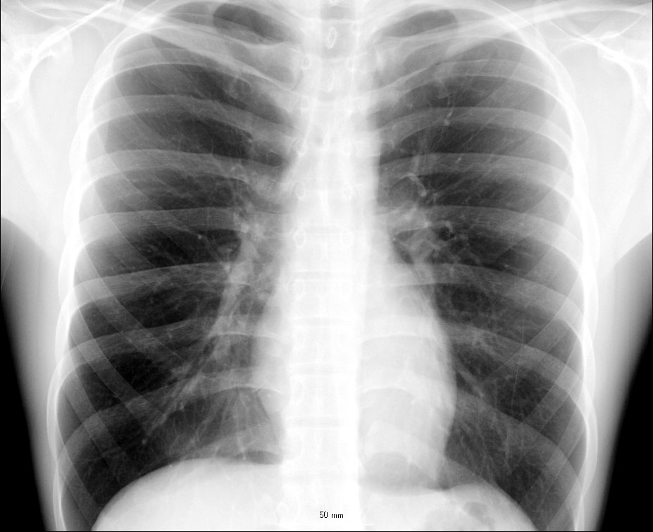
Images
[edit]-
 Volume rendering of a high resolution computed tomography of the thorax. The anterior thoracic wall, the airways and the pulmonary vessels anterior to the root of the lung have been digitally removed in order to visualize the different levels of the pulmonary circulation.
 -
 Thorax. Anterior view.
 -
 Thorax. Anterior view.
 -
 Clearly visible thorax of an artistic gymnast.


_0831.jpg)
Tetrapods
[edit]In mammals, the thorax is the region of the body formed by the sternum, the thoracic vertebrae, and the ribs. It extends from the neck to the diaphragm, and does not include the upper limbs. The heart and the lungs reside in the thoracic cavity, as well as many blood vessels. The inner organs are protected by the rib cage and the sternum. Thoracic vertebrae are also distinguished in birds, but not in reptiles.
Arthropods
[edit]

In insects, crustaceans, and the extinct trilobites, the thorax is one of the three main divisions of the creature's body, each of which is in turn composed of multiple segments. It is the area where the wings and legs attach in insects, or an area of multiple articulating plates in trilobites. In most insects, the thorax itself is composed of three segments; the prothorax, the mesothorax, and the metathorax. In extant insects, the prothorax never has wings, though legs are always present in adults; wings (when present) are restricted to at least the mesothorax, and typically also the metathorax, though the wings may be reduced or modified on either or both segments. In the apocritan Hymenoptera, the first abdominal segment is fused to the metathorax, where it forms a structure known as the propodeum. Accordingly, in these insects, the functional thorax is composed of four segments, and is therefore typically called the mesosoma to distinguish it from the "thorax" of other insects.
Each thoracic segment in an insect is further subdivided into various parts, the most significant of which are the dorsal portion (the notum), the lateral portion (the pleuron; one on each side), and the ventral portion (the sternum). In some insects, each of these parts is composed of one to several independent exoskeletal plates with membrane between them (called sclerites), though in many cases the sclerites are fused to various degrees.
See also
[edit]References
[edit]- ^ "Definition: Thorax". Oxford Learner's Dictionaries.
- ^ "thorax" at Dorland's Medical Dictionary
- ^ Thorax at the U.S. National Library of Medicine Medical Subject Headings (MeSH)
- ^ θώραξ, Henry George Liddell, Robert Scott, A Greek-English Lexicon, on Perseus Digital Library
- ^ "thorax, n.". Oxford English Dictionary. Oxford University Press.
- ^ Safarini, Omar A.; Bordoni, Bruno (2024), "Anatomy, Thorax, Ribs", StatPearls, Treasure Island (FL): StatPearls Publishing, PMID 30855912, retrieved 2024-08-21
- ^ Shahani, Rohit, MD. (2005). Penetrating Chest Trauma. eMedicine. Retrieved 2005-02-05.
- ^ Chest Diseases Retrieved on 2010-1-26
- ^ Atelectasis Lung and Airway Disorders. Retrieved on 2010-1-26
- ^ Pleurisy Lung Diseases. Retrieved on 2010-1-26
External links
[edit]- Sam Gon III. "A guide to the Orders of Trilobites". Retrieved August 23, 2005.
| International | |
|---|---|
| National | |
| Other | |
Thorax
View on GrokipediaGeneral Overview
Etymology
The term "thorax" derives from the Ancient Greek word θώραξ (thōrax), which denoted a "breastplate" or "cuirass," referring to the protective armor encasing the chest in warfare.[1] This metaphorical sense evoked the ribcage's role as a natural shield for vital organs.[11] The word was borrowed into Latin as thorax, retaining its dual connotations of armor and the chest itself, before spreading to European languages during the medieval and Renaissance periods.[12] In English, it first appeared in Middle English around 1400 as a medical term for the body's chest, transitioning from its armored imagery to anatomical precision.[1] This evolution culminated in the 16th century, when anatomists like Andreas Vesalius employed "thorax" in De humani corporis fabrica (1543) to describe the specific upper trunk region, solidifying its role in scientific nomenclature.[13] Cognates persist in modern Romance and Germanic languages, such as Italian torace, directly borrowed from Latin thōrax to mean the chest cavity, and German Thorax, adopted from Greek via Latin for the same anatomical structure.[14][15] These terms underscore the enduring legacy of the Greek origin in describing the protective thoracic enclosure.[1]Definition and Scope
The thorax is defined anatomically as the intermediate region of the trunk situated between the neck superiorly and the abdomen inferiorly, encompassing a cavity that houses vital organs essential for respiration and circulation.[2] In vertebrates, the boundaries of the thorax are precisely delineated: superiorly by the thoracic inlet at the level of the first thoracic vertebra (T1) and the root of the neck, inferiorly by the diaphragm which separates it from the abdominal cavity, posteriorly by the thoracic vertebrae, anteriorly by the sternum, and laterally by the ribs along with the overlying intercostal muscles that form the thoracic wall.[5][2] This configuration establishes the thorax as a protective enclosure for thoracic contents while facilitating mechanical functions such as breathing.[16] The scope of the thorax extends beyond human anatomy to broader taxonomic contexts, including its role as a protective structure in other vertebrates and as a locomotor tagma in arthropods (detailed in comparative anatomy).[2][17] Historically, anatomy texts from the Hellenistic era onward have emphasized the thorax's role as a central structure in body topography, with early dissections by figures like Herophilus revealing its internal compartments and vascular networks, influencing subsequent understandings of organismal architecture.[18]Anatomy of the Human Thorax
Skeletal Framework
The skeletal framework of the human thorax, also known as the thoracic cage, is primarily composed of the 12 thoracic vertebrae (T1–T12), 12 pairs of ribs, and the sternum.[2] The thoracic vertebrae form the posterior aspect, each characterized by a heart-shaped body, a circular vertebral foramen, and demifacets on the body and transverse processes for rib articulation.[19] The ribs are classified into three categories based on their anterior attachments: true ribs (pairs 1–7), which connect directly to the sternum via individual costal cartilages; false ribs (pairs 8–10), which connect indirectly to the sternum through shared costal cartilages; and floating ribs (pairs 11–12), which lack any anterior sternal connection and end freely in the musculature.[20] The sternum, a flat bone in the anterior midline, consists of three parts: the superior manubrium, the elongated body (mesosternum), and the inferior xiphoid process.[21] These bony elements articulate through specialized joints that provide stability and limited mobility to the thoracic cage. Posteriorly, the ribs articulate with the thoracic vertebrae through the costovertebral joints, which connect the head of each rib to the bodies of one or two adjacent vertebrae, and the costotransverse joints, which connect the tubercle of the rib to the transverse process of the corresponding vertebra (present in ribs 1–10). The costovertebral joints are synovial plane joints; for ribs 2–9, the head has two articular facets (superior and inferior) that articulate with the costal facets of two adjacent vertebrae, forming a single joint. Ribs 1, 10, 11, and 12 have only a single articular facet on the head, articulating with one vertebra.[20][22] The costochondral junctions unite the anterior ends of the ribs with their respective costal cartilages via synchondroses, allowing flexibility during respiratory movements.[6] Anteriorly, the sternocostal joints link the costal cartilages of the true ribs to the sternum, with the first seven ribs forming synovial or synchondrotic connections, while the false ribs articulate indirectly through cartilage.[6] Distinct anatomical features enhance the structural integrity and functionality of the thoracic skeleton. Each rib exhibits a characteristic curvature, with the angle of the rib marking the site of greatest posterior bend, and superior and inferior angles defining the curved borders that accommodate muscle attachments and protect underlying structures.[23] The thoracic vertebrae feature long, slender spinous processes that project downward and overlap, contributing to the posterior reinforcement of the cage. On the sternum, the suprasternal (jugular) notch at the superior margin of the manubrium serves as a palpable landmark between the clavicles.[5] Developmentally, the thoracic skeleton arises through endochondral ossification from mesenchymal precursors during fetal life. The ribs begin to ossify from primary centers near their angles starting in the seventh fetal week, progressing to secondary centers at the head, tubercle, and sternal end postnatally.[25] Ossification centers for the thoracic vertebrae emerge around the tenth to eleventh fetal week, initially in the neural arches of the upper thoracic region and vertebral bodies of the lower thoracic area, with fusion occurring progressively into childhood.[26] The sternum develops from paired sternal bars that fuse by the ninth fetal week, with initial ossification centers appearing in the manubrium and upper body around 19 weeks' gestation, followed by sternebrae in the body and xiphoid process later in fetal and postnatal periods.[27]Muscular Framework
The muscular framework of the human thorax consists of layered skeletal muscles that form the flexible thoracic wall, providing structural support, elasticity, and dynamic stability to enclose and protect vital organs. These muscles are arranged in superficial, intermediate, and deep layers, primarily comprising the intercostal muscles that span the interspaces between the 12 pairs of ribs. The diaphragm serves as the inferior muscular boundary, separating the thoracic and abdominal cavities. Accessory muscles such as the pectoralis major and minor, serratus anterior, and latissimus dorsi contribute to the anterolateral and posterolateral aspects of the thoracic wall, enhancing its integrity through attachments to the bony framework.[28] The external intercostal muscles form the most superficial layer of the thoracic wall, consisting of 11 thin, digitated sheets located between the ribs from the first to the eleventh. Each external intercostal originates from the lower border of a rib and inserts onto the upper border of the rib immediately below, with fibers running obliquely downward and forward; they attach to the costal grooves, tubercles, and angles of the ribs. These muscles elevate the ribs during contraction, contributing to thoracic expansion. The internal intercostal muscles lie deep to the external layer, originating from the lower border of a rib and inserting onto the upper border of the rib below, but with fibers oriented obliquely downward and backward; they depress the ribs, aiding in thoracic compression. The innermost intercostal muscles represent the deepest layer, similar in orientation and attachments to the internal intercostals but separated by a neurovascular bundle; they are incomplete posteriorly and absent anteriorly near the sternum.[29][28][30] Accessory muscles reinforce the thoracic wall's anterolateral and posterior surfaces. The pectoralis major, a thick fan-shaped muscle, originates from the medial half of the clavicle, sternum, and costal cartilages of ribs 1–6, inserting into the lateral lip of the intertubercular sulcus of the humerus; it overlies the anterior thoracic wall. The pectoralis minor, triangular and beneath the major, arises from the third to fifth ribs near their costal cartilages and inserts onto the coracoid process of the scapula. The serratus anterior originates from the upper borders of ribs 1–8 or 9 along their lateral surfaces and inserts along the medial border and superior angle of the scapula, forming part of the lateral thoracic wall. The latissimus dorsi, a broad flat muscle, originates from the spinous processes of thoracic vertebrae T7–T12, thoracolumbar fascia, iliac crest, and inferior ribs 8–12, inserting into the floor of the intertubercular sulcus of the humerus; it covers the posterior thoracic wall. The diaphragm, as the dome-shaped inferior boundary, originates peripherally from the xiphoid process of the sternum, costal cartilages of ribs 7–12, and the lateral arcuate ligaments bridging the lumbar vertebrae, converging to insert into a central tendon.[31][32][33][29][34] Innervation of the thoracic muscles derives primarily from thoracic spinal nerves. The intercostal muscles (external, internal, and innermost) are supplied by the intercostal nerves, which are the anterior rami of spinal nerves T1–T11, running in the costal groove between the internal and innermost layers. The accessory muscles receive innervation from branches of the brachial plexus: pectoralis major via lateral and medial pectoral nerves (C5–T1), pectoralis minor via medial pectoral nerve (C8–T1), serratus anterior via long thoracic nerve (C5–C7), and latissimus dorsi via thoracodorsal nerve (C6–C8). The diaphragm is uniquely innervated by the phrenic nerve, arising from C3–C5 spinal segments and descending through the thorax to supply both motor and sensory functions to the muscle. These neural supplies ensure coordinated activation for maintaining thoracic wall integrity.[35][28][33][32][34]Internal Contents
The internal contents of the human thorax are organized into distinct compartments that house vital organs, vessels, nerves, and lymphatic structures, all enclosed within the protective skeletal framework of the rib cage and sternum. The thoracic cavity is primarily divided into the two bilateral pleural cavities, which contain the lungs, and the central mediastinum, a midline compartment extending from the thoracic inlet to the diaphragm.[2] The pleural cavities are paired potential spaces, each lined by a serous membrane consisting of visceral pleura covering the lung surface and parietal pleura adhering to the thoracic wall, with a thin layer of serous fluid maintaining lubrication between them. The left and right lungs occupy these cavities, with each lung extending superiorly from its apex, which projects above the level of the first rib into the root of the neck, to its base, which rests upon the diaphragm. The lungs are separated by the mediastinum and are anchored at their hila, where bronchi, vessels, and nerves enter.[36][37][38] The mediastinum, situated between the pleural cavities, is subdivided into the superior mediastinum above the plane from the sternal angle to the intervertebral disc between the fourth and fifth thoracic vertebrae, and the inferior mediastinum below, which further divides into anterior, middle, and posterior compartments. The anterior mediastinum, the smallest division, lies anterior to the pericardium and contains the thymus gland, loose connective tissue, lymph nodes, and small vessels. The middle mediastinum encompasses the heart, enclosed within the pericardial sac—a double-layered serous membrane with a potential space filled with serous fluid—and includes the ascending aorta, superior vena cava (SVC), and portions of the trachea and main bronchi. The posterior mediastinum, located behind the pericardium and in front of the vertebral column, houses the descending thoracic aorta, esophagus, thoracic duct, vagus nerves, and the sympathetic chain.[39][40][41][42] Major vascular elements within the thorax include the aorta, which arches from the heart in the middle mediastinum before descending in the posterior mediastinum, and the SVC, which drains venous blood into the right atrium from the upper body. The lymphatic system is represented by numerous lymph nodes scattered throughout the mediastinum, particularly in the anterior and posterior divisions, and the thoracic duct, the main lymphatic vessel that ascends in the posterior mediastinum to drain lymph from the lower body and left upper body into the venous system at the junction of the left internal jugular and subclavian veins. Neural structures include the bilateral vagus nerves (cranial nerve X), which descend through the superior and posterior mediastinum providing parasympathetic innervation, and the sympathetic chain, a paired autonomic structure running along the vertebral column in the posterior mediastinum.[39][43][2]Surface Anatomy and Landmarks
The surface anatomy of the human thorax encompasses the external features and palpable structures that provide essential orientation for clinical examination, imaging, and procedural guidance. These landmarks are primarily derived from the underlying skeletal framework, including the sternum, clavicles, and ribs, allowing for reliable identification of intercostal spaces and regional boundaries.[44] Prominent anterior landmarks include the suprasternal notch, also known as the jugular notch, which is a palpable depression at the superior border of the manubrium sterni, situated between the medial ends of the clavicles and corresponding to the level of the second thoracic vertebra. Immediately inferior to this is the sternal angle, or angle of Louis, formed by the junction of the manubrium and the body of the sternum; this readily palpable ridge lies at the level of the fourth thoracic vertebra (T4) anteriorly and the disc between T4 and T5 posteriorly, serving as a critical reference for counting ribs and identifying the second intercostal space. The clavicles form the superior lateral boundaries, easily palpated as curved bony ridges extending laterally from the sternum to the shoulders. Inferiorly, the costal margins are formed by the articulated lower borders of ribs 7 through 10, converging toward the midline at the xiphoid process, the cartilaginous or bony tip of the sternum located at the level of the ninth thoracic vertebra.[45][45][46] Vertical reference lines further delineate the thorax for topographic description. The midclavicular line (also called the mammary line) runs vertically from the midpoint of each clavicle, passing through the nipple in males and approximately the fourth intercostal space in females. The midaxillary line descends through the midpoint of the axilla, while the scapular line is a posterior vertical line drawn through the inferior angle of the scapula when the arms are at rest. These lines divide the thorax into regions, including the sternal region over the sternum, the mammary region encompassing the anterior chest over the breasts (bounded laterally by the midclavicular lines), and the axillary regions flanking the lateral chest walls.[44][47][31] Palpation of these landmarks facilitates clinical assessment, such as tracing the sternal angle to locate intercostal spaces for needle insertion or percussion. Auscultation points are standardized relative to these features: for cardiac sounds, the aortic area is at the second right intercostal space along the sternal border, the pulmonic area at the second left intercostal space, the tricuspid area at the fourth left intercostal space near the sternum, and the mitral (apical) area at the fifth left intercostal space in the midclavicular line. Lung fields for respiratory auscultation extend across the anterior and posterior thorax, bounded superiorly by the clavicles, inferiorly by the costal margins (approximately the sixth rib anteriorly and eighth rib posteriorly), and laterally by the midaxillary line, with symmetric assessment from apex to base.[48][48] Anatomical variations in thoracic surface landmarks can influence clinical interpretation. The normal slight leftward asymmetry arises from the heart's position in the left hemithorax, displacing the apical impulse laterally. In rare cases, such as situs inversus totalis, a congenital mirror-image transposition of thoracic organs reverses this asymmetry, with the heart positioned in the right hemithorax and landmarks appearing inverted on the right side.[49]Physiology and Function of the Human Thorax
Respiratory Mechanics
The respiratory mechanics of the human thorax facilitate breathing through coordinated changes in thoracic volume and pressure gradients. During inspiration, the primary process involves the contraction of the diaphragm, which descends and flattens, increasing the vertical dimension of the thoracic cavity. Simultaneously, the external intercostal muscles contract, elevating the ribs and expanding the anteroposterior and transverse dimensions of the thorax, thereby increasing overall thoracic volume. This volume expansion creates a subatmospheric pressure within the lungs, drawing air inward, with the intrapleural pressure typically measuring around -5 cmH₂O at rest to maintain lung expansion against the chest wall.[50][51][52] Expiration contrasts with inspiration by relying primarily on passive mechanisms during quiet breathing. As the inspiratory muscles relax, the elastic recoil of the lungs and chest wall reduces thoracic volume, elevating intrapleural pressure and expelling air without active muscular effort. In forced expiration, such as during exercise or coughing, accessory muscles including the abdominal muscles and internal intercostals contract to further compress the thorax, accelerating the process and increasing expiratory flow rates.[51][53][54] The thorax expands through specific rib motions that enhance volume changes. Upper ribs (ribs 1-5) exhibit a pump-handle movement, where elevation lifts the sternum anteriorly, increasing the anteroposterior diameter. Lower ribs (ribs 6-10) demonstrate a bucket-handle motion, rotating laterally to widen the transverse diameter, with floating ribs (11-12) contributing minimally. These movements, driven by intercostal and other respiratory muscles attached to the rib cage, collectively optimize thoracic capacity during inhalation.[55][56] Lung-thorax compliance refers to the ease with which the combined system distends under pressure, characterized by a compliance curve that plots volume changes against transpulmonary pressure. In healthy adults, total respiratory compliance is approximately 100-200 mL/cmH₂O, reflecting the balance between lung elasticity and chest wall stiffness; reduced compliance, as in fibrosis, requires greater pressure for equivalent volume shifts, while increased compliance, as in emphysema, allows easier expansion but impairs recoil. Airway resistance, influenced by bronchial diameter and flow rates, complements compliance by opposing airflow, with total resistance typically 1-2 cmH₂O/L/s during normal breathing. These properties ensure efficient gas exchange without excessive energy expenditure.[57][58]Protective and Circulatory Roles
The thorax serves a critical protective role for vital organs, primarily through its bony framework comprising the rib cage, sternum, and thoracic vertebrae. The rib cage, formed by 12 pairs of ribs articulating with the thoracic vertebrae posteriorly and the sternum anteriorly, encases and shields the heart and lungs from external trauma. This structure acts as a semi-rigid barrier that dissipates impact forces, with the costal cartilages providing flexibility to absorb shocks during physical activities or injury.[50][59] The sternum and vertebrae further reinforce this protection, forming an anterior and posterior wall that safeguards the mediastinal contents, including the heart and major vessels, while allowing limited expansion for physiological functions.[60][61] In addition to protection, the thorax plays an essential role in supporting circulation by housing the heart and great vessels within the mediastinum. The heart, positioned centrally and slightly leftward, pumps blood through the aorta, pulmonary trunk, superior and inferior vena cavae, and pulmonary veins, all of which are anchored and protected by the thoracic structures.[62][63] Venous return to the heart is augmented by the thoracic pump mechanism, where intrathoracic pressure changes during respiration facilitate blood flow from the systemic veins into the right atrium, enhancing overall cardiac output without relying solely on skeletal muscle pumps.[64][65] The lymphatic system within the thorax contributes to circulatory homeostasis via the thoracic duct, the largest lymphatic vessel, which originates in the cisterna chyli and ascends through the mediastinum to empty lymph into the venous circulation at the junction of the left internal jugular and subclavian veins. This duct drains approximately 75% of the body's lymph, including fluid from the lower limbs, abdomen, and left thorax, returning it to the bloodstream to maintain fluid balance and immune surveillance.[66][67] Furthermore, the thorax provides postural stability, functioning as a central pillar that aligns the spine and supports upright posture. The thoracic vertebrae and rib cage create a stable scaffold that distributes the weight of the upper body, maintaining the natural kyphotic curve of the thoracic spine and preventing excessive lordosis or scoliosis through muscular and ligamentous attachments.[68][69] This structural integrity ensures efficient load transfer from the head and shoulders to the pelvis, contributing to overall balance and mobility.[70]Clinical Significance of the Human Thorax
Trauma and Injury
Trauma to the human thorax encompasses a range of injuries primarily resulting from blunt or penetrating forces, which can compromise the skeletal framework and underlying structures such as the lungs, heart, and major vessels. Blunt thoracic trauma is the most prevalent form, often occurring in high-energy events like motor vehicle collisions (MVCs), and accounts for approximately 90% of chest injuries, with rib fractures being the most common manifestation. These fractures typically involve 1-3 ribs in elderly patients due to osteoporosis-related bone fragility, though multiple fractures can occur across age groups. In MVCs, rib fractures are reported in 10-20% of cases, serving as a marker for potential severe associated injuries.[71][72][73][74] A severe variant of blunt injury is flail chest, characterized by fractures of three or more consecutive ribs at two or more sites each, leading to a free-floating segment of the chest wall that exhibits paradoxical inward movement during inspiration. This instability disrupts normal respiratory mechanics by allowing the flail segment to move independently of the rest of the thorax. Penetrating thoracic trauma, comprising about 10% of cases, typically arises from stab or gunshot wounds that directly violate the chest wall and may involve the pleura or mediastinum, with gunshots causing more extensive tissue disruption due to cavitation and fragmentation compared to low-velocity stabs.[75][76] Mechanisms of thoracic injury include deceleration forces, which shear the aorta at fixed points like the ligamentum arteriosum, potentially resulting in transection, and are common in rapid stops during MVCs or falls. Compression mechanisms, such as direct anterior chest impact from steering wheels or falls, frequently produce sternal fractures by deforming the rigid sternum against the vertebral column. These injuries highlight the thorax's role in protecting vital organs, as referenced in its anatomical framework.[77][78][79][80] Immediate complications from these injuries include hemothorax, often from lacerated intercostal vessels in rib fractures, occurring in up to 24% of blunt chest trauma cases and leading to significant blood accumulation in the pleural space. Pulmonary contusions, bruising of lung parenchyma from shear forces, affect 30-75% of blunt thoracic trauma patients and impair gas exchange. Cardiac contusions, involving myocardial bruising, arise in blunt trauma scenarios and can cause arrhythmias or reduced contractility, with incidence tied to the proximity of impact to the heart.[71][81][82][83]Pain and Sensory Aspects
Pain in the thorax arises from diverse sources, categorized as musculoskeletal, visceral, or neuropathic, each with distinct characteristics and implications for clinical evaluation. Musculoskeletal pain commonly originates from costochondritis or Tietze syndrome, involving inflammation of the costal cartilages connecting the ribs to the sternum. Costochondritis typically presents as localized sharp or dull pain in the upper anterior chest wall, reproducible by palpation at the costochondral junctions (often the second to fifth ribs), and worsened by deep breathing, coughing, or upper extremity movements.[84] Tietze syndrome shares similar pain features but is distinguished by associated swelling at the affected site.[85] Visceral pain in the thorax frequently stems from cardiac or pleural conditions, such as angina pectoris due to myocardial ischemia or pleuritis from parietal pleural inflammation. Angina manifests as a sensation of pressure, tightness, or heaviness in the chest, often lasting several minutes and relieved by rest or nitroglycerin in stable cases.[86] Pleuritis, in contrast, produces sharp, stabbing pain localized to the chest, neck, or shoulder, intensified by respiratory movements like inhalation, coughing, or sneezing due to friction between inflamed pleural layers.[87] Neuropathic pain, exemplified by intercostal neuralgia, results from irritation or damage to the intercostal nerves and is characterized by burning, stabbing, or radiating sensations following a dermatomal band-like distribution along the ribs, chest, or abdomen. This pain may include paresthesia or allodynia and is commonly exacerbated by trunk movements, coughing, or sneezing.[85] Referred pain is a prominent feature of thoracic disorders, occurring when visceral afferents converge with somatic pathways in the spinal cord, leading to pain perception in distant sites. Cardiac ischemia often refers dull, aching pain to the left arm, shoulder, or jaw through sympathetic fibers entering the spinal cord at T1-T4 dermatomes.[88] Gallbladder pathology, such as cholecystitis, can similarly refer pain to the right shoulder via irritation of the phrenic nerve, which shares C3-C5 roots with shoulder innervation.[89] Sensory innervation of the thoracic region is mediated primarily by the intercostal nerves (T1-T11), which provide somatic sensory input to the skin, intercostal muscles, and parietal pleura along the lateral and anterior chest wall.[35] The phrenic nerve, arising from C3-C5 roots, supplies sensory fibers to the central diaphragm, pericardium, and mediastinal pleura; diaphragmatic irritation thus often results in referred pain to the ipsilateral shoulder rather than the abdomen.[90] Diagnosis of thoracic pain emphasizes a thorough patient history to differentiate pain quality and triggers, such as sharp, breath-aggravated pain suggesting pleural or musculoskeletal etiologies versus dull, exertional discomfort pointing to visceral cardiac sources. Physical examination, including palpation of surface landmarks like the sternal border and intercostal spaces, aids in localizing and reproducing symptoms for targeted assessment.[91]Atelectasis
Atelectasis refers to the partial or complete collapse of lung tissue within the thorax, resulting in diminished lung volume and impaired gas exchange. This condition primarily affects the alveoli, the tiny air sacs responsible for oxygen and carbon dioxide transfer, and is a common postoperative complication that disrupts normal thoracic respiratory function.[92][93] The condition is classified into several types based on underlying mechanisms. Resorption atelectasis, also known as obstructive atelectasis, occurs when an airway blockage—such as a mucus plug, tumor, or foreign body—prevents air from reaching the alveoli, leading to gradual absorption of trapped gas by surrounding blood flow. Compression atelectasis arises from external pressure on the lung, often due to accumulations like pleural effusion, pneumothorax, or abdominal distension that compress the thoracic cavity. Passive atelectasis, frequently observed after surgery, results from reduced surfactant production or shallow breathing, causing the lung to deflate without obstruction or compression.[94][95][96] Pathophysiologically, atelectasis involves the collapse of alveoli, which reduces ventilation while perfusion continues, creating intrapulmonary shunting where deoxygenated blood bypasses functional lung units. This leads to ventilation-perfusion (V/Q) mismatch, hypoxemia, and potential hypercapnia if extensive. Risk factors include general anesthesia, which promotes atelectasis through loss of diaphragmatic tone, reduced functional residual capacity, and impaired mucociliary clearance; the incidence reaches approximately 90% in patients undergoing general anesthesia. Other contributors encompass immobility, smoking, and underlying lung diseases that exacerbate alveolar instability.[92][93][94] In the thorax, atelectasis alters pleural space dynamics by reducing negative intrapleural pressure on the affected side, potentially causing compensatory overexpansion of the contralateral lung or mediastinal shift toward the collapsed area in severe cases. This shift can strain thoracic structures, including the heart and great vessels, though it is typically subtle unless the collapse is lobar or greater.[92][93] Diagnosis relies on clinical symptoms and imaging. Patients often present with dyspnea, tachypnea, or cough, though pain is minimal unless associated with underlying causes; asymptomatic cases are common in mild postoperative scenarios. Chest X-ray (CXR) is the primary diagnostic tool, revealing signs of volume loss such as opacification of the affected lung, elevation of the hemidiaphragm, crowding of ribs, or tracheal deviation.[95][94][96]Pneumothorax
Pneumothorax refers to the abnormal accumulation of air in the pleural space, the potential space between the visceral and parietal pleura surrounding the lungs, leading to partial or complete lung collapse.[97] It is classified into three main types based on etiology: spontaneous, traumatic, and tension pneumothorax, with the latter representing a severe subset often arising from the others.[98] Spontaneous pneumothorax occurs without apparent trauma and is subdivided into primary (in individuals without underlying lung disease) and secondary (in those with preexisting pulmonary conditions).[99] Traumatic pneumothorax results from direct chest injury, while tension pneumothorax involves progressive air buildup under pressure, creating a one-way valve effect.[100] Primary spontaneous pneumothorax predominantly affects tall, thin young males, typically between 15 and 34 years old, with an annual incidence of 7.4 to 18 cases per 100,000 males and 1.2 to 6 cases per 100,000 females.[101] Secondary spontaneous pneumothorax is more common in patients with chronic obstructive pulmonary disease (COPD), cystic fibrosis, or other interstitial lung diseases, where the incidence can reach 12.10 per 10,000 person-years in COPD patients compared to 6.68 per 10,000 in those without.[102] Traumatic pneumothorax arises from blunt or penetrating injuries, such as rib fractures from motor vehicle accidents or iatrogenic causes like central line placement or thoracentesis.[98] Tension pneumothorax develops when air enters the pleural space but cannot escape, often complicating traumatic or iatrogenic cases, leading to mediastinal shift, tracheal deviation, and hemodynamic instability.[100] The pathophysiology involves disruption of the visceral pleura, allowing air to enter the pleural space and equalize the normally negative intrapleural pressure with atmospheric pressure, resulting in lung collapse and impaired ventilation.[97] In the thorax, this disrupts respiratory mechanics by reducing lung compliance and vital capacity, causing respiratory distress, hypoxemia, and, in tension cases, compression of the great vessels that decreases venous return to the heart and precipitates cardiovascular collapse.[98] The overall incidence of pneumothorax varies by type but is estimated at 18 to 28 cases per 100,000 males annually for spontaneous forms, with higher rates in at-risk populations.[103] Management depends on the type, size, and clinical stability. Small, asymptomatic primary spontaneous pneumothoraces may be observed with supplemental oxygen to promote air resorption, while larger or symptomatic cases require chest tube thoracostomy to evacuate air and re-expand the lung.[98] Tension pneumothorax demands immediate needle decompression in the second intercostal space at the midclavicular line to relieve pressure, followed by definitive chest tube insertion.[100] Traumatic pneumothoraces often necessitate similar drainage, with surgical intervention like video-assisted thoracoscopic surgery considered for recurrent or persistent cases, particularly in secondary spontaneous pneumothorax associated with COPD.[97]Comparative Anatomy
In Tetrapods
The thorax in tetrapods, four-limbed vertebrates that emerged during the Late Devonian period approximately 375 million years ago, represents a key evolutionary adaptation from the pectoral girdle and fin structures of lobe-finned fishes, enabling terrestrial locomotion and enhanced respiratory efficiency.[104][105] This transition involved the development of a robust rib cage that encloses vital organs such as the lungs and heart, providing structural support and protection while facilitating the shift from aquatic to aerial respiration.[106] In basal tetrapods like early amphibians, the thorax features loosely articulated ribs without a bony sternum, allowing flexibility for movement in semi-aquatic environments but limiting rigid enclosure of the thoracic cavity.[107] This contrasts with more derived reptiles, where the rib cage becomes increasingly ossified; for instance, in turtles, the dorsal ribs fuse with dermal plates to form the carapace, while the ventral plastron provides a bony shield, creating a highly armored thoracic enclosure that immobilizes the ribs but enhances protection of internal organs.[108] Among amniotes, mammals exhibit a fully developed sternum connected to true ribs, forming a dynamic cage that supports the diaphragm—a muscular partition unique to this group that enables negative-pressure breathing by expanding the thoracic volume during inspiration.[109] Birds, as sauropsid amniotes, possess a prominent keel on the sternum for anchoring powerful flight muscles, with an extensive system of air sacs that extend throughout the thorax and connect to the lungs, optimizing unidirectional airflow for high metabolic demands during flight.[110][111] Functionally, the tetrapod thorax primarily serves to protect thoracic viscera from mechanical damage, with ribs acting as a skeletal framework that evolved initially for locomotor support before being co-opted for respiratory roles in later lineages.[106] In respiration, amphibians rely on buccal pumping, where the mouth and throat generate positive pressure to force air into the lungs, as their loose thoracic structure precludes significant rib movement.[107] In contrast, amniotes employ thoracic aspiration, utilizing rib cage expansion and associated musculature—such as the diaphragm in mammals or costal movements in reptiles and birds—to create negative intrathoracic pressure for efficient lung ventilation, a adaptation that supported fully terrestrial lifestyles.[112] This mammalian mechanism is exemplified in humans, where the diaphragm's contraction lowers intrathoracic pressure to draw air into the lungs.[109]In Arthropods
In arthropods, the thorax represents a distinct tagma, or functional body region, situated between the head and abdomen, primarily specialized for locomotion through jointed appendages. This segmentation reflects the phylum's characteristic tagmosis, where ancestral body segments fuse or specialize for specific roles. The thoracic exoskeleton consists of chitinous plates, including dorsal tergites and ventral sternites, which form rigid sclerites providing structural support, protection, and sites for muscle attachment. These plates are often interconnected by flexible membranes, allowing movement while maintaining the body's integrity. Among major arthropod groups, thoracic structure varies significantly. In insects, the thorax comprises three segments—the prothorax, mesothorax, and metathorax—each bearing a pair of legs, with the mesothorax and metathorax additionally supporting wings in pterygote species for flight. Crustaceans typically feature a thorax with 8 segments, from which arise maxillipeds for feeding and pereopods for walking or swimming, often covered by a carapace that may fuse with the head to form a cephalothorax. In arachnids, the thorax fuses with the head into the prosoma, a consolidated unit bearing four pairs of walking legs, pedipalps, and chelicerae adapted for various functions like prey capture. Myriapods, such as centipedes and millipedes, lack a clearly defined thorax; instead, their elongated trunk consists of numerous leg-bearing segments following the head, with diplopods (millipedes) having two pairs of legs per segment and chilopods (centipedes) one pair for predatory locomotion. Internally, the arthropod thorax accommodates powerful muscles that drive appendage movement, with indirect flight muscles in insects deforming the exoskeleton to power wingbeats. The circulatory system is open, featuring a dorsal tubular heart typically positioned along the midline of the thorax and abdomen, which pumps hemolymph into the hemocoel cavity for nutrient and oxygen distribution before its return via ostia. Respiratory structures vary: terrestrial forms like insects and myriapods rely on tracheae—branched air-filled tubes entering through thoracic spiracles to deliver oxygen directly to tissues—while aquatic crustaceans utilize gills on thoracic appendages for gas exchange, and some arachnids employ book lungs or tracheae within the prosoma. Evolutionary adaptations in the thorax highlight arthropod diversity, with segment fusion enhancing efficiency in locomotion and sensory integration, as seen in the cephalothorax of chelicerates and crustaceans, contrasting with the more segmented myriapod form. In insects, thoracic tagmosis has facilitated the evolution of powered flight, a key factor in their ecological success.References
- https://en.wiktionary.org/wiki/thorax
- https://en.wiktionary.org/wiki/torace
- https://en.wiktionary.org/wiki/Thorax#German
- https://anatomylab.class.[virginia](/page/Virginia).edu/MSI/Lab01/PowerPointHandout_ExtrinsicBack_VertOsteology_Radiology.pdf